General information about what nephrosclerosis is
The kidney is a paired organ responsible for filtering and removing excess fluid and waste from the body. blood clots are dissolved .
Nephrosclerosis is a chronic disease characterized by the replacement of renal tissue (parenchyma) with connective tissue that is unable to perform all basic functions.
With nephrosclerosis, the walls of arterioles and arteries thicken, which negatively affects the blood supply to the kidneys. With prolonged lack of nutrition, the death of the tubules and glomeruli begins, which are responsible for the formation of urine, which leads to the formation of renal failure .
The outcome of the disease can be very different and directly depends on how timely the diagnosis was made and how timely and correctly the treatment was started. With active therapy in the early stages, recovery occurs. In advanced cases, deaths occur. A synonym for nephrosclerosis is a wrinkled kidney .
Mechanism of development of kidney nephrosclerosis
| GreenFlames09 Flickr |
Nephrosclerosis is not an independent disease and, as a rule, it is provoked by other pathologies.
But, despite the cause of development, nephrosclerosis has a common pathogenesis. Under the influence of provoking factors, destruction of the renal epithelium occurs with damage to some nephrons. Since the cells of the organ are not able to regenerate, the damaged areas are replaced by connective fibers, and a gradual compaction of the organ occurs.
In unaffected areas, filtration and blood circulation increase, as a result of which the volume of urine excreted increases, but its density decreases. Due to disruption of blood flow that regulates glomerular filtration, arterial hypertension develops or worsens.
Pathogenesis
Long-term exposure to negative factors leads to persistent vasospasm and impaired trophism. This leads to irritation of histocytic elements, and as a consequence, to local or diffuse proliferation of connective tissue, which cannot perform the functions of the renal system.
The affected buds first become denser, and then shrink and decrease in volume. As the connective tissue grows, it compresses the tubules and glomeruli, causing their atrophy. Part of the glomerular apparatus leaves the systemic circulation, and capillaries in the affected areas cease to function. As a result of impaired blood flow, the formation and release of angiotensin and renin into the blood increases, which leads first to hypertension , and then to hypertrophy of the heart chambers . As the process progresses, the death of tubules and glomeruli is noted.
Causes
Nephrosclerosis is a complication of diseases such as arterial hypertension , glomerulonephritis , diabetes mellitus , gradually leading to the development of chronic kidney disease.
It is customary to highlight:
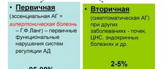
- primary (hypertensive) nephrosclerosis;
- secondary nephrosclerosis.
The primary form (primarily wrinkled kidney) develops against the background of a pathology that is not related to the functioning of the renal system, but disrupts normal blood circulation in the kidney. A primary wrinkled kidney can be caused by:
- hypertonic disease;
- thrombosis;
- atherosclerosis.
Types of primary nephrosclerosis
- Atherosclerotic nephrosclerosis . When atherosclerotic plaques form on the walls of renal vessels, blood flow decreases, which leads to the death of renal tissue. The characteristic localization of atherosclerotic plaques is the branching of the renal artery. This type of nephrosclerosis is considered the safest compared to others, because it is characterized by only partial kidney damage. The left kidney is most often affected; bilateral nephrosclerosis .
- Hypertensive nephrosclerosis . With constantly elevated blood pressure , blood vessels lose their elasticity and there is a tendency to spasm. With insufficient oxygen and nutrition, cell death occurs. Types of hypertensive nephrosclerosis: arteriolonecrotic - observed in malignant hypertension and leads to the death of capillaries and arterioles; arterilosclerotic – there is an excessive proliferation of connective tissue on the vascular wall.
- Involutive nephrosclerosis . Changes are formed as a result of involutional, age-related changes upon reaching a certain age. After 45-50 years, more calcium is deposited on the walls of veins and arteries, the number of muscle fibers increases, which negatively affects blood circulation. The cause of the pathology may be atrophy of the elements of the urinary tubules, which also manifests itself with age.
- Diabetic nephrosclerosis . A complication of diabetes mellitus is necrotization of the kidney tissue, which prevents the organ from fully functioning and doing its job. Patients note swelling and increased blood pressure. Deviations in the general blood test are recorded.
- Infarction nephrosclerosis . It is formed as a result of blockage of the renal artery by an embolus or thrombus, which causes necrosis of the feeding area of the renal tissue.
- Venous nephrosclerosis . With venous congestion of the kidneys, blood flow becomes difficult, venous stagnation is formed, which creates optimal conditions for an increase in the number of tropocollagen molecules.
Secondary nephrosclerosis (secondary wrinkled kidney) is formed as a result of the progression of pathology of the renal system.
Diseases that provoke the development of a secondary wrinkled kidney:
- Glomerulonephritis . In inflammatory diseases of the throat (pharyngitis, sore throat , laryngitis ), hyaline is deposited on the vessels of the kidneys, impairing their elasticity.
- Nephritis , pyelitis , pyelonephritis . Inflammatory disease of the kidney tissue or parts of the kidney. Infectious agents penetrate the tubules and glomeruli, affecting the parenchyma and forming blood clots.
- Systemic lupus erythematosus . A disease in which the body produces antibodies against its own organs and systems, damaging the structure and disrupting their functioning.
- Nephropathy of pregnancy. A specific change in hormonal levels leads to instability of blood pressure, vasospasm, and fluid retention in the body.
- Nephrolithiasis. Concretions and stones are most often found in the right kidney.
- Tuberculosis . The tuberculosis bacillus, circulating throughout the body, affects the renal system.
- Diabetes . High sugar levels in uncontrolled diabetes negatively affect the condition of the vascular wall.
- Traumatic injury to renal tissue . Blockage of blood vessels occurs as a result of the detachment of particles of parenchyma, which negatively affects the blood circulation of the organ and leads to the death of nephrons and tubules.
- Ionizing radiation . It negatively affects the functioning of the entire body, including the renal system.
Drug therapy for kidney damage caused by disorders of purine metabolism
Purine metabolism is a complex cascade of biochemical reactions in which many enzyme systems take part. The content of purines in the body consists of their intake from food and endogenous synthesis. Most of the salts of uric acid - urates - are formed endogenously during the metabolism of nucleic acids, but there are other ways of biosynthesis of these substances. In all variants, the most important intermediate is inosinic acid, which subsequently undergoes hydrolysis. The resulting hypoxanthine is converted into xanthine and uric acid under the influence of the enzyme xanthine oxidase. From a biochemical point of view, disorders of purine metabolism represent various types of imbalance between the enzyme systems responsible for the synthesis and transport of uric acid and its precursors. The intake of a significant amount of purines from food is also essential.
It is believed that the body of an adult healthy person contains about 1000 mg of uric acid. If purine metabolism is impaired, this figure can increase several times. The content of uric acid in the body is not a rigid parameter and does not have any diagnostic value. Even the main indicator of the state of purine metabolism - the concentration of uric acid in the blood serum is not particularly harsh. The minimum and maximum normal values differ by approximately 2.5 times - 200–450 µmol/l in men and 160–400 µmol/day in women. In healthy people, approximately 750 mg or 2/3 of the total volume of uric acid is excreted and synthesized again per day. Of this amount, about 80% or 600 mg is excreted by the kidneys. The remaining 20% is excreted through the gastrointestinal tract. According to P. M. Klimenko et al. (2010) the normal clearance of uric acid is 5.4–9.0 ml/min [1].
Renal excretion of urate is a complex and multistep process. Filtration of plasma urate occurs in the glomeruli. The urates that enter the ultrafiltrate are almost completely reabsorbed in the proximal tubule and then secreted into the lumen of the nephron. Some of the secreted urate is reabsorbed. The process of active secretion of urates is very sensitive to various chemical agents. It is believed that renal secretion of urates is increased by orotic acid, losartan, estrogens, and tetracycline breakdown products (expired tetracyclines are highly toxic!); renal excretion of urate is reduced by ethambutol, thiazides and thiazide-like diuretics, and to a lesser extent by furosemide and acetazolamide [2]. It is quite obvious that the severity of the observed effects varies greatly from drug to drug and does not always have clinical application. In particular, the uricosuric properties of estrogens are not significant. Losartan has recently begun to appear in treatment regimens for gouty tubulointerstitial nephritis in patients who do not have nephrolithiasis [3]. The tendency of thiazides and indapamide to reduce the renal excretion of urates and increase their serum concentration is quite pronounced, which makes these drugs at least undesirable for articular gout and, especially, for gouty nephropathy.
Clinical variants of kidney damage due to impaired purine metabolism
Diseases associated with disorders of purine metabolism are relatively common, which makes issues related to their treatment relevant. Urology specialists, as well as most general practitioners, are well aware of the features of urate nephrolithiasis. At the same time, these specialists often have no idea at all about the existence of other, sometimes more serious, diseases caused by disorders of purine metabolism. Meanwhile, they all occur with varying frequency in hospitals, as well as in the provision of outpatient medical care.
The most significant consequence of disorders of purine metabolism is an increase in the level of uric acid in the blood - hyperuricemia, which is the main etiological factor of various pathological conditions. Depending on the etiology, hyperuricemia is divided into primary (without an obvious cause) and secondary to a disease.
The clinical consequence of primary hyperuricemia is gout in the broad sense of the term. This includes classic acute microcrystalline arthritis, and various variants of gouty nephropathy, one of which is urate nephrolithiasis, and tophi of various locations, and complications of all these conditions.
In the group of diseases associated with primary hyperuricemia, genetically determined disorders of purine metabolism stand somewhat apart. Among them are Lesch-Nychen syndrome, Gierke's disease, various variants of hereditary defects in the transport systems of the renal tubules and others. Distinctive signs of hyperuricemia inherited as a monogenic type (that is, associated with a defect in a specific gene that determines the development of the entire symptom complex) are manifestation in early childhood, high overproduction of uric acid, rapid, sometimes even “malignant” progression of the disease up to the formation of end-stage renal failure , often very moderate effectiveness of treatment measures, despite the most active therapy [4].
Clinical diagnosis of disorders of purine metabolism inherited in a polygenic manner is currently difficult. The manifestations and course of the disease in this case vary greatly depending on external factors, and the biological effect of a significant part of the genes is still not completely clear [3, 4].
In nephrological and general therapeutic practice, the concept of “gouty kidney” was introduced several decades ago to determine kidney damage due to hyperuricemia, which in modern medicine has been transformed into “gouty nephropathy”. Considering the experimentally proven damaging effect of uric acid salts on renal structures, the term “urate nephropathy” was also proposed. All these concepts are generalizing and combine several processes that are quite different in their pathogenesis: acute uric acid nephropathy, urate nephrolithiasis and chronic tubulointerstitial nephritis. Some authors also note the possibility of immune complex glomerulonephritis, the triggering factor of which is overproduction of uric acid [5].
In urological practice, patients with urate nephrolithiasis are most often encountered. Up to 80% of such patients had an episode of acute arthritis at least once in their lives, and not necessarily of the classical localization - the first metatarsophalangeal joint. Recently, atypical variants of gouty arthritis, for example, gout, have become increasingly common. In addition, the widespread and uncontrolled use of non-steroidal anti-inflammatory drugs often blurs the clinical picture, increasing the proportion of arthritis with less activity of the inflammatory process. It can be noted that the combination of arthritis and urate nephrolithiasis is not mandatory, but rather characteristic [5, 6].
The clinical picture of a kidney or ureteral calculus is well known, so there is no point in describing it in detail again. The only thing worth noting is that in the most severe, “malignant” course, along with the formation of urate stones in the lumen of the urinary tract, there may also be deposition of urate crystals in the renal interstitium, which is called “nephrocalcinosis”. Unlike nephrolithiasis, nephrocalcinosis in gout is always bilateral. Nephrocalcinosis does not have any specific symptoms. Clinical manifestations are reduced to the progression of renal failure due to nephrosclerosis. Nephrocalcinosis is in most cases detected by ultrasound scanning and requires specific therapy.
Chronic tubulointerstitial nephritis is a characteristic and common variant of gouty nephropathy. However, due to the less vivid clinical picture, it is known mainly to nephrologists and rheumatologists.
In the initial stages of tubulointerstitial nephritis, the pathological process mainly affects the tubules and renal interstitium, so the leading symptom is a violation of the concentration function of the kidneys - polyuria with low urine density (hyposthenuria). Proteinuria does not exceed 1 g/day or is completely absent - it is associated with impaired protein reabsorption by the tubules. Gouty interstitial nephritis is characterized by persistent uraturia, as well as persistent or episodic microhematuria, especially after a respiratory viral infection.
The level of blood urate is also naturally increased, but it must be remembered that the very fact of the presence of chronic renal failure is also the cause of hyperuricemia. With an obvious clinical picture of chronic tubulointerstitial nephritis, its connection with disorders of purine metabolism is beyond doubt with the following ratios of blood urate and creatinine levels: respectively > 536 µmol/l and < 132 µmol/l; > 595 µmol/l and 132–176 µmol/l; > 714 µmol/l and > 176 µmol/l [7].
In an immunohistochemical study of renal biopsy specimens, some patients with a clinical picture of gouty tubulointerstitial nephritis showed fluorescence of the C3 fraction of complement and IgG, which is characteristic of immune complex glomerulonephritis. This made it possible to identify chronic glomerulonephritis as a separate variant of gouty nephropathy [6].
With the progression of gouty tubulointerstitial nephritis, the development of arterial hypertension and nephrosclerosis is natural.
Acute uric acid nephropathy (acute gouty kidney) is based on obstruction of the renal tubules by urate crystals, which leads to acute renal failure. The disease begins with oliguria. Some patients simultaneously complain of renal colic-type pain and gross hematuria, which may be explained by the migration of large urate crystals through the ureter. Pathognomonic is high uraturia, which is not typical for acute renal failure of other etiologies, as well as a significant increase in the level of uric acid in the blood (above 850–900 µmol/l). In modern nephrological practice, it is believed that the diagnosis of acute uric acid nephropathy is beyond doubt when the ratio of blood urate and creatinine levels (in mg) is > 1 [8].
The assumption of acute uric acid nephropathy is based on a combination of three clinical signs - highly active arthritis with characteristic localization, a sharp decrease in diuresis and brick-brown urine. The diagnosis is all the more likely if the patient indicates hypohydration of any origin - from visiting a bathhouse and physical work at high temperatures to inadequate infusion therapy and overdose of diuretics, as well as consuming significant amounts of meat products and/or alcohol. In the natural course of the disease, oliguria almost always progresses to anuria with a detailed clinical picture of acute renal failure.
The problem of acute uric acid nephropathy is closely related to secondary hyperuricemia. The reasons for increased levels of uric acid in the blood serum are quite numerous and varied. Among them: chronic renal failure, regardless of etiology, obesity, especially high degrees, poorly compensated diabetes mellitus, acromegaly, hypothyroidism, hypoparathyroidism, toxicosis of pregnancy, myeloproliferative diseases, sarcoidosis, chronic lead intoxication, chronic alcoholism. There is a clear connection between the increased risk of urate nephrolithiasis and the presence of severe psoriasis in the patient, especially articular psoriasis. In most cases, the severity of hyperuricemia in these diseases is mild, less often moderate. Thus, disorders of purine metabolism rarely significantly affect the clinical picture of the disease.
The most striking and clinically significant variant of secondary hyperuricemia is “tumor lysis syndrome” (“tumor disintegration syndrome”), which develops during chemotherapy and radiotherapy for lymphoproliferative diseases, less often for tumors of other localizations. A key component of this syndrome, along with hyperphosphatemia and hyperkalemia, is the overproduction of uric acid, leading to the development of acute uric acid nephropathy, often in intact kidneys [11]. However, severe hyperuricemia caused by genetic disorders extremely rarely leads to acute uric acid nephropathy [3, 4].
Drug therapy for kidney diseases caused by disorders of purine metabolism
Conservative therapy of any variant of gouty nephropathy is based on reducing the level of hyperuricemia, and therefore hyperuricuria, as well as increasing the solubility of urate in urine.
All patients are required to be prescribed a diet, the purpose of which is to reduce the intake of purines into the body from food. This is achieved by completely excluding meat from young animals, offal, meat broths, sausages, etc. from the diet; meat from full-aged animals and fish are allowed to a limited extent. Patients are recommended to have a predominantly plant-based diet, plenty of alkaline drinks, citrus fruits and drinks based on them, as well as complete abstinence from alcohol.
In the presence of renal failure, arterial hypertension, circulatory failure, and obesity, additional restrictions are introduced. First of all, it is recommended to reduce the consumption of table salt, since the effectiveness of ACE inhibitors, especially indicated for nephropathies complicated by arterial hypertension, and indeed all antihypertensive therapy directly depends on the amount of sodium entering the body. With severe filtration deficiency, there is a need to limit protein intake. In case of obesity, reduce the total caloric intake of the diet.
In a number of patients, for example, with rarely recurrent urate nephrolithiasis without renal failure, with sufficient motivation on the part of the patient, it is generally possible to confine oneself to the correction of diet and drinking regimen, without resorting to the prescription of medications.
Medicines used for the pathogenetic treatment of gouty nephropathy are divided into:
- drugs that affect purine metabolism (allopurinol, febuxostat);
- drugs that increase the renal excretion of purines (probenecid, benzbromarone);
- drugs that increase the solubility of uric acid and its salts (citric acid and its salts - citrates).
The basic drug that affects purine metabolism is allopurinol, which is an inhibitor of the enzyme xanthine oxidase. Under the influence of this enzyme, the last stage of uric acid synthesis occurs. The urate precursors xanthine and hypoxanthine have almost 10 times higher solubility in water compared to uric acid. Stopping purine metabolism at this stage reduces the risk of crystal formation, and therefore microcrystalline arthritis and nephropathy, to almost zero.
Allopurinol is indicated for gouty tubulointerstitial nephritis, acute uric acid nephropathy, urate nephrolithiasis in combination with hyperuricemia, as well as chemotherapy for malignant neoplasms to prevent the development of secondary hyperuricemia and acute renal failure. The minimum effective dosage is 200 mg/day, the average therapeutic dosage is 300–400 mg/day. Chemotherapy for malignant neoplasms requires high, close to maximum, dosages of allopurinol - 600–900 mg/day [2].
Allopurinol tends to cause dyspeptic disorders and skin rashes, which occur in almost every fifth patient. The side effects of this drug are often unpleasant, but not dangerous, and due to the almost complete (until recently) lack of alternatives to this drug, most patients still continue treatment.
Recently, a new xanthine oxidase inhibitor febuxostat has appeared on the domestic market, which differs from allopurinol in higher selectivity [3]. Domestic experience with febuxostat is still extremely limited, but foreign researchers note its higher effectiveness against hyperuricemia [9]. However, it can already be noted that this drug is a complete replacement for allopurinol in conditions of intolerance, allergies, etc.
In conclusion, xanthine oxidase inhibitors are contraindicated in patients receiving azathioprine and 6-mercaptopurine, as this enzyme is involved in their metabolism. When administered together, the risk of toxicity, primarily bone marrow toxicity, increases sharply.
Recombinant urate oxidase, rasburicase, is also used abroad. The drug is significantly more effective than allopurinol in reducing hyperuricemia and is used mainly in hematological practice for the prevention of acute urate nephropathy [3].
Medicines that increase the renal excretion of purines - uricosuric drugs - inhibit the process of reabsorption of urate from the lumen of the renal tubules. In modern clinical practice, this group of drugs is used very limitedly. Not all patients demonstrate sufficient effectiveness. In addition, the direct pharmacological effect of increasing renal excretion of urate results in an increased risk of nephrolithiasis. The most famous uricosuric drug, probenecid, is currently practically absent from the domestic market. Benzbromarone is registered in Russia, but is available only in very small quantities. All uricosuric drugs undergo hepatic metabolism in the body and have some hepatotoxicity. Another feature of these drugs is the huge number of drug interactions, which complicates their use as part of multicomponent regimens.
Citrate therapy is an integral part of the drug treatment of gouty nephropathy. The effect of citric acid salts on the process of crystal formation in urine is multifaceted. The solubility of uric acid varies significantly depending on the reaction of the medium. In an acidic environment, urates have very poor solubility and easily pass into the solid phase - they crystallize. With a neutral or alkaline reaction, the solubility of these salts increases. The main effect of citrates is the ability to alkalize urine, which prevents crystallization of urates and creates conditions for the dissolution of already formed crystals. This is the basis of litholytic therapy. However, with an alkaline reaction of the environment, the solubility of phosphates decreases. The layering of a phosphate film on the urate stone makes the process of further litholysis practically hopeless. This dictates the need for careful monitoring of the urine reaction throughout the course of treatment. In modern conditions, the empirical use of plant materials rich in citric acid and its salts has been replaced by drugs that include chemically pure citrate and a set of test strips for monitoring urine reactions.
Research 1980–90s demonstrated the effectiveness of litholysis of urate stones using citrate mixtures in monotherapy in the order of 75–80% [10, 11]. Currently, as a result of improving the technique, the efficiency of litholysis has been increased to 85–90%, depending on the characteristics of the chemical composition of the stones [1, 12, 13].
In recent years, studies have appeared indicating the advisability of including citrate preparations in multicomponent treatment regimens. In particular, with urate stones of the ureter, especially in its distal third, combination therapy, including citrate and tamsulosin, led to spontaneous passage of 84.8% of stones, which significantly differs from the groups of patients receiving monotherapy with these drugs (68.8% and 58. 8%, respectively), as well as from patients receiving placebo (26.1%) [14].
There is convincing evidence for the effectiveness of the combination of allopurinol and citrate in gouty interstitial nephritis. A twelve-week course of combination therapy, including citrate 3 g/day and allopurinol 100–200 mg/day, led to an increase in glomerular filtration rate by an average of 15 ml/min compared with the control group. The clearance of uric acid also increased significantly. Note the low dosage of allopurinol. 200 mg/day is considered minimally effective, and 100 mg/day is generally a subclinical dosage; nevertheless, it turned out to be effective. An assumption can be made about the possible potentiation of the effects of allopurinol and citrate. An additional positive consequence should be a reduction in the frequency of side effects of allopurinol, which is a significant limiting factor in the drug treatment of gouty nephropathy. Unfortunately, the authors did not focus on this [15].
A more striking effect of citrate on renal function was noted in the treatment of chronic interstitial nephritis caused by hyperuricemia in obese patients [16].
The mechanism of action of citrate is not limited to alkalinization of urine. Citrate is one of the physiological inhibitors of crystal formation. Since urine is normally a supersaturated saline solution, the presence of crystal formation inhibitors in it is a necessary condition for the adequate functioning of the entire urinary system. Hypocitraturia is one of the factors contributing to stone formation. This may explain the effectiveness of citrate mixtures not only for urate, but also for calcium-oxalate nephrolithiasis [17–19].
Along with the mechanisms of action described above, citric acid salts additionally have antiseptic, cytoprotective and metabolic effects, which can also be used in clinical practice. In particular, C. Strassner and A. Friesen reported the disappearance of candiduria in 16 out of 18 patients during therapy with citrate mixtures, which is likely due to a change in urine reaction [20]. The conclusion about the cytoprotective effect of citrate was made based on the successful attempts of P. Bruhl et al. with its help, prevent chemical injury to the bladder mucosa during therapy with drugs from the oxazaphosphorine group - cyclophosphamide and ifosfamide [21] (in modern oncological and nephrological practice, a drug from the mucolytic group - mesna, which has practically no effect on the acid-base state, is used for this purpose). In addition, the use of citrate to correct acidosis due to ureterosigmostomy has been reported [22].
The main difficulty in citrate therapy for urate nephrolithiasis is selecting an adequate dosage of the drug. N.K. Dzeranov, who has studied and developed this aspect for many years, recommends starting with prescribing a diet and assessing the urine reaction for 5 days at a strictly defined time of day. Based on the obtained average values of urine pH level, the initial dose of the drug and, most importantly, its distribution during the day are determined. After 5 days of treatment, the average urine reaction values are determined again at a strictly similar time of day and, if necessary, the dosage of the drug is adjusted [23]. “Interactive”, that is, in real time, changing the dosage of citrate is ineffective and even unsafe, as it leads to jumps in pH levels, which can cause phosphate crystallization.
Due to the fact that citrate is normally present in the body, drugs based on it are practically free of toxicity. However, there are clinical situations where the use of these drugs requires caution. The use of citrate mixtures is undesirable for acute uric acid nephropathy and, in general, for acute renal failure of any etiology. The limiting factor here is not the citrate ion, but potassium, the removal of which is difficult in this clinical situation. In acute uric acid nephropathy, it is advisable to administer a 4% sodium bicarbonate solution, saline solution, etc. in combination with loop diuretics. It is necessary to maintain diuresis at a level of at least 100–150 ml/hour, and urine pH at least 6.5. If possible, xanthioxidase inhibitors are prescribed. Citrate mixtures are advisable when diuresis is restored and the glomerular filtration rate reaches 25–30 ml/min, when the risk of hyperkalemia is practically absent [5, 6].
In severe circulatory failure, the limiting factor is the increased intake of sodium into the body, also contained in citrate mixtures. Sometimes acetazolamide is preferable in this situation. This drug from the group of diuretics - carbonic anhydrase inhibitors strongly, and most importantly, uncontrollably alkalinizes the urine, which makes it uncompetitive compared to citrate in the drug therapy of urate nephrolithiasis. However, acetazolamide is practically the only way to increase the pH level of urine without resorting to the introduction of salts, which is extremely undesirable in conditions of severe heart failure.
Thus, drug treatment of patients with kidney diseases caused by disorders of purine metabolism, despite the very limited choice of drugs and the apparent simplicity of their choice, is a complex and multifaceted problem that requires an interdisciplinary approach.
Literature
- Klimenko P. M., Chabanov V. A., Akinshevich I. Yu. Possibilities of conservative treatment of patients with urate nephrolithiasis // News of medicine and pharmacy. 2010. No. 3. P. 5–7.
- Federal guidelines for the use of medicines (formulary system). Issue X. 2009. Ed. Chuchalina A.G., Belousova Yu.B., Yasnetsova V.V.M.: JSC RIC “Man and Medicine”.
- Shcherbak A., Bobkova I., Kozlovskaya L. Prevention and treatment of kidney damage in patients with urate dysmetabolism // Doctor. 2013. No. 6. P. 6–10.
- Doherty M. New insights into the epidemiology of gout // Rheumatology. 2009; 48 (2): 2–8.
- Nephrology. Guide for doctors. Edited by I. E. Tareeva. M.: Medicine. 2000. 688 p.
- Nephrology. National leadership. Ed. N. A. Mukhina. M.: GEOTAR-Media. 2009. 716 p.
- Kenny J., Goldfarb D. Update on the pathophysiology and management of uric and renal stones // Curr. Rheumatol. Rep. 2010. 12: 125.
- Coffier B., Altman A., Pui CH Guidelines for the management of pediatric and adult tumor lysis syndrome: an evidence-based review // J. Clin. Oncol. 2008; 26:27–67.
- Becker M., Kisicki J., Khosravan R. Febuxostat (TMX-67), a novel, non-purine, selective inhibitor of xanthine oxidase, is safe and decreases serum urate in heathy volunteers // Nucleos. Nucleic Acids. 2004; 23:1111.
- Chugtai MN, Khan FA, Kaleem M., Ahmed M. Management of uric acid stone // J Pak Med Assoc. 1992, July; 42 (7): 153–155.
- Petritsch PH Uric acid calculi: results of conservative treatment // Urology. 1977 Dec; 10(6):536–538.
- Eliseev M. S., Denisov I. S., Barskova V. G. Use of Uralit-U citrate in patients with gout and nephrolithiasis // Modern rheumatology. 2012. No. 3. pp. 13–15.
- Pasechnikov S.P., Mitchenko M.V. Modern aspects of citrate therapy for urolithiasis. Experience of using the drug Uralit-U // Men's health. 2007. No. 3. pp. 109–113.
- El-Gamal O., El-Bendary M., Ragab M., Rasheed M. Role of combined use of potassium citrate and tamsulosin in the management of uric acid distal ureteral calculi // Urological Research. 2012, June, Vol. 40, Issue 3, p. 219–224.
- Saito J., Matsuzawa Y., Ito H., Omura M., Ito Y., Yoshimura K., Yajima Y., Kino T., Nishikawa T. The alkalizer citrate reduces serum uric acid levels and improves renal // Endocr Res. . 2010; 35 (4): 145–154.
- Saito J., Matsuzawa Y., Ito H., Omura M., Kino T., Nishikawa T. Alkalizer Administration Improves Renal Function in Hyperuricemia Associated with Obesity // Japanese Clinical Medicine. 2013: 4.
- Butz M. Oxalate stone prophylaxis by alkalinizing therapy // Urologe A. 1982, May; 21 (3): 142–6.
- Ito H. Combined administration of calcium and citrate reduces urinary oxalate excretion // Hinyokika Kiyo. 1991, Oct; 37(10):1107–1110.
- Berg C., Larsson L., Tiselius HG Effects of different doses of alkaline citrate on urine composition and crystallization of calcium oxalate // Urological Research 1990, February, Vol. 18, Issue 1, p. 13–16.
- Strassner C., Friesen A. Therapy of candiduria by alkalinization of urine. Oral treatment with potassium-sodium-hydrogen citrate. https://www.ncbi.nlm.nih.gov/pubmed/7498850.
- Bruhl P., Hoefer-Janker H., Scheef W., Vahlensieck W. Prophylactic alkalization of the urine during cytostatic tumor treatment with the oxazaphosphorine derivatives, cyclophosphamide and ifosfamide // Onkologie. 1979, Jun; 2 (3): 120–124.
- Sasagama I., Nakada T., Ishgooka M., Kubota Y., Sawamura T. Effect of standardized mixture of potassium and sodium citrate and citric acid (Uralit-U) on the correction of postoperative acidosis in patients who suffered uriterosigmostomy // Nephron . 1994; 66:477–478.
- Dzeranov N.K., Rapoport L.M. Litholytic therapy. Practical recommendations. M.: LLC "Informpoligraf". 2011. 16 p.
S. K. Yarovoy1, Doctor of Medical Sciences R. R. Maksudov
Federal State Budgetary Institution Research Institute of Urology, Ministry of Health of the Russian Federation, Moscow
1 Contact information
Symptoms of nephrosclerosis
In the initial stages of the disease, specific symptoms may be completely absent. Pathology is often an incidental finding during routine diagnostics or medical examination based on the results of urine and blood tests. As the pathology progresses, the death of nephrons begins to affect the functioning of the renal system, which is manifested by certain, specific symptoms. The intensity and severity of the clinical picture depends on the degree of necrosis in the renal tissue.
Main symptoms:
- attacks of pain in the lumbar spine;
- increased blood pressure levels;
- swelling of the face and lower extremities;
- appetite disorders;
- pronounced, unreasonable weakness;
- aversion to meat dishes;
- decrease in daily urine volume;
- frequent urge to urinate;
- severe headaches that do not respond to analgesics;
- frequent night urination;
- weight loss;
- constant presence of thirst;
- change in urine color (appearance of cloudy sediment or blood);
- attacks of angina pectoris ;
- disturbance of visual perception;
- fragility of bones.
Tests and diagnostics
At the first signs of deterioration in the renal system, you should contact a nephrologist. The doctor will help you correctly collect anamnesis, take tests and assess the severity of clinical symptoms.
Lab tests:
- Blood chemistry . With nephrosclerosis, there is an increase in urea and creatinine , as well as a decrease in protein. In more advanced stages, the levels of magnesium, phosphorus, potassium and sodium increase.
- General urine analysis . A decrease in specific gravity is recorded, casts, red blood cells and protein are detected.
- General blood analysis . There is a decrease in platelet and hemoglobin .
Hardware diagnostics of nephrosclerosis:
- ultrasound examination of the kidneys;
- excretory urography;
- scintigraphy;
- kidney tissue biopsy;
- Dopplerography of the renal vessels.
Types of vascular kidney diseases
Diseases of the vessels of the kidneys and urinary system are classified into groups:
- atheroembolic disease;
- thrombosis of renal vessels, stenosis;
- arterial blockage;
- cortical necrosis;
- nephrosclerosis;
- vascular tumors of the kidney;
- developmental anomalies.
Congenital anomalies in the development of kidney vessels include an extra, accessory vessel. Changes in the number of arteries interfere with the blood flow necessary for proper functioning.
Procedures and operations
If there is no effect from the drug therapy, the progression of the nephrosclerotic process and the inability to restore the functional functioning of the renal system, they resort to hemodialysis or a kidney transplant.
The essence of hemodialysis is to filter the patient’s blood through a special artificial kidney apparatus, thanks to which the body is cleansed of decay products and the water-salt balance is normalized. At the same time, it is recommended to take antihypertensive medications and vitamin complexes .
In the most extreme cases, they resort to kidney transplantation. After surgery, medications are prescribed that suppress the work of the patient’s own immune system to prevent rejection of the transplanted donor organ.
Prevention
There are no specific preventive measures that are guaranteed to prevent the development of nephrosclerosis. If you follow certain recommendations, you can significantly reduce the risk of developing nephrosclerosis:
- drink at least 1.5-2 liters of water daily;
- consume meat dishes and salt in moderation;
- completely eliminate canned products;
- engage in physical education;
- monitor your weight;
- complete cessation of bad habits;
- regular monitoring of blood pressure levels;
- undergo medical examination with examination of the renal system;
- Do not take medications without a doctor’s prescription.
Complications of nephrosclerosis
Complications of nephrosclerosis occur when the initial number of nephrons dies by 70-75%. The patient develops chronic renal failure.
The affected kidney stops producing the hormone erythropoietin, which is involved in the process of maturation of red blood cells in the bone marrow, as a result of which the patient develops iron deficiency anemia.
Also, in people diagnosed with nephrosclerosis, the risk of developing nephrogenic arterial hypertension increases, vitamin D metabolism is disrupted, which threatens the development of osteoporosis, characterized by a tendency to fractures.
Diet for nephrosclerosis
Diet for kidney failure
- Efficacy: therapeutic effect after 2 weeks
- Timing: constantly
- Cost of products: 1200 - 1300 rubles per week
Diet after kidney transplant
- Efficacy: healing effect
- Timing: constantly
- Cost of products: 1450-1550 rubles. in Week
To achieve maximum results, in addition to therapeutic measures, it is recommended to adhere to a balanced and rational diet if possible. A diet for renal pathology involves reducing the load on the renal system, depending on the degree of nephrosclerosis. Basic recommendations:
- Restriction in protein diet, because it affects the level of urea in the blood. Poultry, rabbit, fish and lean beef are allowed.
- Reduce salt intake. In the early stages of the disease, you can consume a small amount of salt (up to 15 g per day) to control the appearance of swelling in the lower extremities. In more advanced cases, the amount of salt consumed is reduced to 7 g. Salt cannot be completely excluded from the diet, because this can cause dehydration.
- The diet must include legumes, raisins, bananas, green vegetables and whole grain flour products rich in calcium, potassium, and phosphorus.
- The food consumed must be high in calories, because... During illness, the body consumes its own proteins, which can cause an increase in urea levels. The diet must include fruits, potatoes, and butter.
Forecast
Shriveled kidney is a chronic disease that lasts a long time and is characterized by periodic alternation of stages of remission and exacerbation. The number of exacerbations directly depends on proper nutrition and treatment tactics in the fight against the underlying disease.
The prognosis is considered favorable if the pathology is detected at an early stage. The functional state of the renal system deteriorates, and the prognosis is considered unfavorable in the malignant course of nephrosclerosis. The disease can lead to the development of chronic renal failure.
In the most severe cases, the work of the renal system is replaced by hemodialysis or a kidney transplant. Lack of adequate therapy and failure to take measures to maintain the functioning of vital organs and systems can lead to death.
Treatment of the disease
When diagnosed with renal nephrosclerosis, treatment depends on the manifestations of the disease. If nephrosclerosis is not accompanied by obvious signs of renal failure, but is manifested by an unstable increase in blood pressure, then treatment consists of limiting the intake of table salt and liquid and using antihypertensive drugs. In addition, diuretics, anabolic drugs, enterosorbents, and vitamins are used.
In case of severe renal failure, antihypertensive drugs should be prescribed with great caution, since a sharp decrease in blood pressure can lead to impaired renal blood flow and deterioration of the organ.
Important: if you have azotemia, you should follow a protein-restricted diet, this will reduce the formation of nitrogenous toxins in the body.
Surgical treatment is removal of the kidney (nephrectomy). Can be done in several ways:
abdominal surgery - removal of the kidney is performed through a large incision in the anterior abdominal wall; laparoscopic surgery - several small punctures are made in the anterior abdominal wall, into which tubes are inserted, and the kidney is removed with special instruments; percutaneous endoscopic retroperitoneal nephrectomy - removal of the kidney through a small puncture in the lumbar region, bypassing the abdominal organs.