Fibrosis is a diagnosis indicating the formation of scars, pulmonary fibrosis is the formation of scars in the lungs. In patients with this diagnosis, the elastic properties and extensibility of the lung tissue are reduced, and the passage of oxygen through the walls of the alveoli is difficult. The disease is extremely difficult to treat and can lead to irreversible consequences. Pathology is found in most cases in people over 60 years of age.
Symptoms of fibrosis
Among the pronounced symptoms of pulmonary fibrosis:
- shortness of breath, which worsens after physical activity,
- bouts of dry cough,
- persistent pain in the chest area,
- wheezing,
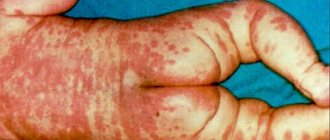
- change in color of lips and limbs,
- increase in phalanges,
- weight loss,
- fatigue and general weakness.
Other diseases of the respiratory system also have similar symptoms, so if unpleasant signals appear, it is best to immediately consult a doctor. An accurate diagnosis will help you quickly make the most correct decision on a treatment regimen and begin to implement it.
Diagnostic measures
Diagnosis of pulmonary fibrosis begins with collecting complaints, studying anamnesis and an objective examination of the patient. It is important to identify the presence of chronic diseases that can provoke the development of this pathology, as well as pay attention to typical clinical manifestations.
To confirm the diagnosis, the specialist prescribes a number of laboratory and instrumental studies, namely:
- General and biochemical blood tests.
- General urine analysis.
- Spirography. This research method allows you to evaluate the peak expiratory air flow rate, as well as the vital capacity of the lungs.
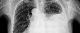
- Plain radiography of the chest organs, in at least two projections. In the image, the doctor reveals deformation and strengthening of the pulmonary pattern, especially in the lower sections.
- Computed and magnetic resonance imaging. Like the previous method, they can reveal structural changes in the lungs. However, with their help it is possible to conduct a more detailed and clear assessment of the pathological process.
Causes of the disease
Pathology can be caused by various reasons:
- hereditary predisposition,
- inflammatory processes in the lungs,
- infectious diseases,
- negative impact of the environment,
- work in hazardous industries,
- the effects of radiation therapy,
- chronic autoimmune diseases,
- complication of lung diseases,
- treatment with various drugs.
Bad habits, such as smoking, can also cause dangerous pathology. However, there are cases of the disease developing without any clear cause. Each individual patient must undergo a full examination to identify the factors that influenced the occurrence of the pathology, this will help to choose the right treatment that will significantly improve the condition.
Pathogenesis
The final sections of the respiratory tract consist of bronchioles (terminal and respiratory), acini and lobules. Terminal bronchioles are air-conducting, and respiratory bronchioles are involved in gas exchange, since they are connected to the alveoli through pores. The acini is the structure of the lung where gas exchange occurs and includes several respiratory bronchioles. The acini (from 3 to 24) are united into pulmonary lobules. The framework of the lobule includes interlobular septa, periarterial, peribronchial connective tissue, connective tissue of the alveolar and acini septa.
Connective tissue consists of collagen, reticular and elastin fibers. Interweavings of collagen and elastin fibers are present around the bronchi and in the alveolar walls, which determines the elastic properties of the lungs. Collagen provides rigidity to the intrapulmonary framework.
Connective tissue cells (smooth muscle and fibroblasts) synthesize elastin and collagen. Its primary role is to maintain the tone of the terminal sections of the respiratory tract. At the same time, connective tissue cells also synthesize enzymes (proteases) responsible for the destruction of elastin and collagen . The enzyme and antienzyme systems are in equilibrium under normal conditions. Under conditions of infectious inflammation, they continue to have an elastolytic (destructive) effect due to the high production of elastase (an enzyme that destroys elastic fibers, elastin, collagen). This causes the destruction of the normal connective tissue matrix, the destruction of the alveolar walls and the formation of common cavities. Excessive production of elastase or the absence of enzymes for its destruction is observed in a number of lung diseases ( COPD , emphysema , cystic fibrosis ).
In parallel, during inflammatory processes, there is an activation of fibroblasts that produce collagen. As a result, there is an enhanced process of development of connective tissue in the lung parenchyma and the formation of fibrosis. The development of fibrotic changes is preceded by edema and infiltration of the lung tissue. When the alveolar membrane is destroyed, intra-alveolar fibrosis develops at this site - growths of connective tissue spread into the alveoli. Then the connective tissue around the arteries and bronchi is involved in the pathological process.
Currently, a more acceptable theory of the pathogenesis of silicosis and other pneumoconioses is the immunological one, which states that the development of these occupational diseases is impossible without phagocytosis of dust particles by alveolar macrophages. The rate of their death depends on the fibrogenic aggressiveness of the dust and is proportional to it. The death of alveolar macrophages is a mandatory step in the formation of a silicotic nodule. The formation of a nodule is possible under the condition of repeated phagocytosis of dust. The resulting pro-inflammatory mediators ( cytokines ) provoke the accumulation of inflammatory cells in the septa of the alveoli, and oxygen radicals, damaging lung tissue, cause the death of macrophages.
Proteolytic enzymes (metalloproteinase and elastase) are released from destroyed macrophages and destroy lung tissue. The inflammatory phase is accompanied by recovery processes in which growth factors stimulate the formation of cells, epithelium and new vessels in damaged tissues. Uncontrolled epithelization processes lead to the development of fibrosis. Growth factors play one of the key roles in fibrogenesis . Their stimulating effect on the proliferation of fibroblasts and the destruction of collagen and fibronectin (high molecular weight protein of connective tissue) has been proven. In idiopathic fibrosis, fibrosis predominates with mild inflammatory changes. The main mechanism for the progression of pneumofibrosis is constant damage to the alveolar epithelium with impaired regeneration and excessive deposition of connective tissue due to the activation of fibroblasts.
Treatment of fibrosis
Treatment of the disease is prescribed by a professional specialist, taking into account the results of the examination and the characteristics of the patient’s disease. There are a number of drugs that help prevent the occurrence of various infections and the development of inflammation. Treatment of fibrosis is supported by therapeutic breathing exercises and the complete exclusion of provoking factors. In case of the total form of the disease, surgical intervention is recommended.
In the early stages of the disease, therapy is used to help reduce scarring. You can consult with a pulmonologist and make an appointment by calling our clinic or using the form on the website.
FIND OUT PRICES
Medicines
- Bronchodilators: Atrovent , Ipratropium-native , Ipratropium Air , Spiriva , Tiotropium-native , Ultibro Breezhaler .
- Mucolytics: Acetylcysteine , Eucabal , Bronchomucin , Fluditec , Ambrol , Ermucin , Lazolvan .
- Glucocorticoids: Prednisolone .
- Antitumor drugs (cystatics): Cyclophosphamide .
List of sources
- Ivanova A. S. Fibrosing processes / A. S. Ivanova, E. A. Yuryeva, V. V. Dlin. M.: Overlay, 2008. 196 p.
- Idiopathic fibrosing alveolitis / M. M. Ilkovich [et al.] // In the book: Disseminated lung diseases / Ed. M. M. Ilkovich. M.: GEOTAR-Media, 2011. pp. 24–84. 3.
- Avdeev SN. Idiopathic pulmonary fibrosis: modern approaches to therapy. Practical Pulmonology 2015;1 22–31.
- Tsvetkova O.A., Voronkova O.O. Modern approach to the treatment of patients with idiopathic pulmonary fibrosis. Clinical Med 2017;95:281–5. 31.
- Aisanov Z.R., Kokosov A.N., Ovcharenko S.I., Khmelkova N.G., Tsoi A.N., Chuchalin A.G., Shmelev E.I. Chronic lung diseases. Federal program. RMJ, 2001; No. 1: p. 9 – 33.
Forecast
The prognosis and life expectancy for pulmonary fibrosis depends on the disease against which it developed, the degree of progression of the fibrosis itself, its extent and the degree of involvement of the bronchial tree (meaning the formation of bronchiectasis, which aggravates the course of the disease). Among the prognostic criteria, the main one is the type of formation of pulmonary fibrosis.
Favorable types include fibrous changes in the form of cords in the central and peripheral interstitium of the lungs, as well as the atelectatic type of fibrosis. The latter is formed in places of organized pneumonia or at the site of long-term collapse of lung tissue as a result of closure of the bronchi. The occurrence of these types of pulmonary fibrosis does not affect perfusion and diffusion in the lungs and does not lead to the development of respiratory failure. Unfavorable types include changes in the form of acinar fibrosis and honeycomb lung, which always lead to impaired perfusion and severe respiratory failure. It is this factor that reduces the patient’s life expectancy.
Honeycomb lung and its significant prevalence are considered the most unfavorable prognostic sign. At the same time, importance is attached to the size of the “honeycombs” - large “honeycombs” or a mixed type of structure with alternating small and large cysts are considered more unfavorable. As for their localization, the lower zonal location is considered unfavorable.
The natural history of idiopathic fibrosis is associated with gradual changes in lung tissue and progressive fibrotic damage, which at the final stage takes on the appearance of a “honeycomb lung”. Increasing fibrosis progressively worsens the patient's condition and affects life expectancy.
It is difficult to answer the question of how long people live with this disease, since it depends on the state of pulmonary function. Terms can range from 2 to 6 years. Thus, in acute cases, patients live no more than 2 years, in subacute cases - from 2 to 4 years, and in chronic cases - from 4 to 6 years. The situation is aggravated by concomitant diseases that arise with age. Death is caused not only by the development of acute respiratory failure, but also by ischemic heart disease, pulmonary embolism or lung cancer .
With slowly progressing silicosis, the prognosis for life is positive, since the process of transition from one stage to another can last decades. There are cases when the progression of fibrosis is not detected at all - in such cases the prognosis for life is favorable. A severe complication of silicosis is spontaneous pneumothorax , but it is very rare in modern forms of silicosis. tuberculosis develops against the background of silicotic fibrosis , then the course of the disease is unfavorable and, in general, the prognosis depends on the severity of tuberculosis and silicosis, and the form of tuberculosis.
Procedures and operations
During any exercise, the heart and lungs work actively. Special breathing exercises are aimed at increasing the functionality of the lungs, reducing shortness of breath and increasing exercise tolerance. The complex should include exercises for training diaphragmatic breathing and exercises with forced exhalation. You can take breathing exercises according to the method of P. A. Buteyko as a basis. The duration of breathing exercises is 10 minutes, and you need to perform it 3-5 times during the day. The ideal option is individually selected gymnastics. If the condition of the respiratory system allows, you can engage in race walking.
Among the procedures, oxygen replacement therapy is indicated, which is carried out when the blood oxygen pressure decreases to less than 60 mm Hg. Art. In case of chronic respiratory failure, oxygen therapy is carried out at home for a long time (18 hours a day) in a low-flow (2-5 l/min) oxygen supply mode. In case of severe respiratory failure, helium-oxygen mixtures are used. Oxygen concentrators are used at home.
To remove circulating immune complexes from the blood (especially in bronchial asthma ), hemosorption .