The fallopian tubes are named after the Italian physician Gabriel Fallopius, who first described their structure. These are paired hollow tubes through which fertilized eggs move into the uterine cavity. Each tube lies in a double fold of the peritoneum - the mesentery of the tube. The length of the pipe is approximately 10-12 cm. Normally, the right pipe is slightly longer than the left. Width – about 4-6 mm. The inside of the fallopian tube consists of a mucous membrane with ciliated epithelium. Thanks to the muscular activity of the tube and the oscillatory directed movements of the cilia of the epithelium, the fertilized egg is pushed towards the uterus.
Sections of the fallopian tube
There are several sections in the pipe:
The uterine (interstitial) part, the mouth of the fallopian tube , is a section of the canal adjacent to the wall of the uterus. It opens into the uterine cavity with a hole measuring about 2 mm.
The isthmus is the narrowest section with a diameter of about 2-3 mm.
Ampoule - this section accounts for almost half the length of the pipe. The ampulla follows the isthmus, gradually increasing in diameter to 8 mm. In this section, the sperm meets the egg. In the ampullary section, the folds of the mucous membrane are well defined. They are large in size and form secondary and tertiary folds.
The funnel is a continuation of the ampoule; it is a funnel-shaped extension of the pipe, along the edges of which there are numerous irregularly shaped fimbriae. One of the largest fimbriae stretches in the fold of the peritoneum all the way to the ovary. At the top of the funnel there is a round hole that opens into the abdominal cavity. Through it, the fertilized egg, with the help of peristaltic movements of the tube, enters the ampoule. The cilia of the fimbria epithelium have ciliated movements towards the uterus, thus they are able to attract an egg from the abdominal cavity and propel it towards the uterus.
The fallopian tubes are supplied with blood by the ovarian and tubal branches of the uterine arteries and branches of the ovarian arteries.
Paired organ structure
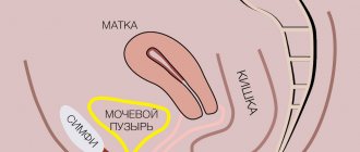
If we talk about the structure of the fallopian tube, it includes 4 sections along its entire length. They extend to the sides, starting from the body of the uterus in an almost horizontal position and ending in an expanded part that has a fringed structure and is called the funnel.
Recalling the structure of the fallopian tube, it should be said that these funnels are located very close to the ovary, where the egg is born, which subsequently collides with the sperm.
The funnels are followed by the ampullary part of the tube, after which the fallopian tube begins to gradually narrow. This section of the isthmus is called in medicine the isthmic part.
The anatomical features of the fallopian tubes are such that they end in the part of the same name. And it is there that the pipes make the transition to the muscular organ.
The importance of the fallopian tubes in reproduction
The fallopian tubes play an important role in a woman's reproductive function. Thanks to peristaltic movements, the tubes ensure the transport of the fertilized egg into the uterine cavity. Peristalsis is best expressed at the time of ovulation and several days after it. The function of the fallopian tube is also to create conditions favorable for conception and development of the embryo as it moves towards the uterus. Tubal secretions, containing glycoproteins, prostaglandins and various biologically active substances, provide nutrition to the embryo in the first days of its existence.
Clinical picture
Symptoms of inflammation of the fallopian tubes are varied. The clinical picture of the disease is influenced by the location of the pathological focus, its type, as well as concomitant diseases.
- Pain is one of the main signs of the disease. Painful sensations are localized mainly in the lower abdomen, although they can radiate to the groin and lumbar region and even to the lower extremities. In addition, pain often intensifies during urination, defecation, during sexual intercourse, and also during menstruation. As a rule, intense pain, aggravated by physical activity, is characteristic of an acute process. Dull pain indicates a chronic form.
- Irregular menstruation
- Mucopurulent discharge with an unpleasant odor appears, as a rule, in an acute form. In a chronic process, there may be no discharge.
- In acute illness, body temperature often rises to 38.5°C, accompanied by chills and intense sweating. With a chronic disease, low-grade fever is noted, but in some cases there is no increase in temperature.
- Weakness, muscle pain, nausea, vomiting, lack of appetite, etc. are also possible - these signs are caused by the effect of toxins on the body.
Tubal obstruction
Obstruction can result from:
- Inflammatory processes in the pelvic cavity, which can occur after difficult childbirth or abortion and with complicated endometriosis.
- Surgical interventions in the abdominal cavity, leading to the formation of adhesions in the pelvis.
- Tubal infection due to complications of sexually transmitted diseases. In this case we are talking about ureaplasmosis, chlamydia, mycoplasmosis.
- Tubal ligation (female sterilization).
- Sometimes congenital underdevelopment occurs when the pipes are too short, or, conversely, long and crooked.
Obstruction can be organic, when the lumen of the pipe is closed by a film of connective tissue, and functional, when the functioning of the pipe is disrupted. Complete blockage occurs when the lumen of the canal is blocked along its entire length. In partial, the lumen in one part of the oviduct is closed. Blocked tubes usually do not affect a woman’s well-being. People usually learn about the need to check the function of the fallopian tubes only if there are problems with conception.
Using folk recipes
Traditional medicine is not officially recognized. One of these is borovaya uterus, which was previously widely used, especially in the Siberian region. It is generally accepted that this plant has anti-inflammatory, antimicrobial, and diuretic effects.
Alcohol and water infusions using boron uterus in the treatment of tubal infertility have survived to this day. In the first case, to prepare the product you will need 50 g of a medicinal plant and half a liter of vodka. In the case of a water infusion, you need to brew 1 teaspoon of crushed medicinal herbs in 1 glass of boiled water, and then leave it in a water bath for 15 minutes.
The alcohol infusion must be taken by dissolving 30-40 drops in a small volume of water, 3 times a day before meals. The course of treatment should continue for 6 months. In this case, one week of taking the drug should be followed by a 3-week break. During menstrual bleeding, taking boron uterus is unacceptable.
We must not forget that while taking boron uterus, an allergic reaction may develop. Therefore, before starting treatment, you should consult your doctor.
How to check the fallopian tubes?
There are instrumental diagnostic and treatment methods for checking the patency of pipes:
Hysterosalpingography (HSG) of the fallopian tubes . This procedure is also called metrosalpingography. After local anesthesia, a contrast liquid is injected into the uterus under X-ray control. It should penetrate both pipes and flow into the abdominal cavity. X-ray images show which areas the contrast has passed through and where the obstacle is located. If adhesions are detected, the doctor can immediately recanalize the pipe. HSG has a drawback: in 20% of cases, a false result is possible due to muscle spasm of the tube.
Fallopian tube laparoscopy is a diagnostic operation. Under general anesthesia, punctures are made in the abdomen and air is pumped into it. A laparoscope is inserted into the incision under the navel, and special instruments are placed into other punctures to perform the operation. As with HSG, a contrast agent is poured into the uterus and seen whether it flows through the fallopian tubes into the abdominal cavity. If adhesions are detected, they are immediately removed by a doctor.
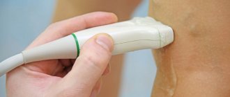
Echohysterosalpingography is a study using ultrasound. This method is less accurate. Instead of a radiopaque agent, saline solution is used.
In Vitro Fertilization
If drug therapy does not give the desired effect, that is, pregnancy still does not occur, then the procedure of in vitro fertilization is recommended. For this manipulation, an egg is collected from women, and sperm from men, after which fertilization is carried out in the laboratory.
After 3-5 days, the embryo is placed in the patient’s uterus for subsequent gestation. This method of reproductive technology is recognized as the most effective. It is resorted to in case of complete obstruction of the tubes or in the presence of serious disorders at the cellular or chemical level in the man’s sperm.
In conclusion, it should be added that obstruction of the fallopian tubes cannot in any way be considered a serious pathology for women, but it is still fraught with the development of infertility. If the necessary actions are not taken in time to correct the indicated condition, then an ectopic pregnancy is possible, as a result of which the patient may lose one of the tubes, which significantly reduces the chances of becoming pregnant. Here you cannot rely only on traditional medicine or self-medicate, since such actions can only aggravate the situation. In order to receive adequate help, you need to contact specialists.
What is hydrosalpinx?
Hydrosalpinx occurs when there is excessive accumulation of pathological fluid in the cavity of the tubes during salpingitis due to impaired lymph and blood flow. This transudate has a toxic effect on the embryo and endometrium of the uterus, leading to infertility and the risk of ectopic pregnancy. The sluggish process does not affect the woman’s well-being in any way. It can be detected during an ultrasound of the pelvic organs. Untreated hydrosalpinx can lead to rupture of the fallopian tube. Treatment is mainly surgical; in parallel, anti-inflammatory treatment, physiotherapy and antibiotic therapy are carried out. If it is impossible to restore the function of the tube, it is recommended to remove it and then refer the woman to IVF.
Since the fallopian tubes play an important role in fertilization, it is necessary to carefully monitor your health to prevent the development of inflammatory diseases. If a source of infection is identified, sanitize it promptly. This way you will keep your fallopian tubes healthy and conceiving a baby will not be difficult for you!
Symptoms of fimbriae immobility
Fimbriae immobility is one of the most difficult causes of infertility to diagnose. A routine examination does not reveal any pathologies of the reproductive system. You can suspect fimbriae dysfunction if you have the following picture:
- the ovaries do not have cysts and function normally;
- the monthly cycle is not disrupted, the follicle matures and comes out monthly;
- the fallopian tubes have a normal diameter and are completely patent;
- the endometrium has an optimal thickness for embryo attachment;
- analysis of sex hormones does not reveal abnormalities;
- The man is also healthy and was examined by a urologist.
When a woman cannot become pregnant within a year in good reproductive health, there is a high probability that the cause is immobility of the fimbriae.
Necessary examinations before the HSG procedure under sedation (during sleep)
- complete blood count + platelets + ESR - valid for 2 weeks;
- biochemical blood test (total protein, glucose, bilirubin, ALT, AST, alkaline phosphatase - valid for 1 month;
- coagulogram (APTT, PTI, fibrinogen, INR) - valid for 1 month;
- blood type, Rh factor (on a stamped form, a stamp in the passport is not accepted) - valid for an indefinite period;
- HIV - valid for 3 months;
- Wasserman reaction - valid for 3 months;
- HBs antigen - valid for 3 months;
- HCV antibodies - valid for 3 months;
- ECG - valid for 2 weeks;
- Flora smear is valid for 2 weeks.
How to check patency if for some reason an HSG is not possible? Read about such methods below, but they are outdated and uninformative
. Therefore, we do not recommend resorting to these types of diagnostics, but we consider it necessary to mention them.
Perturbation hydroturbation
This method of medical correction is quite outdated, but in some institutions it is still carried out. It can also be quite painful for the patient.
The essence of the manipulation is that a woman in a gynecological chair is inserted into the uterus and air or a special liquid is supplied through the catheter. Under strong pressure, the fallopian tubes straighten, and the adhesions spontaneously break. The manipulation is carried out under the control of an ultrasonic sensor.
The disadvantage of this method of treatment is that severe stretching of the fallopian tubes may occur and displacement from their usual place.
Preparation
Given that most procedures are not simple, they require appropriate preparation on the part of the patient, especially if the use of general anesthesia is envisaged.
The most important points remain:
- Complete preliminary examination (consultation with a doctor and passing all tests).
- 2 days before almost all procedures, you need to abstain from sexual intercourse. The doctor may stop taking certain medications if they affect the final result.
- A week before the relevant procedure, you should stop using vaginal suppositories, douching, and the like.
Thus, it will be possible to obtain the most reliable results of any of the above examinations. You can do sonohysterosalpingoscopy at the AltraVita clinic. Here the examination will be as safe and comfortable as possible for the patient. Our specialists use cutting-edge diagnostic equipment and have extensive experience in this field, which allows us to achieve highly accurate research results.
Consequences of the operation
The uterus and fallopian tubes share common nerve fibers, blood and lymphatic vessels. In addition, the condition of the mammary glands and the neuroendocrine system as a whole depends on their work. Therefore, disruption of these connections negatively affects the functioning of the adrenal glands and thyroid gland.
Hormonal imbalance is one of the consequences of surgery to remove the fallopian tubes.
Women complain of symptoms such as:
- Headache and dizziness;
- Nervousness, irritability, tearfulness;
- Painful sensations in the heart area;
- Increased sweating;
- Rush of blood to the upper half of the body.
Symptoms tend to intensify before the next menstruation, and they do not bother all women (observed in approximately 42% of cases).
About 35% of patients notice menstrual irregularities 2-3 months after removal of the appendage. During an ultrasound, they are diagnosed with an enlarged ovary on the side where the fallopian tube was removed. Over time, it undergoes sclerotic changes, which is caused by disruption of the flow of lymph and blood.
There is also an alternation of normal menstrual cycles with disrupted ones. There may be a decrease in the performance of the luteal body and cessation of ovulation. However, such conditions are rarely observed.
The following changes occur in the mammary glands:
- The glands become rough in 6% of patients;
- The breasts become larger due to diffuse expansion of the lobules in 15% of patients;
- The thyroid gland increases in size and its functioning is disrupted in 26% of patients;
- The following symptoms may also develop: excess weight gain, the appearance of body hair, and the formation of stretch marks on the skin.
These symptoms are especially pronounced in those women who have undergone surgery to remove both appendages.
Diagnostic features
The mandatory task of every woman is to carry out diagnostic measures. The main problem during pregnancy is obstruction. In this regard, care must be taken to carry out examinations if violations are suspected.
Lack of patency may be due to natural or purposeful reasons. In the first case, the pathology develops due to factors beyond the control of the woman, in the second case, the patency is disrupted by special intervention to prevent conception.
If the tubes are not passable in the isthmic department, treatment will not be effective and IVF becomes the only chance for motherhood. Moreover, if the intramural section of the fallopian tube is subjected to formed adhesions, the woman is diagnosed with infertility.
Only if the mouths of the fallopian tubes are visualized, the possibility of giving birth to a child remains.
The following disorders can lead to an ectopic pregnancy:
- adhesions;
- kinks;
- narrowing.
If the mouths of the fallopian tubes are free, this means the possibility of conception with further correct development of the fetus. If the fallopian tubes are not visualized, this means that the girl is experiencing pathological processes and additional intervention is required.