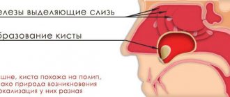
A sinus cyst is a fluid-filled formation with elastic walls; the location of the cyst affects the symptoms of the disease. Most often, cysts form in the maxillary sinuses. The reasons for the development of cysts in the nasal sinuses are varied; there are a number of reasons for the appearance of a neoplasm:
- Blockage of the ducts of the mucus-secreting glands.
- Dental diseases, tooth roots protruding into the paranasal sinuses.
- Allergic diseases of the nose.
- Frequent inflammatory processes.
- Anatomical features of the structure of the nose.
- Mechanical injuries of the nose.
In the oncology department of the Yusupov Hospital, you can undergo examination using MRI, CT, ultrasound, diagnosis of diseases of the paranasal sinuses if a nasal tumor is suspected. The patient will be able to undergo a full examination at the diagnostic center, take tests in the hospital laboratory, and receive advice from an otolaryngologist, oncologist and other specialists.
Cyst in the sinus: symptoms and consequences
Sinus cyst, symptoms of the disease depend on the location of the cyst, there are false and retention. The cyst can increase in size and eventually block the sinus cavity. A false cyst in the sinuses can develop due to an inflammatory, allergic process, while a true cyst develops due to blockage of the ducts of the mucus-secreting gland. A cyst of the paranasal sinuses may not manifest itself as pronounced symptoms for a long time, then nasal congestion, headache, and soreness in the face, on the side of the affected sinus cyst, begin to bother you. The pain can intensify when diving, and inflammatory processes in the nose often develop.
As the cyst of the maxillary sinuses develops, it manifests itself in the following disorders:
- Constant nasal congestion.
- Respiratory dysfunction.
- Pain in the area of the sinus affected by the cyst.
- Unpleasant sensations and false symptoms of increased intraocular pressure.
- Severe headaches.
- Purulent discharge (concomitant development of sinusitis).
- Partial or complete loss of smell.
The consequences of cyst development are negative:
- A growing cyst provokes an inflammatory process and can lead to the development of sinusitis.
- Reduces the patient’s quality of life and causes constant severe headaches.
- A large sinus cyst can lead to destruction of nasal tissue, reach the nasopharynx, impair respiratory function, and deform the face.
- Some sinus cysts can develop into a malignant tumor.
Treatment of maxillary cysts
There are no conservative methods for treating cystic formations in the maxillary sinus. The doctor chooses one of the options for surgical intervention, which depends on the type of cyst, its location, the professional qualities of the specialist and the individual characteristics of the patient.
In modern medicine, the following operations are widely used to treat cysts:
- Open surgery. A manipulation that involves general anesthesia and has many complications and risks. The anterior wall of the maxillary sinus is opened, and the cyst is removed through the resulting hole. The main problem is the overgrowing of the access with scar tissue. This completely contradicts the physiology of the body and does not allow sinus lifting and dental implantation in this area in the future.
- Endoscopic treatment. One of the most advanced and successful in modern medicine. Using a special instrument, the doctor enters the maxillary sinus through its natural opening without damaging the bone tissue. The cyst is then sucked out or desquamated.
- Classic removal. A typical open sinus lift approach is performed, through which the cyst is removed. The operation is performed in cases where the cause of the development of the formation is a diseased tooth.
Since cystic formations are most often discovered during the preliminary examination for sinus lift, the doctor must choose a treatment method that will not interfere with further bone grafting. The final decision always belongs to the patient, so he needs to be told the advantages and disadvantages of each therapeutic technique.
Main sinus cyst: symptoms, treatment
A cyst of the main (sphenoid) sinus develops more often in young people and rarely in older people. The cavity of the main sinus of the nose is covered with mucous membrane. The sheath glands secrete mucus; disruptions in functioning lead to blockage of the gland ducts and the formation of a cyst of the sphenoid sinus. Inflammatory processes, injuries, and allergic reactions have a negative effect on the mucous membrane. A growing cyst of the sphenoid sinus is accompanied by a feeling of fullness in the nose, nausea and dizziness may occur, a headache that radiates to the back of the head, and visual disturbances rarely develop.
If a sinus cyst is detected, treatment is carried out after a complete examination. The doctor prescribes diagnostic tests for the patient using:
- X-rays to determine the location of the cyst, changes in the condition of the facial bones and nasal septum.
- Magnetic resonance imaging, which determines the condition of the skull bones, tissues, and blood vessels. A more informative study than radiography. Computed tomography has the same capabilities. Research is carried out as prescribed by a doctor.
- Research using an endoscope allows you to examine the nasal cavity and perform a biopsy of nasal tissue.
After the studies, conservative or surgical treatment may be prescribed. Conservative treatment is aimed at pain relief, relieving an allergic reaction, and treating the inflammatory process. If conservative treatment is ineffective or the cyst is large, the patient is referred for removal of the formation.
Diagnostics
Figure 1. Cyst in the maxillary sinus.
Source: Contemporary clinical dentistry / Open-i (Attribution-NonCommercial-ShareAlike 3.0 Unported) Before surgery to remove a sinus cyst, you need to take tests (for HIV, syphilis, hepatitis B and C, general blood test).
Usually, to make a diagnosis, the doctor only needs to collect an anamnesis and a general physical examination, including the nose.
Other diagnostic tests include:
Endoscopy of the nose. The doctor performs a detailed examination of the nose and sinuses using a narrow tube with a lighted magnifying lens or small camera.
Visualization methods. Images obtained from a computed tomography (CT) scan can help the doctor accurately determine the size and location of cysts in the deeper areas of the sinuses, and assess the degree of swelling and inflammation.
Test for cystic fibrosis. If a child needs diagnosis, the doctor may suggest testing for cystic fibrosis. This is an inherited disease that affects the glands that produce nasal mucus, tears, sweat, saliva and digestive juices. This is usually a non-invasive sweat test that can determine whether your sweat is more or less salty than most people.
Removal of sinus cyst without surgery
If a sinus cyst is detected, treatment without surgery is carried out using decongestant sprays, antibiotics, painkillers, mucolytics, steroids and antihistamines. They treat concomitant diseases - allergies, sinusitis, inflammatory processes of the gums, teeth, nasal mucosa. In combination with these drugs, various means for rinsing the nasal cavity, regenerating and restorative sprays are used. Treatment is prescribed by the doctor based on the results of the patient’s examination.
What to do?
When deciding on treatment for a cyst, an ENT specialist usually takes a wait-and-see approach. The fact is that the growth of education has been going on for decades. Therefore, as long as it is small and does not cause any discomfort, there is no need to touch it. In addition, although infrequently, a maxillary sinus cyst can empty on its own. There is no pain, and the contents of the “ball” flow out through the nose.
But if the pathology causes concern, then the only effective way to treat it is surgery. Many patients, fearing surgical intervention, try to get rid of the problem with the help of traditional medicine: they do rinses, instill herbal decoctions into the nose or lubricate the mucous membranes with them. There is no point in such procedures, since they only temporarily make breathing easier, but do not reduce the size of the formation.
Cyst in the sinus: surgery, reviews
Patient reviews give preference to the endoscopic method of cyst removal. When performing the classical method and laser vaporization, an incision is required under the upper lip to gain access to the cyst. Classic removal of a sinus cyst involves dissection of the soft tissue under the upper lip from the frenulum to the first molar (Caldwell-Luc method) or using the Denker method, which is also carried out through the facial part. Classic operations are more traumatic, with long postoperative recovery, but these techniques allow you to gain access to a cyst located in a hard-to-reach place. Endoscopic removal is carried out using an endoscope, the cyst is removed through a small burr hole, the operation is performed under local anesthesia.
At the Yusupov Hospital, patients will be able to undergo a full examination using modern diagnostic equipment. The hospital hosts doctors from various fields, there is a clinical laboratory, and a rehabilitation center. In the hospital you can recover from surgery or illness, and patients have a 24-hour inpatient facility.
Possible complications
After surgery to remove a cyst from the maxillary sinus, swelling of the mucous membrane, a feeling of dryness or profuse nasal discharge, and pain in the area through which the sinus was accessed may appear. These symptoms are not complications of surgery and usually resolve within a few days. If they persist and intensify, you should consult a doctor.
The nature of complications after surgery depends on the technique used:
- if access to the sinus was carried out through the alveolus of the tooth, an incision or puncture in the upper jaw, a fistulous tract may appear;
- with micromaxillary and radical maxillary sinus there is a risk of damage to the infraorbital or trigeminal nerve with the development of neuritis. In this case, areas of the skin, nasal mucosa and mouth become numb, which requires additional treatment;
- inflammation and/or suppuration of the wound develops if it becomes infected (with poor quality oral care, in cases where the patient does not follow the doctor’s orders).
Comments
The other day I visited LORA, because... I'm worried about constant nasal congestion. HE examined and diagnosed “odontogenic sinusitis” and prescribed symptomatic treatment. He said that in order for everything to return to normal, you need to go to the dentist and look for the cause. No cyst was found. Explain what could be the reason and why this is dangerous?
Tanyusha (06/03/2020 at 17:53) Reply to comment
- Dear Tanyusha, the diagnosis of odontogenic sinusitis indicates that the problem should be looked for in the oral cavity. The cause of its development is often periodontitis, perforation of the sinuses during dental treatment, impacted and dystopic teeth. If the exact cause is not identified and eliminated, then the formation of a cyst will not take long to occur.
Editorial staff of the portal UltraSmile.ru (06/08/2020 at 09:16) Reply to comment
Hello. Today I had an MRI about pain on the right side of my face. A cyst was discovered in the maxillary sinus on the right. Tell me where to run and what to do? Thank you.
Victoria (08/17/2021 at 04:57 pm) Reply to comment
Write your comment Cancel reply
Why do jaw cysts form?
Odontogenic cysts (developing or inflammatory) get their name from the nature of their origin. Most jaw cysts are lined by epithelium, which is derived from odontogenic epithelium.
The occurrence of such cysts is usually associated with unerupted teeth (third molars of the lower or upper jaw, second premolars of the lower jaw, and canines of the upper jaw). They can also form near additional teeth and in combination with odontomas. The occurrence of odontogenic cysts is rarely associated with baby teeth.
Most often, jaw cysts are discovered between the ages of 10 and 30 years. Men, especially white-skinned men, suffer from them more often.
In most cases, dental cysts do not manifest themselves in any way and are an accidental finding obtained during an X-ray examination. But, in some cases, they can reach significant sizes, which lead to the expansion of bone tissue, but even they may not give pronounced symptoms until a secondary infection occurs.
What is the prognosis for the disease?
How successfully the situation in a patient with an odontogenic cyst will be resolved depends on at what stage the cyst was discovered, how severe the symptoms were and how it was treated.
As a rule, the use of surgical treatment gives a positive prognosis. Therapeutic treatment provides a positive prognosis only if it is started at the initial stage of the disease.
A negative prognosis may be associated with detection of the disease at a late stage: odontogenic cysts can provoke the development of serious pathologies that cause deformation of the maxillofacial tissues.
Our services in otorhinolaryngology
The administration of CELT JSC regularly updates the price list posted on the clinic’s website. However, in order to avoid possible misunderstandings, we ask you to clarify the cost of services by phone: +7
| Service name | Price in rubles |
| X-ray of the paranasal sinuses | 2 200 |
| MSCT of the paranasal sinuses | 5 000 |
| Endonasal maxillary sinusotomy | 40 000 — 75 000 |
All services
Make an appointment through the application or by calling +7 +7 We work every day:
- Monday—Friday: 8.00—20.00
- Saturday: 8.00–18.00
- Sunday is a day off
The nearest metro and MCC stations to the clinic:
- Highway of Enthusiasts or Perovo
- Partisan
- Enthusiast Highway
Driving directions
Conservative treatment: when possible
Conservative treatment is only possible for small root cysts. At this time, the canals of the tooth are treated, on the roots of which a cyst has formed. The treatment consists of thorough cleaning and filling of the dental canals and is carried out under a microscope, which ensures the accuracy of all manipulations and allows you to find all the canals in the tooth and fill them. Most often, after endodontic treatment, the cyst disappears, which is recorded on an x-ray taken a few weeks after the operation.
A small cyst can only be detected by x-ray
When the neoplasm is small, less than 10–15 mm, it is almost impossible to find out about its existence, since no alarming signs are observed. At this stage, the only option to identify pathology is to do a computed tomography scan. A dentist may also suspect its presence during a routine examination or other doctors if a person needs to visit them.
At the initial stage, the problem can only be detected on an x-ray
Types of cystic formations
There are several types of cystic formations of the upper jaw - true (retention), false, odontogenic.
| Variety | Description |
| True (retention) | They are formed from the glands of the mucous membrane, the exit ducts of which are clogged. Their wall is lined with ciliated epithelium. |
| False (cyst-like) | The main difference between cyst-like formations is the absence of an epithelial lining. The reasons for their development are not fully understood; the main etiological factor is considered to be exposure to an allergen. |
| Odontogenic | Odontogenic cysts are associated with dental diseases. They are always localized at the bottom of the VChP, while other varieties can arise in any area. |
Separately, there are 3 forms of cystic formation depending on the nature of the contents:
- mucocele (mucus);
- pyocele (pus);
- hydrocele (serous contents).
How are odontogenic cysts diagnosed?
The leading method for identifying odontogenic cysts is radiography, which is capable of visualizing jaw cysts at an early stage of their development. On an x-ray image, the cyst is distinguished by the presence of clear boundaries, and the formation itself gives a characteristic shadow of a round or oval shape, immersed in the sinus of the tooth root.
Ultrasound examination also helps to recognize odontogenic cysts.
As already mentioned, pronounced symptoms are characteristic of the late stage of development of a pathological formation, therefore it is difficult to diagnose a cyst at the initial stage, relying only on symptoms.
The final diagnosis is made on the basis of histological examination. It is important to differentiate an odontogenic cyst from other pathologies (adenomatoid odontogenic tumor, ameloblastic fibroodontoma and calcifying epithelial odontogenic tumor).
The CT scan method is widely used in the diagnosis of jaw cysts to confirm the presence of calcifications along the wall of the cyst, as well as tiny spots that are usually not found on x-rays. In addition, computed tomography is necessary during the surgical planning stage.