Table of contents
- Etiology and pathogenesis
- Clinical manifestations
- Principles of treatment
Deep-lying vessels are pathologically altered capillaries that feed hemangiomas and malformations, as well as enlarged venules located in the skin at a depth of up to 5 mm.
In our company you can purchase the following equipment for removing deep-lying vessels:
- M22 (Lumenis)
Conditions in which deep-lying vessels are present in the skin include the following:
- Infantile hemangiomas
- Capillary hemangiomas
- Cavernous hemangiomas
- Vascular malformations
- Phlebeurysm
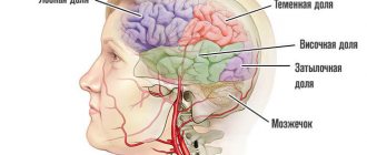
The heart is the basis of the circulatory system
The heart is a muscular organ about the size of a human fist that is located on the left side of the chest, just in front of the lungs. This organ is actually a powerful double pump with four chambers that pumps blood and keeps it moving throughout the body.
The right side of the heart consists of an upper (atrium) and lower (ventricle) chamber. The atrium receives processed venous blood, saturated with carbon dioxide, and then sends it to the ventricle. From there it enters the pulmonary arteries, where it is again saturated with oxygen. “Fresh” blood circulates to the left upper chamber (atrium), from where it enters the aorta and begins renewed transportation throughout the body.
The heart muscle beats more than 3 billion times during a lifetime.
Etiology and pathogenesis
Infantile hemangiomas
They are the most common vascular tumors in children, occurring in 3–10% of fair-skinned newborns. Neither the causes nor the specific cells from which infantile hemangiomas arise have been definitively established to date. There are many theories on this subject, in which placental tissue, endothelial progenitor cells and mesenchymal stem cells are “suspected”.
There is evidence that during the third trimester of pregnancy, immature fetal endothelial cells coexist with immature pericytes, which remain proliferative for a limited period after birth. Angiogenic peptides (fibroblast and vascular endothelial growth factors) stimulate the division of these immature cells, which leads to the development of hemangioma. As endothelial cells differentiate, an influx of mast cells, myeloid cells, and tissue inhibitors of metalloproteinases (TIMPs) begins. Together they stop division and trigger the involution of the hemangioma.
Infantile hemangiomas consist of large and actively proliferating vascular endothelial cells. At the beginning of proliferation, they randomly accumulate in a certain area, but over time they organize into special vascular spaces with channels filled with blood cells ( Fig. 1 ). Because infantile hemangiomas are benign, they cannot divide indefinitely. Over time, the activity of this process begins to fade, and hemangiomas acquire a lobular structure. Mast cells influence the slowdown of proliferation; they also participate in the spread of arterioles and venules feeding the tumor.
Rice. 1. Histological picture of infantile hemangioma with large endothelial cells in the dermal layer of the skin (Medscape)
Capillary hemangiomas
Common benign tumors of the orbit and periorbital region in children. They are composed of endothelial cells and are characterized by rapid growth followed by spontaneous involution ( Fig. 2 ). This distinguishes capillary hemangiomas from vascular malformations, which persist into adults.
Capillary hemangiomas are believed to be of placental origin. They usually show two phases of growth - proliferative and involutive. The proliferative phase is characterized by rapid tumor growth and lasts from 8–18 months or more. At the same time, the number of endothelial and mast cells increases - the latter stimulate the growth of blood vessels feeding the hemangioma.
The involutive phase is characterized by slow regression of hemangioma - about 50% of tumors disappear by 5 years, and 75% by 7 years. At this time, the number of mast cells decreases to normal, and a decrease in endothelial cell activity is observed.
Rice. 2. Histological picture of capillary hemangioma with characteristic dermal clusters in which thin-walled capillaries are visible (Ilovepathology)
Cavernous hemangiomas
They are the result of the formation of vascular channels in the surrounding soft tissues ( Fig. 3 ). A local change in hemodynamics contributes to the appearance of new vessels in a given tumor, which allows it to spread even more. A fibrous capsule appears at the border of cavernous hemangioma with normal tissues. Visual impairment may occur due to compression of the optic nerve, eye muscles, or surrounding blood vessels.
Rice. 3. Histological picture of cavernous hemangioma with large spaces filled with blood (Ilovepathology)
Vascular malformations
The most common capillary malformation is port-wine stain (congenital capillary angiodysplasia, nevus flamingo). It is present immediately after the birth of the child and increases in proportion to his growth ( Fig. 4 ). Its distinctive feature is the absence of spontaneous regression. Sometimes a port-wine stain may appear on the skin in adulthood (acquired capillary angiodysplasia).
Rice. 4. Capillary malformation in a 3-month-old child with a port-wine stain on the cheek - randomly located capillaries in the papillary dermis are visible (Merrow C., et al. Revised classification of vascular lesions from the International society for the study of vascular anomalies: Radiologic-pathologic update Radiographics 2016;36(5):150197)
Venous malformations occur in 1–4% of children—like capillary malformations, they are present from birth. Venous malformations consist of a mass of small venules, each of which has a wall one layer of endothelial cells thick. These venules randomly intertwine with each other, forming a disordered structure ( Fig. 5 ). These malformations grow in proportion to the child's height, but can increase significantly during puberty, hormonal changes, or when infection occurs.
Rice. 5. Histological picture of venous malformation ( a ) and venous malformation with intravascular endothelial hyperplasia ( b ) (North P. E Histology of vascular malformations. In: Mattassi R., Loose D., Vaghi M. (eds) Hemangiomas and vascular malformations. Springer, Milano 2015)
Lymphatic malformations are interlacing of single-layer vessels and vesicles filled with lymphatic fluid ( Fig. 6 ). About 75% of malformations occur in the cervicofacial region. In the past they were called lymphangiomas, which is a misnomer because there is no cellular hyperplasia. Lymphatic malformations never regress, but can expand and contract depending on the volume of lymph, the presence of injury, inflammation and other factors.
Rice. 6. Histological picture of lymphatic malformation (Pathology Outlines)
Phlebeurysm
This is an external manifestation of chronic venous insufficiency, which is associated with a violation of the reverse flow of blood from the extremities to the heart. Mild forms (venous telangiectasias) cause only cosmetic defects, while severe forms can lead to the formation of ulcers, necrosis and malnutrition of the limb.
Varicose veins are recorded in 23% of the adult population, and if mild forms of the disease are taken into account, the figure increases to 80% in men and 85% in women. Among its causes, it is worth mentioning increased pressure in the superficial veins, weakness of the valve apparatus and venous walls, pregnancy and prolonged standing.
Clinical manifestations
Infantile hemangiomas
The earliest sign of infantile hemangioma is blanching of the skin (possibly with small telangiectasias), which later transforms into a red or crimson spot ( Fig. 7 ). A characteristic feature is the rapid growth of the tumor in the first 4 weeks of a child’s life. It usually grows faster than the newborn itself, which distinguishes it from vascular malformations, which increase in proportion to the growth of the infant.
Infantile hemangioma may become convex or dome-shaped, acquire a lobulated structure, or remain flat. During the first year of a child’s life, the phase of tumor proliferation occurs, with its greatest increase recorded in the first 4–6 months.
The involutive phase of infantile hemangioma can be short or long, and this is impossible to predict in advance. But it is reliably known that of all lesions that have resolved by the 6th year of a child’s life, 38% leave behind scars, telangiectasia, or anetodermic (atrophic) skin. If the hemangioma resolves after 6 years, these residual effects are recorded in 80% of cases. It has also been observed that infantile hemangiomas, which take longer to develop, have a higher incidence of residual skin lesions.
Rice. 7. Infantile hemangioma on the forehead ( a ), scalp ( b ), cheek ( c ) and upper lip ( d ) (Itinteang T., et al. Biology of infantile hemangioma. Frontier Surg 2014; 1)
Capillary hemangiomas
Typically these are red spots of progressive size in the periorbital area. Hemangiomas turn white when pressed, unlike port-wine stains, whose color does not change. Due to the extent of the tumor, the child may be unable to open the affected eye ( Figure 8 ). A decrease in visual acuity on the affected side is typical - amblyopia is observed in 43–60% of patients.
Rice. 8. Capillary hemangioma of the lower eyelid (Kavoussi SC, et al. Spontaneous periocular ecchymosis in children: Differential diagnosis and current trends in evaluation and manangement. J Clin Exp Ophthalmol 2014; 5: 1–10)
Cavernous hemangiomas
They are neoplasms located deep in the skin, which appear as a bluish swelling on the arm or other area of the body ( Fig. 9 ). They can have a small diameter or reach large sizes.
Rice. 9. Cavernous hemangioma in the periauricular area (Skintags Removing)
Vascular malformations
A port-wine stain ( capillary malformation ) first appears as a light pink or pink area that, upon closer inspection, consists of individual vascular branches. Most malformations are located on the face or neck, in 45% occupying from 1 to 3 areas innervated by the trigeminal nerve, and in 55% they can move to the opposite side of the face.
As the child grows, the port-wine stain increases in size, becoming light red or completely red ( Figure 10 ). At the same time, it looks like a continuous network of vessels, where it is impossible to identify individual branches. Malignancy of a port-wine stain is possible with degeneration into a cancerous tumor.
Rice. 10. Port-wine stain as an adult (Instagram.com/birthmarksbeauty)
Venous malformations appear as soft, bluish-colored lesions that typically occur on the face, extremities, or trunk ( Figure 11 ). They are dependent lesions, i.e. expand and contract depending on the patient's position. Venous malformations are prone to thrombosis, resulting in phleboliths, which in this case are a pathognomonic symptom.
Lymphatic malformations occur from birth and appear as small, dome-shaped nodules that are reddish in color due to slight bleeding. Based on the size of the lymphatic chambers, they are divided into macrocystic (> 2 cm), microcystic (<2 cm) and mixed. Macrocystic lymphatic malformations can significantly increase in size ( Fig. 12 ), leading to bone deformation and disfigurement of the child's face.
Rice. 11. Venous malformation of the face (Craniofacial team of Texas)
Rice. 12. Lymphatic malformation in the neck (Dr. P. Marazzi/Science Photo Library)
https://www.merckmanuals.com/
Phlebeurysm
Clinical manifestations are extremely varied - from small venous telangiectasias (“spider veins”) to large protrusions of large veins under the skin, with ulcers and necrosis. If we talk about a mild degree of the disease, which can be cured non-surgically, venous telangiectasias are divided into tree-like (branching), arachnid (stellate), linear (simple), spotted (pantiform). In the absence of therapy, they gradually spread throughout the skin ( Fig. 13 ), after which large and deep vessels can be affected with the addition of various complications.
Rice. 13. Telangiectasia on the thigh (Primary Care Dermatology Society)
Principles of treatment
The choice of treatment method should begin with a thorough assessment of the patient's condition. Based on this, it should be determined whether a particular method can solve the identified problem.
To treat deep-lying vessels, including those feeding hemangiomas and malformations, sclerotherapy - the introduction of a special substance into the vascular lumen, which causes destruction of the endothelium with the subsequent formation of a thin fibrous cord. In this way, vessels with a diameter of 4 mm or more can be eliminated.
The “gold standard” for removing small superficial vessels with a depth of up to 5 mm is the Nd : YAG laser . Its radiation has an optimal penetration depth and is well absorbed by blood hemoglobin. Laser therapy procedures are carried out without compromising the integrity of the skin, are well tolerated and do not require long-term rehabilitation. Nd:YAG laser can be combined with sclerotherapy or surgical removal of large and deep-lying vessels.
In the case of deep-lying vessels, they are often “covered” by a network of surface capillaries, which absorbs part of the laser radiation. Therefore, a combination of intense pulsed light (IPL) and a 1064 nm Nd:YAG laser is used for effective therapy. Both of these modules are part of the universal M22 platform from Lumenis.
Questions from our users:
- deep vessels removal
- remove blood vessels with laser
- vessels after laser resurfacing
- blood vessels treatment
Some numbers
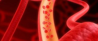
There are more than 95 thousand kilometers of blood vessels in the body of a healthy adult. More than seven thousand liters of blood are pumped through them every day. The size of the blood vessels varies from 25 mm
(aortic diameter)
to eight microns
(capillary diameter).
Down with atherosclerosis!
Atherosclerosis is one of the most common vascular diseases. It is characterized by the formation of fatty plaques on the walls of the arteries. Find out how to prevent atherosclerosis.