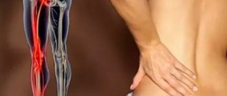
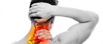
How does a pinched nerve manifest?
Intense, stabbing or shooting (like an electric shock) pain in the neck, lower back or chest. The pain can spread to the arm, to the back of the head, along the rib into the chest, often to the heart area, or to the leg. In the compression zone, the muscles are tense, spasmed, and there may be swelling of the soft tissues. Muscle tension leads to curvature of the spine; on the side of pain, the shoulder, shoulder blade, and thigh rise. This “distortion” of the back muscles is called a painful (algic) pose. If proper treatment is not given, a pinched nerve can persist for several weeks and significantly impair the patient's quality of life.
Causes of pinched nerve:
- The most common cause is osteochondrosis. In this case, the roots of the spinal nerves are pinched in the intervertebral foramina at the site of protrusion (bulging) of the intervertebral disc. An additional cause of pinched nerves is bone growths of the vertebrae (osteophytes).
- A herniated disc is a complication of osteochondrosis and, as a rule, leads to persistent pinching of the nerve root. In this case, the pain spreads along the arm or leg and is accompanied by decreased reflexes, impaired skin sensitivity, a feeling of “tingling needles” or weakness in the fingers or toes.
- Constant intense physical activity leads to compression of the nerve between the muscles and tendons. These are the so-called “tunnel” syndromes. They occur in people of certain professions: athletes, tennis players (pinched nerve in the elbow), typists and other “typing” people (compression in the wrist).
Causes
There are several reasons that cause pinching of the sciatic nerve. The occurrence of sciatica is often associated with the spine, which is not surprising - throughout life it experiences enormous stress. People work physically, carry heavy loads, do not watch their weight and lead a sedentary lifestyle. Without dosing the load and carrying out prevention, the condition of the spine worsens. So, among the causes of sciatica associated with the spine, we can highlight:
- osteochondrosis;
- inflammatory processes in the spinal column;
- intervertebral hernia;
- displacement of vertebral discs;
- spinal injury.
There are several possible causes of sciatica
Diabetes, hypothermia, any tumor processes, and radiculitis can also trigger the development of sciatica. The disease can also be caused by spasms of muscle fibers (usually piriformis syndrome), which can compress the nerve.
Important! To begin effective treatment for sciatica, it is important to identify the underlying cause. Only a doctor can do this by conducting a series of diagnostic studies. Otherwise, only the pain syndrome will be relieved, but the pain will return over time and, possibly, with greater severity.
What examinations are necessary for a pinched nerve?
An important diagnostic method is electromyography (EMG). It is this study that objectively assesses the degree of compression (damage) of the nerve. An EMG records the conduction of electrical impulses along the nerves of an arm or leg and gives the doctor an assessment of the state of the peripheral nervous system.
Magnetic resonance imaging (MRI) and computed tomography (CT) can assess the condition of the vertebrae and intervertebral discs, the presence of hernias and protrusions, and spinal cord damage.
X-ray of the spine
informative in case of poor posture - scoliosis, kyphosis, as well as injuries: fractures and subluxations of the vertebrae.
Pathophysiology
By its nature, nerve damage can be either ischemic or mechanical.
Repeated injury and trauma to the nerve can lead to microvascular (ischemic) changes, swelling, damage to the outer layers of the nerve (the myelin sheath) that facilitate the transmission of nerve impulses, and structural changes in membranes at the organelle level (both the myelin sheath and the nerve axon) . Focal segmental demyelination in the area of compression is a common feature of compression syndromes. Full restoration of function after surgical decompression is associated with remyelination of the injured nerve. Incomplete recovery occurs due to Wallerian axonal degeneration and persistent fibrotic changes at the neuromuscular junction, which may prevent complete reinnervation and restoration of function.
Effective treatment for pinched nerves
Traditional treatments for pain symptoms include anti-inflammatory painkillers (NSAIDs), muscle relaxants (drugs that relax muscles), drugs that improve blood circulation, B vitamins, and physical therapy. These methods reduce symptoms, but do not affect the causes and mechanisms of the disease.
Blockades of pressure points and paravertebral blockades are a classic effective method of treating muscle-articular pain. The doctor specifically introduces a complex of pain medications (into the pain “trigger” zone or along the pinched nerve). Pain relief occurs a few minutes after the injection.
An alternative modern method for treating osteochondrosis, scoliosis and herniated intervertebral discs is shock wave therapy. The principle of UVT is the effect of low-frequency sound waves on tense muscles and ligaments, joints, and bones. The acoustic wave reduces inflammation of the pinched nerve, restores blood circulation and starts the process of cell regeneration. There are known cases of restoration of bone structure after the onset of bone necrosis.
The First Neurology clinic actively uses more than 20 types of treatment for pain syndromes in the back and limbs, as well as headaches. Modern high-tech methods relieve pain after 2-3 sessions and reduce the drug load (the amount and duration of taking pain medications).
These methods include, first of all, carboxytherapy. The introduction of small amounts of carbon dioxide into the projections of the spine and joints causes blood flow and stimulates blood circulation in the area of the pinched nerve. Carboxytherapy has a rapid analgesic effect, improves trophism (nutrition) of intervertebral discs and nerves. The undoubted advantage of the method is the absence of side effects.
Mesoinjection therapy has been used at the First Neurology clinic for more than 10 years. This is a method of introducing drugs into the deep layer of skin - mesoderm. Locally - in the projection of the pinched nerve, the neurologist administers muscle relaxants (muscle relaxers) or vascular, anti-inflammatory drugs. The absorption and effect of the drug last more than 2 weeks, so mesotherapy is performed once every 14 days.
Hi Top high-tone therapy is the method of choice in the treatment of a pinched nerve in an arm or leg. By applying pulsed current to the sensory endings of the peripheral nerve, Hi Top stimulates the nerves, muscles and spinal cord.
Dosed traction of the spine is carried out at the First Neurology clinic using the specialized orthorpedic complex ORMED-Professional. Traction eliminates pinched spinal nerves, reduces back muscle tone, and quickly relieves pain in the spine.
A little about massages
If you combine various treatment methods with massage, you can achieve improvement quite quickly. The main thing is to first eliminate the pain, and then attend sessions with a massage therapist, since if there is discomfort, the desired effect will not be achieved.
It is important to choose the right massage therapist. He must have an appropriate document confirming the availability of the necessary skills. If the massage technique is incorrect, it can seriously harm your health.
Massotherapy
Usually the course of massages is quite long, but it needs to be completed completely to get the desired effect. As a rule, you will have to visit a massage therapist at least 10 times . Massage stimulates blood circulation, enhances tissue nutrition and helps release the sciatic nerve, and therefore improve the condition. Further, even after healing, it is necessary to periodically repeat the course to avoid relapses.
Our specialists
Tarasova Svetlana Vitalievna
Expert No. 1 in the treatment of headaches and migraines. Head of the Center for the Treatment of Pain and Multiple Sclerosis.
Somnologist.
Epileptologist. Botulinum therapist. The doctor is a neurologist of the highest category. Physiotherapist. Doctor of Medical Sciences.
Experience: 23 years.Derevianko Leonid Sergeevich
Head of the Center for Diagnostics and Treatment of Sleep Disorders.
The doctor is a neurologist of the highest category. Vertebrologist. Somnologist. Epileptologist. Botulinum therapist. Physiotherapist. Experience: 23 years.
Palagin Maxim Anatolievich
The doctor is a neurologist. Somnologist. Epileptologist. Botulinum therapist. Physiotherapist. Experience: 6 years.
Romanova Tatyana Alexandrovna
Pediatric neurologist. Experience: 24 years.
Temina Lyudmila Borisovna
Pediatric neurologist of the highest category. Candidate of Medical Sciences.
Experience: 46 years.
Zhuravleva Nadezhda Vladimirovna
Head of the center for diagnosis and treatment of myasthenia gravis.
The doctor is a neurologist of the highest category. Physiotherapist. Experience: 16 years.
Mizonov Sergey Vladimirovich
The doctor is a neurologist. Chiropractor. Osteopath. Physiotherapist. Experience: 8 years.
Bezgina Elena Vladimirovna
The doctor is a neurologist of the highest category. Botulinum therapist. Physiotherapist. Experience: 24 years.
Drozdova Lyubov Vladimirovna
The doctor is a neurologist. Vertebroneurologist. Ozone therapist. Physiotherapist. Experience: 17 years.
Read also
Gliomas
What is a glioma?
Glioma is a neuroepithelial tumor, a type of tumor of the central nervous system, the source of growth of which is glial tissue. In turn, glial tissue (glia)… Read more
Brain concussion
A concussion is a mild traumatic brain injury without damage to the bones of the vault and base of the skull. In this condition, there are no pronounced disturbances in the functioning of the brain and there are no changes...
More details
Chiari malformation
What is Chiari Malformation? Chiari malformation (formerly Arnold-Chiari malformation) is a congenital defect of brain development that involves the dislocation of the cerebellar tonsils into the spinal canal through the large…
More details
Vertebral artery syndrome
The main vascular highways carrying blood to the brain pass in the neck - these are two carotid and two vertebral arteries. A decrease in blood flow through the vertebral arteries will lead to the development of acute...
More details
Encopresis
Encopresis is a condition in which a person does not control or feel the urge to defecate, and also cannot control the act of defecation itself; Fecal incontinence significantly impairs the patient’s quality of life,...
More details
Etiology
Pinched nerve syndrome can result from chronic damage to the nerve as it passes through the osseous-ligamentous tunnel. Compression usually occurs between the ligamentous canal and adjacent bony surfaces.
In cases of nerve entrapment, at least one part of the compressive surface is movable. This results in either repeated compression or friction/sliding against sharp or hard edges, resulting in chronic trauma. Immobilizing the nerve with a splint or lifestyle modifications may relieve symptoms. Compression neuropathies can also be caused by systemic disorders such as rheumatoid arthritis, acromegaly, or hypothyroidism.