The clavicle is a curved S-shaped bone, consisting of a body and having two ends with articular surfaces - the acromial (external) and sternum (internal). Together with the scapula, it forms the girdle of the upper limbs. The clavicle is the only bone that connects the trunk (axial skeleton) to the upper limb. Due to the fact that the clavicle is involved in the formation of two joints (sternoclavicular and acromioclavicular), dislocations occur in two types: the sternal end of the clavicle and the acromial end of the clavicle. Dislocations of the acromial end most often occur; dislocations of the sternal end of the clavicle are much less common. Dislocation of the scapular (acromial) end can be complete or incomplete (depending on which ligament is torn). Dislocation of the sternal end is divided into suprasternal, presternal and retrosternal (depending on the trajectory of the clavicle displacement).
Treatment.
To treat clavicle dislocations, two methods are used: conservative and surgical intervention. A conservative treatment method is used to reduce dislocations of the acromial end of the clavicle using a closed method using anesthesia. Then the specialist fixes the reduced collarbone using a plaster cast and special splints with constant pressure and support (orthopedic splints). In case of complete dislocation and rupture of ligaments, urgent surgical treatment is required. For sternal dislocations, only surgical treatment is indicated (open reduction using metal structures). After the operation, the arm is fixed in a plaster cast. The immobilization period lasts 4-6 weeks.
Causes of pain in the collarbone
As already mentioned, the causes of pain in the collarbone can be caused by injuries to the bone itself, as well as pathological processes in the spine or shoulder joint.
Clavicle injuries
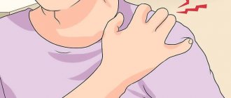
Patients often complain that their left or right collarbone in the shoulder joint area hurts. If the appearance of such symptoms was preceded by a blow to the bone, to the sternum, or a fall on an outstretched arm, then the cause may be a fracture of the collarbone. With this injury, there is a displacement of the central bone fragment back and up, and the peripheral one - inward and downward. Painful sensations are accompanied by:
- hemorrhage;
- local swelling;
- visually visible displacement of fragments.
Joint displacement
Pain between the collarbones can occur due to misalignment of the clavicular joints. Since the clavicle has two ends (acromial and sternal), dislocations can occur at both ends:
- dislocations of the acromial end are much more common and are felt by painful symptoms, which intensify when trying to put pressure on the protruding end of the clavicle;
- Dislocations of the sternal end are usually characterized by pain in the area of the sternoclavicular joint, which is deformed, and swelling. There may be a protrusion of the end of the clavicle when dislocated forward or its retraction when dislocated backward.
Spinal diseases
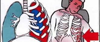
Spinal diseases are often accompanied by compression of the nerve roots of the cervical spine. If the 3rd root is pinched, pain symptoms are accompanied by numbness in the area behind the ear and enlargement of the tongue. Compression of the 4th root is characterized by pain in the heart, difficulty swallowing, hiccups, and a feeling of a lump in the throat.
Brachial plexus neuralgia
Pain under and in the collarbone on the left side can be caused by diseases such as neuralgia of the ribs or brachial plexus. It is felt during inhalation/exhalation and is accompanied by:
- intense heartbeat;
- false pain in the heart.
Shoulder periarthritis
Inflammatory processes in the capsule of the shoulder joint, as well as in nearby tendons and ligaments, lead to pain directly in the joint and its irradiation to the collarbone. As a rule, unpleasant sensations occur for no reason at night.
More about shoulder periarthritis
Diseases of internal organs
In addition to the above, projection pain in the right clavicle can occur due to liver or gallbladder diseases. Pathological processes in the stomach and pancreas may be accompanied by radiating pain in the left collarbone. With angina pectoris and myocardial infarction, bursting pain occurs, radiating to the left collarbone
Rehabilitation.
Rehabilitation measures are aimed at speedy recovery of the patient after the injury.
- A mandatory set of exercises to develop an injured collarbone (selected by a traumatologist, taking into account the type of dislocation and the complexity of the injury)
- Physiotherapeutic treatment (UHF therapy, therapeutic massage, acupuncture, electrophoresis)
- Balanced nutrition (including foods containing essential microelements and vitamins)
The recovery period is approximately 1.5-2 months, after which normal performance returns.
If, in case of a dislocated collarbone, you do not seek help from a specialist in time or do not follow his recommendations, then the consequences of such an attitude may be damage to nerves, blood vessels and dysfunction of the hand.
Diagnosis of pain in the collarbone
Specialists at the CELT Pain Clinic do not recommend self-medication, as it can cause irreparable harm to your body. In order for the treatment to be successful, it is necessary to identify the reason why the collarbone hurts and direct all efforts to eliminate it.
In addition to being examined by a doctor and taking a medical history, diagnosing collarbone pain includes the following:
- radiography of the collarbone;
- CT scan;
- Magnetic resonance imaging.
Anatomy and structure
In its shape, the clavicle is a tubular bone , and in structure it is a spongy bone (its cavity contains spongy substance, and not bone marrow, as in most other bones of the human skeleton).
The bone is distinguished by a rounded body and two ends: sternal and acromial . At its sternal end there is a saddle-shaped sternal articular surface designed for articulation with the sternum. The upper surface of the clavicle is completely smooth, and on the lower there are two tubercles: cone-shaped and elongated (trapezoidal line). Muscles and ligaments are attached to these formations, allowing movement of both the collarbone itself and the bones of the upper limbs.
Muscles attached to the clavicle and their places of attachment:
- Deltoid – deltoid tuberosity;
- Sternum - hyoid - medial third of the bone;
- Trapezius - lateral third of the clavicle;
- The sternum major is the rounded edge of the medial third;
- Subclavian – subclavian groove;
- Sternocleidoid – mastoid – medial third.
Ligaments that have a connection to the bone and the place of attachment:
- Trapezoidal – trapezoidal line.
- Conical - a cone-shaped tubercle.
There are many blood vessels and nerves around the collarbone, including the brachial plexus, which is responsible for normal innervation and movement of the upper limb.
The structure of the collarbone in men and women is slightly different - in men it is more curved than in the fair sex. This is due to the greater tension force of the muscles attached to this bone.
Links[edit]
- Shoulder complex. In: Prentice WE. ed. Principles of Athletic Training: A Guide to Evidence-Based Clinical Practice, 17e
. McGraw-Hill; Accessed October 30, 2021. https://accessphysiotherapy-mhmedical-com.libaccess.lib.mcmaster.ca/content.aspx?bookid=2992§ionid=250962289 - "Busy Bones". 2011-05-13. Retrieved December 2, 2021.
- ^ abc Moore, Keith L.; Dally, Arthur F. (1999). Clinically Oriented Anatomy (4th ed.). Lippincott Williams and Wilkins. ISBN 978-0-683-06141-3.
- "medind.nic.in" (PDF). Archiving (PDF) from the original on 2014-03-27. Retrieved February 13, 2012.
- Bernat, A., Huysmans, T., Van Glabbeek, F., Sijbers, J., Gielen, J. and Van Tongel, A. (2014). "Anatomy of the clavicle: a three-dimensional cadaveric study." Clinical Anatomy
.
27
(5): 712–723. DOI: 10.1002/ca.22288. PMID 24142486. S2CID 23982787. CS1 maint: multiple names: authors list (link) - ^ abc Romer, Alfred Sherwood; Parsons, Thomas S. (1977). Vertebrate body
. Philadelphia, PA: Holt-Saunders International. pp. 184–186. ISBN 978-0-03-910284-5. - Larson, Susan G. (2009). "Evolution of the Hominin Shoulder: Early Homo
".
In Greene, Frederick E.; Fleagle, John J.; Leakey, Richard E. (ed.). The first people - the origin and early evolution of the genus
Homo. Paleobiology and paleoanthropology of vertebrates. Springer. paragraph 66. DOI: 10.1007/978-1-4020-9980-9. ISBN 978-1-4020-9979-3. - Martin, A. J. (2006). Introduction to the Study of Dinosaurs. Second edition. Oxford, Blackwell Publishing. pp. 299-300. ISBN 1-4051-3413-5.
First aid for acromial dislocation
When providing first aid to a victim who has suffered a dislocation of the collarbone, first of all, one should ensure fixation and calmness of the injured limb. This can be done using a bandage or a rag scarf. The arm is suspended in a bent state, and a small soft cushion is placed in the armpit.
Since the injury is very painful, it is permissible to apply a cold compress to the painful area. Ice should be wrapped in cloth to prevent the skin from freezing. This kind of compress can be applied for no more than a quarter of an hour with an interval of thirty minutes between procedures.
It is important to know!
Attempting to straighten a dislocation on your own is strictly prohibited, since inexperienced actions can lead to negative consequences. Also, until the ambulance arrives, you should not give the patient strong painkillers, which can change the clinical picture.
Functions, features
The clavicular formation is the connection between the sternum and the shoulder girdle and has several important responsibilities. The clavicle for the body is:
- A solid, reliable support for the shoulder blade and free limb, also suspends the shoulder blade from the body, and connects the arm to the body.
- Conductor of impulses from the hand to the skeleton.
- Protector for the armpit-neck channel (under the collarbone there are paths for the movement of lymph, blood flow, and signals from nerve endings).
The clavicle itself becomes a bone already in the 5th week of development of the human embryo - this is its uniqueness. It is also one of the last parts of the skeleton, completing complete ossification by the age of 23-25.
Pathologies in bone anatomy are formed from mechanical damage (fractures, bruises, dislocations) and non-mechanical causes (diseases of varying duration and severity). We must not forget that neighboring bones and organs can hurt, and the echoes of their ailments will be reflected in the structure of the collarbone.
Symptoms of types of dislocations
At the same time, there are symptoms characteristic of each individual type of dislocation.
Prosternal type of dislocation
Thus, the first, prosternal type of dislocation is the most common and is easily determined by the protrusion of the inner end of the clavicle forward.
Suprasternal type of dislocation
In the case of a suprasternal dislocation, the collarbone protrudes forward and upward.
Substernal type of dislocation
For the third type of dislocation, according to its name, there is a retraction of the inner end of the clavicle. Retrosternal dislocation of the inner end of the clavicle is considered to be the most dangerous, since in this case there is a serious risk of damage to important anatomical structures.
Also helping to avoid confusion is the fact that in the event of a fracture, in addition to swelling and deformation, there is limited mobility of the shoulder, bruising and rupture of soft tissues from fragments of the broken bone. In addition, displacement during a fracture, unlike a dislocation, usually occurs forward and downward. However, an x-ray should be taken to rule out the presence of a bone fracture.
Attention!
In people who are obese, the outward signs of a dislocated collarbone may be less noticeable.
Prevention of scoliosis
To ensure that the spine remains straight and all parallel bones/limbs of the body are positioned correctly, preventive measures should be taken from an early age.
- Maintain good posture both when walking and while sitting. Correct it if the slightest violations are noticed in the early stages.
- Avoid carrying excessive weights. Especially in one hand or on one shoulder.
- When sitting for a long time, your legs (knee bend) should form a 90-degree angle. The back remains straight. Every 15-20 minutes you need to get up and stretch.
- It is important to strengthen your back and abdominal muscles. Therefore, daily gymnastics is mandatory.
- You should lead a normal active lifestyle. Hiking, running, swimming, cycling will do excellent service to the spine. Refusal of prolonged sitting, standing, and excessive loads.
- It is advisable to take a general massage course at least once a year.
- Watch your weight. In thick, obese people, the load on the joints and spine increases significantly.
- If you experience prolonged discomfort in your back, you must seek qualified help in a timely manner.
Following these simple rules will keep your spine strong and healthy. At the same time, a girl, a man, a teenager, and a child will always be able to walk beautifully.