Treatment of AV and SA blockades in Tver (including laser) is carried out by a cardiovascular surgeon, candidate of medical sciences - Ilya Borisovich Lukin.
To make an appointment with a vascular surgeon, as well as to find out the cost of the appointment, you can call:
- 8 (Research Institute “Phlebology”, Tver, 2nd Krasina St. 49)
- 8 (University Clinic, Tver, Peterburgskoye Highway, 115 building 2)
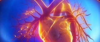
The human heart consists of 2 atria and 2 ventricles (right and left). First, the atria contract and push blood into the ventricles, then the ventricles contract and the blood spreads throughout the body. For this to happen synchronously at regular intervals, there is a conduction system of the heart. The sinus node (SA, SA, sinoatrial, sinoauricular, sinoatrial) generates electrical impulses at a frequency of 60-80 per minute. Electrical impulses are transmitted through nerve fibers like wires to the atria and they contract. At the same time, the impulse is transmitted to the atrioventricular node (AV, AV, atrioventricular), then along the nerve fibers (bundle branches) the impulse passes to the ventricles, as a result of which they contract.
Introductory part
Bradycardia is a decrease in heart rate less than 55 beats per minute.
Sometimes bradycardia is a variant of the norm, for example, for a trained cardiovascular system of athletes. But still, bradycardia is more often a consequence of cardiac dysfunction. Bradycardia can also be observed in healthy people, but in any case, pathological causes of its occurrence should be excluded, since, becoming constant, the reduced heart rate leads to the fact that the internal organs begin to suffer from insufficient blood circulation.
Clinical picture
With a first-degree blockade, as a rule, the patient does not present any specific complaints. Changed health is associated with the underlying disease. In the second or third degree, circulatory disorders of a compensatory and adaptive nature occur: each contraction of the ventricles becomes larger in volume, which leads to myocardial hypertrophy. Cardiac pathology is usually accompanied by a number of symptoms:
- Bradycardia of 30 beats per minute causes insufficient blood flow to the brain, dizziness occurs, and short-term loss of consciousness is possible.
- Patients feel rare strong impulses (beats) of the heart into the chest. This is caused by the overlap of the rhythm of contractions of the atria and ventricles and the formation of single complete regular systoles.
- When listening to the patient's heart, they have the characteristic of a “cannon shot”. During examination of the neck, a pronounced pulsation of the veins is detected due to the return wave of blood into the jugular vein.
- For diagnosis, an important point is the absence of acceleration of the pulse after physical activity, or any fluctuations when holding the breath while taking a deep breath.
The distances between atrial and ventricular contractions are not changed, but are not connected into a single complex
If the blockade is caused by an inflammatory process or unfinished scarring, then all the signs are inconsistent.
In patients with pathological menopause and vegetative-vascular dystonia, the influence of the vagus nerve is pronounced. This is determined by conducting a test with Atropine. After a small dose is administered subcutaneously, the blockade is removed.
A severe manifestation of complete atrioventricular block is Morgagni-Adams-Stokes syndrome, caused in half of the cases by short-term ventricular asystole with preserved atrial flutter. The other half is represented by the outcome of flutter or ventricular fibrillation (hyperdynamic form).
Causes of bradycardia
The cause of bradycardia is changes in the conduction system of the heart that disrupt the propagation of the electrical impulse from the sinus node, causing the heart to contract. Such disorders can be caused by any processes that lead to changes in the heart muscle - myocarditis, atherosclerosis of the coronary vessels leading to coronary heart disease, cardiosclerosis, post-infarction scars, etc.
Risk factors for the disease include:
- diseases of the cardiovascular system, including inflammatory processes
- congenital anomalies
- disorders of the central nervous system
- increased intracranial pressure
Minor and intermittent bradycardia occurs, for example, when wearing a tightly tied tie or other pressure on the carotid artery, since in this case the blood supply to the brain is disrupted and, as a result, feedback to the heart.
In addition, due to pressure on the thyroid gland, temporary dysfunction may occur, namely, a slowdown in the release of hormones, of which, in particular, thyrioedin is directly responsible for the heart rate.
All cases of bradycardia can be divided into 3 groups:
- toxic bradycardia,
- bradycardia of central origin and
- bradycardia of mechanical and degenerative origin
Toxic bradycardia occurs from exposure to the sinus and atrioventricular nodes of so-called cardiac poisons, such as. digitalis, pilocarpine, morphine, muscarine, nicotine, chloroform, chloral hydrate, etc.
Bradycardia of central origin is observed with meningitis, tumors and hemorrhages in the medulla oblongata, increased intracranial pressure, etc.
A slowing of the heartbeat appears reflexively in many diseases of the abdominal cavity and when sensitive receptors on the skin are irritated.
The largest group of slow heartbeats consists of bradycardias of mechanical and degenerative origin. These include cases of bradycardia in diseases of the heart muscle, fatty degeneration of the heart, sclerosis of the coronary arteries, exudative pericarditis, hereditary heart defects, the aging process, the formation of scar tissue changes after myocardial infarction, infectious diseases; and also for unknown reasons.
Bradycardia is usually caused by damage to the sinus node (sick node syndrome) or diseases of the conduction system of the heart (heart block).
B>Sick sinus syndrome (sinus node disease). In these cases, the impulses in the sinus node are significantly reduced and do not meet the needs.
- sinus bradycardia (normal but very infrequent heartbeats)
- sinus node failure (the source of electrical impulses that causes the heart to contract has stopped working)
- SA (sinoatrial) block (impulses are generated, but cannot exit the sinus node) in the presence of symptoms of circulatory failure
Heart block (atrioventricular conduction disorders)
- in this case, either not all excitation impulses of the sinus node reach the ventricles of the heart (incomplete AV block), or they do not pass at all and the atria and ventricles contract independently of each other.
Heart block can occur in the atrioventricular (atrioventricular) node or conducting tissues.
- 1st degree AV block if the patient has clinically significant symptoms (see below) and electrophysiological examination shows that the lesion is located at the very inferior part of the AV junction
- 2nd degree AV block, type 1 – in the presence of symptoms such as weakness, fatigue, dizziness, lightheadedness, syncope
- 2nd degree and 3rd degree AV block are indications for pacing, regardless of symptoms (poor prognosis)
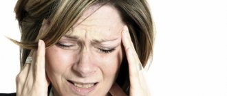
Weakened cardiac function is insufficient to pump the required amount of blood through the circulatory system. The first organ to respond to a lack of blood is the very sensitive brain. Due to insufficient oxygen supply, you may feel dizzy, weak, tired, slow, suddenly short of breath, and even lose consciousness.
In these cases, an artificial pacemaker is used - a pacemaker that can stimulate the heart with electrical impulses. This device sets the heart a constant and adequate rhythm, causing the heart muscle to contract rhythmically. Thus, blood circulation and the supply of oxygen and nutrients to the body are normalized.
to the top of the page
What are the dangers of heart blocks?
During blockades, bradycardia (slow pulse) develops, because not every impulse passes to the heart, the patient may feel interruptions in the functioning of the heart, weakness, etc. For more information about the symptoms of bradycardia, see “Bradycardia: Symptoms and Treatment.”
The most dangerous condition is when a complete block occurs, in which case the heart stops for a few seconds, this may be enough for the patient to lose consciousness. Usually, the heart “starts” on its own, but sometimes this may not happen. Imagine a light bulb with bad wiring and a rusty socket: You turn it on, and it begins to short out - it’s on, then it’s off, it blinks, then it blinks, i.e. works intermittently. Knock - it will light up, then go out again. But at one moment it can go out forever. It’s the same with the heart: the impulse sometimes passes, sometimes it doesn’t, sometimes the heart works normally, sometimes intermittently, sometimes weakness, sometimes fainting, but at one moment it may stop and not start.
You can always buy a new light bulb. Unfortunately, this does not work with the heart; its failure inevitably leads to the death of the patient.
Main symptoms of bradycardia
The clinical picture of bradycardia disease corresponds to the changes in central hemodynamics that develop in this case. Characteristic signs for this pathology are revealed during the initial examination, i.e. during a patient interview. Already on the basis of the most characteristic complaints, a preliminary diagnosis can be made.
The frequency of certain symptoms and the most characteristic complaints depends on the severity of the disease, the severity of hemodynamic changes and the duration of the disease at the time of visiting a doctor.
The patient's age suggests a high probability of developing cardiac arrhythmias. The etiology of the disease may be an important factor in deciding whether pacemaker implantation is necessary.
Main manifestations
- Attacks of loss of consciousness or dizziness during a decrease in pulse (Morgagni-Edams-Stokes attacks or MES prodromes).
- Arterial hypertension or unstable blood pressure, poorly corrected by antihypertensive therapy against the background of bradycardia or periodic decreases in heart rate.
- Decreased tolerance to physical activity, increased fatigue, accompanied by bradycardia.
- Chronic circulatory failure in the large and small circles, not amenable to conservative therapy, with constant or intermittent bradycardia.
- Angina pectoris of exertion and rest in combination with a decrease in heart rate, especially in the absence of evidence of stenotic lesions of the coronary arteries.
- Persistent disability of the patient due to decreased heart rate, disease refractoriness to conservative therapy.
to the top of the page
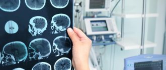
Basic methods for diagnosing cardiac conduction disorders
1. ECG (electrocardiogram)
A standard 12-lead ECG at rest can identify all the main types of cardiac conduction disorders: sinoatrial and atrioventricular block, bundle branch block. Drug tests in combination with ECG are almost never used at present.
2. Holter monitoring (monitoring) ECG
This type of study allows you to record an ECG for a day or more. It allows you to determine whether the patient has significant pauses (cardiac arrest). Pauses longer than 3 seconds are considered significant. If there are no significant pauses, installation of a pacemaker is almost never indicated.
3. Electrophysiological study of the heart (EPS)
This is the most reliable, but complex and expensive method for diagnosing arrhythmias. EPI is performed only in a hospital and requires the installation of several catheters in the veins of the arms and legs. Electrodes are inserted into the heart through these catheters and cardiac stimulation is performed—arrhythmias are caused and eliminated, and their parameters are examined.
To detect the most common types of cardiac conduction disorders, there is a simpler type of EPI - transesophageal EPI. In this case, a thin wire (probe-electrode) is inserted through the mouth or nose into the esophagus and the left atrium is stimulated through it. This type of study is performed on an outpatient basis. In particular, transesophageal EPI makes it possible to determine how long after cessation of stimulation the function of the sinus node (that is, its own pacemaker) is restored - this is necessary in order to make a diagnosis of sick sinus syndrome, one of the most common types of conduction disorders in the elderly.
Diagnostic capabilities
- ECG - allows you to detect bradycardia or transient SA and AV blockades at the time of treatment.
- 24-hour Holter monitoring is the most reliable method for diagnosing incoming heart rhythm disturbances per day of observation (continuous ECG recording).
- EchoCG - a decrease in the ejection fraction below 45%, an increase in the size and volume of the heart that arose against the background of bradycardia, indicates severe myocardial pathology.
- An X-ray examination of the chest allows one to assess the size of the heart shadow and identify signs of venous congestion in the lungs.
- Bicycle ergometry allows you to identify coronary artery disease and assess the adequate increase in heart rate contractions during physical activity.
- Transesophageal examination of the cardiac conduction system is carried out to confirm the diagnosis in cases where transient blockades cannot be detected by conventional methods (ECG, Holter monitoring), in the presence of clinical manifestations. In some cases, it allows one to verify the organic or functional etiology of cardiac arrhythmias.
to the top of the page
Prevention
Prevention of atrioventricular (AV) heart block involves treating the underlying disease. Additionally, the patient is prescribed a cardiological diet, moderate physical and emotional stress, and cessation of smoking and alcohol consumption. It is important to follow the doctor's prescriptions and recommendations for successful therapy.
To prevent the development of the disease, it is necessary to undergo a timely examination by a cardiologist. At the cardiology center of the Federal Scientific and Clinical Center of the Federal Medical and Biological Agency, specialists will select an examination program for you and prescribe adequate treatment.
Take care of yourself and your heart!
Therapy and surgical treatment
AV blockade 1st degree
It is difficult to determine the level of blockade using a regular “electrocardiogram”. PQ interval is greater than 0.20s. Each P wave corresponds to a QRS complex.
Causes:
observed in healthy individuals, athletes, with increased parasympathetic tone, taking certain medications (cardiac glycosides, quinidine, procainamide, propranolol, verapamil), rheumatic attack, myocarditis, congenital heart defects (atrial septal defect, patent ductus arteriosus). With narrow QRS complexes, the most likely level of AV block is a node. If the QRS complexes are wide, conduction disturbances are possible both in the AV node and in the His bundle.
Treatment.
If the PQ interval does not exceed 400 ms and there are no clinical manifestations, no treatment is required.
AV blockade 2nd degree
Increasing prolongation of the PQ interval until the loss of the QRS complex.
Causes:
observed in healthy individuals, athletes, when taking certain medications (cardiac glycosides, beta-blockers, calcium antagonists, clonidine, methyldopa, flecainide, encainide, propafenone, lithium), with myocardial infarction (especially lower), rheumatic attack, myocarditis. With narrow QRS complexes, the most likely level of AV node blockade. If the QRS complexes are wide, disruption of impulse conduction is possible both in the AV node and in the His bundle.
Mobitz I type.
With Holter monitoring, it is detected in almost 6% of healthy individuals, especially often in athletes. Typically, AV blockade appears during sleep, when parasympathetic tone is increased. In lower myocardial infarction, AV blockade type Mobitz I often serves as a precursor to complete AV blockade, which in such cases is accompanied by a stable AV nodal escape rhythm, is well tolerated and does not require a pacemaker.
There are no clinical manifestations:
no treatment required.
In case of hemodynamic disorders:
atropine, 0.5-2.0 mg, IV, then “electrocardiostimulation” (ECS). If AV blockade is caused by myocardial ischemia, then the level of adenosine in the tissues is increased; in such cases, an adenosine antagonist, aminophylline, is prescribed.
Type Mobitz II.
The level of blockade is distal to the AV node (in the bundle of His or its legs). High risk of progression to complete AV block with a slow idioventricular rhythm, which entails a sharp decrease in cardiac output.
Treatment.
Regardless of the clinical manifestations, temporary, then permanent “electrocardiostimulation” (ECS) is indicated.
Complete AV block.
The atria and ventricles are excited independently of each other. The frequency of contractions of the atria exceeds the frequency of contractions of the ventricles. Same PP intervals and same RR intervals, PQ intervals vary. Causes: complete AV block can be congenital. An acquired form of complete AV block occurs with myocardial infarction, isolated disease of the cardiac conduction system (Lenegra's disease), aortic defects, taking certain medications (cardiac glycosides, quinidine, procainamide), endocarditis, Lyme disease, hyperkalemia, infiltrative diseases (amyloidosis, sarcoidosis ), collagenosis, trauma, rheumatic attack. Impulse conduction blockade is possible at the level of the AV node (for example, with congenital complete AV block with narrow QRS complexes), the His bundle, or the distal fibers of the His-Purkinje system.
Treatment
— permanent “electrocardiostimulation” (ECS). If the causes of blockade are reversible (for example, hyperkalemia), if blockade occurs in the early postoperative period or with lower myocardial infarction, temporary pacemaker is limited.
Congenital complete
AV blockade is accompanied by a stable replacement AV nodal rhythm, usually does not cause hemodynamic disturbances, is well tolerated and does not require pacemaker. Tactics while awaiting pacemaker implantation depend on the type of replacement rhythm and its stability.
For hemodynamically significant bradyarrhythmias
- constant ECS. With dual-chamber and atrial pacemaker, the risk of stroke is significantly lower than with ventricular pacemaker
to the top of the page
Pacemakers
Currently, there are many options for permanent pacing. They have one thing in common: under the skin of the anterior chest wall (usually near the collarbone) an electric pacemaker is implanted - a small metal box, the wires from which (electrodes) go through the veins to the right side of the heart. These wires sense the heart's own electrical activity (to synchronize their work with it) and transmit impulses to the heart. Modern pacemakers almost always stimulate both the atria and the ventricles. In addition, they are tuned so that the heart rate increases during exercise and decreases at rest. There are essentially no contraindications for cardiac pacing: pacemaker implantation is a simple and harmless procedure that can be performed at any age.
A separate type of cardiac pacing is the so-called biventricular stimulation. It is performed not for conduction disorders themselves, but to achieve synchronous contraction of all the walls of the left ventricle. Therefore, the installation of a biventricular stimulator is also called resynchronization therapy. Electrodes from the stimulator go to the right ventricle and to the coronary sinus (which is directly adjacent to the left ventricle). This type of treatment greatly helps some heart failure patients.
Some pacemakers also have a defibrillator function: they detect life-threatening arrhythmias and automatically deliver a shock to stop them.
Patients with pacemakers lead normal lives. They just need to avoid the influence of a strong magnetic field. Thus, magnetic resonance imaging (MRI) is contraindicated in patients with implanted pacemakers.
From time to time, patients with implanted pacemakers need to be seen by specialists: to check the serviceability of the stimulator (in particular, its battery capacity), and to adjust the stimulation parameters.
Share: