- How does a hemorrhagic stroke occur?
- What types of hematomas occur in hemorrhagic stroke?
- Symptoms of hemorrhagic stroke
- Recovery time after hemorrhagic stroke
- Rehabilitation objectives
- Recovery points after hemorrhagic stroke
- Types of rehabilitation after hemorrhagic stroke
- Drug treatment
- Motor recovery
- Movement restoration techniques
- Speech restoration
- Restoring breathing and swallowing
- Psychological recovery
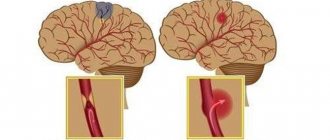
Hemorrhagic stroke is a condition of acute circulatory disturbance in the brain, accompanied by hemorrhage into the cranial cavity and leading to the formation of hematomas.
Today, hemorrhagic stroke is a serious condition leading to disability. The disease mainly affects men, but most often it is women who die. Mortality is very high, with hemorrhage leading to the death of almost 50% of patients. This occurs within 30 days after the attack occurred.
But even after the crisis is over, the struggle must continue, because problems with blood circulation that began in the brain have many consequences. But in this case, rehabilitation after a hemorrhagic stroke, along with the patient’s gradual return to normal life, is possible.
How does a hemorrhagic stroke occur?
Cerebral hemorrhage during a stroke is a spontaneous phenomenon. The functioning of the brain requires large amounts of oxygen from the blood. Nutrition is produced thanks to two carotid and two vertebral arteries, which form a circle between themselves passing through the base of the brain.
When a vessel is damaged, acute blood failure occurs, which is necessarily compensated by healthy vessels. However, over time, the effectiveness of such a mutual assistance system decreases. The appearance of cerebral hemorrhage during a hemorrhagic stroke is caused by sudden ruptures of the vascular walls or their increased permeability due to other chronic diseases.
During a cerebral hemorrhage, the death of brain cells that are damaged by blood contact occurs. In nearby areas of the brain, oxygen deficiency begins, since blood does not reach them and cannot move further through the vascular system.
What types of hematomas occur in hemorrhagic stroke?
Types of hematomas in cerebral hemorrhage or hemorrhagic stroke:
- Parenchymatous with the distribution of blood directly inside or under the membrane of the brain, with the formation of hematomas and with saturation of the nerve brain tissue. With such a hemorrhagic stroke, severe neurological deficits can form.
- Intraventricular with the effusion of blood into the ventricles of the brain, with tissue penetration and the formation of a hematoma. With this type, death especially often occurs within 3-5 days.
- Subarachnoid hemorrhages in the area between the arachnoid and pia mater of the brain.
- Mixed types of strokes, in which focal hemorrhages appear in different parts of the brain.
There is also a classification based on the volume of blood that forms hematomas: small - up to 20 ml, medium - in the range of 20-50 ml, large - from 50 and above.
General principles of treatment
After admission to the hospital, the neurologist assesses the severity of the condition, and then determines whether the patient needs to be operated on or whether it is better to treat him conservatively.
Many doctors agree that the most dangerous are the 3rd day after a vascular accident, and stabilization of the condition occurs on days 5-7-14, depending on the severity of the pathology.
Regardless of the type of stroke, treatment begins with:
- Bed rest.
- Elimination of any physical stress. For this purpose, laxatives and other symptomatic drugs may be prescribed, for example, antitussives for severe dry cough.
- Organization of patient care for the prevention of infectious complications and bedsores.
- Adequate nutrition depending on the level of consciousness: if the patient can swallow on his own, he eats gentle food through the mouth, if he cannot, through a tube.
- Prescription of drugs that regulate blood clotting. This is necessary to stop bleeding or prevent its recurrence.
- The use of neuroprotectors - medications that reduce the “suffering” of brain cells from oxygen starvation.
Medicines are prescribed only when they are necessary and not contraindicated.
Symptoms of hemorrhagic stroke
With hemorrhoidal stroke, symptoms most often appear suddenly, mainly during the daytime and during very heavy physical activity. The following symptoms are typical:
- convulsions;
- Strong headache;
- sudden loss of consciousness;
- nausea and vomiting;
- serious depression of a person's consciousness, similar to or leading to a coma
In addition, general symptoms of a stroke appear:
- decreased facial expressions and muscle strength affecting any part of the body;
- the occurrence of speech disorders and problems with visual perception;
- loss of coordination of movement with impaired ability to move.
During an attack, the patient's skin becomes cold and sometimes acquires a purplish-bluish tint. Breathing becomes more frequent, noisy, with wheezing. Sometimes, on the affected side, the pupil dilates, diverges, or chaotic movements of the eyeballs occur.
An important indicator of the severity of the patient’s current condition is comatose status with loss of consciousness, which can last several days. This makes the prognosis very unfavorable.
Subarachnoid hemorrhage most often results from rupture of a cerebral aneurysm. As a result, the patient experiences severe headaches localized in the back of the head and forehead. Then it spreads to the rest of the head. Later, other symptoms of hemorrhagic stroke appear.
In case of extensive cerebral hemorrhage, neurosurgical microtechnical operations are performed. In this case, removal of the hematoma leads to a decrease in pressure on the brain tissue and prevents the development of brain edema. But surgery is performed strictly for medical reasons.
In case of an aneurysm, surgery is performed to stop bleeding, and the patient is prescribed hemostatic drugs. Often with subarachnoid lesions, narrowing of the vessel occurs and the development of ischemic stroke. In this case, calcium channel blockers are necessarily prescribed to prevent narrowing and spasm of the vascular walls.
In the first 3 weeks after the attack, the patient’s condition remains the most severe due to progressive cerebral edema, as well as a gradual increase in cerebral symptoms. If there are any chronic pathologies in the body, aggravated by a hemorrhagic stroke, the patient may die. A month later, the patient begins to experience regression of cerebral symptoms and blood pressure stabilizes.
This is interesting! It is recommended to always have glycine in your home medicine cabinet. Many qualified doctors classify this drug as a neuroprotector or a substance that protects brain tissue.
If unpleasant weakness in the limbs or speech disorder occurs, you should immediately put 5 glycine tablets under your tongue. To some extent, such prevention can prevent acute brain damage and the development of hemorrhagic lesions.
Many people are interested in the difference between ischemic and hemorrhagic stroke. Two such lesions have almost identical symptoms, but have different reasons for the development of such a serious pathology. With ischemic stroke, the prognosis is favorable and mainly depends on the degree of brain damage that has occurred.
The following symptoms are characteristic of each type of this disease:
- suppression of speech ability;
- partial or complete paralysis of the body and limbs;
- impairment of visual sensitivity or its complete loss;
- impaired coordination of movements;
- complete loss of hearing in a hemorrhagic condition;
- • serious inhibition of the functions of the cerebral cortex;
- • increased blood pressure.
The consequences of a stroke mainly depend on the location and extent of the existing lesion. A hemorrhagic attack has an acute onset and progresses rapidly. The onset of coma in this case is possible within the first few minutes or hours. With ischemic stroke, the symptoms are less severe.
Damage to the left hemisphere leads to paralysis of the right side of the body. Hemorrhage in the cerebellar area deprives the patient of sensitivity, the ability to swallow, see and speak. Often the consequence of a hemorrhagic stroke is the gradual development of dementia or dementia.
Causes of hemorrhage
Hemorrhage due to arterial hypertension
Fig.16
Hemorrhage due to cavernous angioma
Fig.17
Fig.18
Hemorrhage due to cerebral amyloid angiopathy
Fig.19
Recovery time after hemorrhagic stroke
The rehabilitation performed after such a brain injury consists of several stages:
- early period, lasting about six months after the attack;
- later recovery, lasting from six months to 1 year;
- the stage of final recovery, which in its duration occupies the entire subsequent time.
A year after the attack, residual effects begin. The best results during rehabilitation can be obtained in the first year; for this reason, recovery should not be delayed.
Effective results are obtained by strictly observing the following important principles:
- taking action at an early stage of ongoing treatment in a hospital;
- daily implementation by the patient of appropriate recommendations without any delays;
- prescribing special physical activity with appropriate intensity and gradual complication of exercises.
In addition, it is very important to take comprehensive measures: prescribing physiotherapy, drug treatment and the necessary correction of the patient’s psychological state. During this period, a recovering person must protect himself from unnecessary stressful situations, and also be surrounded by the care and love of family and friends.
Rehabilitation objectives
The main objectives of the ongoing rehabilitation are:
- Work on restoring lost everyday and physiological functions of a person suffering from a stroke. Restoring full range of motion, the ability to independently care for oneself and perform simple household chores.
- Restoration of lost professional ability to work with a return to the previous place of activity or assistance in retraining.
- Maintaining the necessary social activity of a person, first of all, restoring his contact with loved ones and the ability to make new acquaintances;
It is very important to prevent a possible relapse and correct the patient’s current lifestyle.
Recovery points after hemorrhagic stroke
Complications for the patient mainly depend on the location of the hemorrhage that occurs, as well as its volume. Today there are the following groups of possible threatening consequences:
- Motor disorders. The appearance of severe weakness and paresis in a person, the patient’s inability to accept and maintain a sitting position. This may be paralysis of one side of the body, high muscle tone or spasms that occur, which seriously complicates self-care, requires mandatory assistance and causes a lot of inconvenience to the person.
- Impaired sensitivity is, for example, numbness of the limbs, a feeling of “insects under the skin” and burning, the inability to control the hands, leading to various everyday problems.
- The appearance of speech pathologies after a stroke is confusion of speech and the inability to correctly pronounce individual sounds or loss of the ability to maintain a conversation. There may be loss of skills such as recognizing the meaning of words, counting, reading, telling time by a clock, and understanding calendar periodicity.
- Problems with swallowing when eating food and various liquids, and in some cases, complete loss of the ability to eat independently.
- Disorders of the excretory system: the appearance of urinary and stool incontinence, problems with the intestines and urinary system that arise on an ongoing basis.
The patient often develops acute and chronic psychological and mental disorders in the form of depression, excessive emotional temper or apathy. Sometimes partial amnesia occurs: the inability to recognize familiar people or objects, the understanding of performing simple everyday actions disappears.
Focal symptoms
Focal manifestations are a group of symptoms characterized by a number of specific neurological signs that occur depending on the location of the lesion.
The most common focal manifestations are:
- weakness in the arm/leg/half of the body, up to paralysis;
- violation of the movement of facial muscles, due to which the eyelid does not rise, the corner of the mouth drops and the cheek “sails” when exhaling;
- impaired skin sensitivity;
- pathology of speech, vision, hearing;
- exotropia;
- swallowing disorder;
- spatial disorientation of varying severity - staggering, tilting and turning to the side when walking, inability to assume a vertical body position.
Sometimes, based on characteristic focal manifestations, the localization of the pathology can be assumed:
- When the lesion is located in the frontal lobe, characteristic symptoms may be a forced rotation of the patient’s head and eyes in the direction of the hemorrhage. The so-called “frontal psyche” often develops, which manifests itself in an inadequate assessment of the state of health and a decrease in criticism of one’s actions.
- The localization of the hematoma in the temporal lobe may be indicated by early epileptic seizures.
- Hematomas of the parieto-occipital localization can sometimes have scant symptoms and are detected by chance during CT or MRI of the brain, which indicates the importance of neuroimaging methods.
- Hemorrhagic stroke in the brainstem often manifests as breathing and heartbeat disturbances.
- Damage to the cerebellum is characterized by loss of consciousness, vomiting, severe dizziness and loss of coordination.
It should be noted that these clinical manifestations can be seen with small hematomas. With its significant size, cerebral edema rapidly increases and the patient’s condition becomes so severe that it becomes impossible to determine focal symptoms.
Types of rehabilitation after hemorrhagic stroke
The necessary medicinal and motor rehabilitation is carried out for the following purposes:
- significant reduction in the risk of recurrence of hemorrhagic stroke;
- gradual return to normal lifestyle;
- return of lost ability to work;
- the patient performs basic skills and actions;
- preserving the patient’s identity through the provision of social and psychological assistance.
Drug treatment
In the human brain, dead neurons are replaced with elements with high activity. But due to the increasing load, additional energy is necessarily required. Neurons that remain alive after damage restore lost functions over time.
Prescribed medications provide additional nutrition and significantly improve recovery processes in the brain. Additional courses of essential drug therapy are recommended once every three months throughout the year.
Drugs that are administered to the patient by injection during treatment:
- nootropics, which include piracetam, Actovegin and other drugs;
- medications to stimulate the necessary impulses in brain cells, for example, proserin or neuromidin;
- selected B vitamins that improve cell metabolism.
Taking the prescribed medications should continue even after completion of the prescribed treatment in the hospital. At home, tablets are taken with food.
Chronically high blood pressure is corrected with medication. If necessary, the doctor prescribes suitable medications for blood pressure, calcium channel blockers in vascular cells, or drugs to lower blood sugar levels.
Motor recovery
It is necessary to restore lost movement already at the stage of inpatient treatment after primary care has been provided. It is imperative to provide physical activity to the limbs to improve blood circulation, which helps in reducing tone and prevents the appearance of congestion in the blood vessels, and also protects the patient from the occurrence of pneumonia due to a recumbent lifestyle.
The position of the limbs in a supine state, if necessary, is corrected using a splint or special weights. Such patients must turn over every 2 hours to prevent the appearance of bedsores on the skin; in addition, this is recommended to stabilize blood pressure.
A few days after the attack, passive gymnastics begins, during which the person is assisted in smooth movement of various parts of the body. Procedures are carried out only if the person does not experience pain. If the condition improves, you should help the patient learn to take a sitting position, and then help him restore an upright position. Before walking, leg training is performed in order to regain the lost sense of position in space. Then the patient needs to begin to move around using supports, walkers and canes.
Movement restoration techniques
Physical therapy plays a significant role in the rehabilitation of lost motor ability. In this case, suitable exercises are developed for different muscle groups, classes are conducted using special simulators, and a device is used to reduce high muscle tone.
Prescribing a special massage is very useful. First, the limbs are stroked with a sufficiently high muscle tone, and then other muscle groups are rubbed. The use of a warm heating pad before the session significantly improves the effect. The massage procedure can take 20 minutes and mainly depends on the current condition of the patient.
Effective methods of physiotherapy include oxygen baths, electrophoresis of blood vessels in the neck, and electrical stimulation of muscles that have lost their functions as a result of a stroke.
Speech restoration
The return of speech is possible even after an attack that occurred more than a year ago. Conversation after a stroke should be slow and words pronounced clearly. At the same time, there is no need to rush to answer and ask difficult questions.
Working with a speech therapist helps a lot in restoring muscle functionality. Practicing in front of a mirror can also be very useful. In the process of noticeable improvements, it is possible to complicate tasks and stimulate a person to pronounce more complex spoken phrases.
Restoring breathing and swallowing
Nutrition of patients in a hospital is most often performed using a special feeding tube. Then comes the time to independently master the skill of eating food. Proper preparation of foods greatly simplifies the rehabilitation of a patient seriously affected by a stroke. Cooked food should be warm, not hard and have a soft texture. Flavorful foods trigger saliva production.
Patients should absolutely not be rushed during the process of eating; this process can take half an hour or more. You may need help holding a utensil or spoon. Restoration of the functions of the swallowing muscles occurs with regular repetition of the nutritional process.
Diagnosis of the disease
During the initial examination, the doctor collects anamnesis and conducts a neurological examination. In some cases, this is enough to make a diagnosis and classify the disease. If localization cannot be established, additional examinations are prescribed.
What diagnostic methods are used:
- emergency general and biochemical blood test;
- ECG;
- coagulogram to assess blood clotting;
- CT;
- MRI.
The images allow you to determine the location of the damaged area, its size and damage to the brain.
If it is necessary to identify the source of hemorrhage, CT angiography is prescribed.
