Under hip dysplasia
(HJ) refers to a deviation from the normal development of the joint, which is a congenital pathology that leads to various types of injuries. Medical practice shows that, often, joint underdevelopment occurs in combination with insufficiently formed connective tissue.
In the early stages, it may manifest itself in visually noticeable asymmetry of skin folds or shortening/limited mobility of the lower limb.
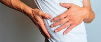
It is customary to note chronic pain, pronounced lameness, and increased fatigue of the affected limb as consequences.
Diagnosis of a pathological disease is carried out taking into account the characteristic signs, as well as the results of instrumental research methods.
In the treatment of hip dysplasia, specialized means of therapeutic exercises, fixation, and a complex of drug therapy are used.
General information
Hip dysplasia is a congenital pathological disease that causes dislocation (subluxation) of the hip in a newborn.
In each clinical case, the existing degree of underdevelopment of the articular joint may vary to some extent (both gross violations and increased mobility can be observed).
To avoid complications, it is important to diagnose and treat this type of disease as early as possible - immediately after birth and in the first months of a person’s life.
According to current data, hip dysplasia is one of the most common congenital pathologies. Specialists in the field of traumatology and orthopedics note that out of 1000 infants, the disorder occurs in 2-3% of cases.
The female half of humanity is more susceptible to the disease than the male half, which accounts for up to 80% of diagnosed cases.
Massage and exercise therapy
Exercises and massage are performed before feeding: these procedures stimulate blood circulation and improve nutrition of the structures of the hip joint. As a result, the growth processes of cartilage and bone tissue are stimulated, nerve conduction is enhanced - and the joint is formed correctly.
Massage movements are performed smoothly and gently. Apply stroking, rubbing and kneading the muscles of the thighs, buttocks, and lower back. The newborn is laid out on both his back and stomach. The duration of the massage is about 5 minutes. After the procedure, you can leave him to lie on his stomach for some time so that his legs hang down to the sides. This hardens and further strengthens the body.
A set of exercises is selected by a physical therapy doctor or pediatrician according to the degree of development of the disease. Most often this is: abduction of bent legs to the sides (contraindicated in slip syndrome), flexion and extension in the hip and knee joints. The movements are performed very smoothly. At first, they are recommended to be done in water, while swimming. The duration of the gymnastics is also about 5 minutes.
To work with a newborn at home, parents need to attend massage and exercise therapy courses at the clinic.
Causes of hip dysplasia in children and adults
The occurrence of a disease can be determined by a combination of several reasons.
A hereditary predisposition is clearly visible - the pathological disorder occurs 10 times more often in patients whose parents had pathological disorders of the hip joint.
There are high risks of giving birth to a baby with the pathology in question during presentation (breech).
The risks of the disease are highest among children whose mothers experienced severe toxicosis or gynecological diseases during pregnancy. Risk factors also include oligohydramnios and relatively large fetal size.
Numerous studies suggest a relationship between the frequency of diseases and the favorable environmental situation in the patient’s region of residence. According to the data obtained, in a polluted environment, the disease occurs several times more often than in regions with a favorable environment.
It is also worth noting that the progression of pathology can also be influenced by traditional features of swaddling newborns. If new parents refuse swaddling and the baby’s legs are in a natural position, the diagnosis is several times less common than where it is customary to swaddle children tightly.
Features of caring for newborns with dysplasia
With the right approach to treatment and care, dysplasia in newborns can be overcome. If your baby has disorders in the development of the hip joints, then he needs daily care and constant observance of special rules when carrying, feeding, and putting to bed.
- Hip dysplasia in newborns eliminates vertical loads on the legs.
- If the child is in a lying position, then his feet should hang slightly, this way better relieves tension from the thigh muscles.
- Transportation by car in a special child seat that does not interfere with the wide spread of the legs.
- The correct position when carrying in your arms: hold the baby in front of you by the back, while his legs should tightly grasp you from behind.
- Make sure that when feeding and sitting down, the hips are separated as much as possible.
The hip joint is an important supporting element of the human skeleton. He is constantly exposed to heavy loads when carrying heavy loads, running, and long walks. It is necessary to monitor the correct full development of this joint from infancy, otherwise in adult life the disease will still make itself felt, but it will be much more difficult to cure than dysplasia in newborns.
Pathogenesis
Considering the design of the hip joint, one can identify two key elements that form it: the femoral head and the acetabulum. In the upper part of the cavity there is a plate of cartilage tissue, due to which the area of contact of the articular surfaces increases.
With normal development, the hip joint of a child is significantly different from that of adults: the acetabulum is flatter, almost vertical, and the ligaments are incredibly elastic.
Violations of the hip joint departments lead to the impossibility of maintaining the articular elements in their natural position, which leads to their inevitable displacement. In a situation where the femoral head moves as high as possible, a dislocation occurs.
A long-term absence of measures to correct the pathologically incorrect position of the bones can cause accumulation of connective and fatty tissues in the acetabulum, which aggravates the situation and can make reduction impossible.
Severity of traffic accidents
- I degree – pre-dislocation. A developmental deviation in which the muscles and ligaments are not changed, the head is located inside the beveled cavity of the joint.
- II degree – subluxation. Only part of the femoral head is located inside the articulation cavity, as it moves upward. The ligaments are stretched and lose tension.
- III degree – dislocation. The head of the femur comes completely out of the socket and is located higher. The ligaments are tense and stretched, and the cartilaginous rim fits inside the joint.
Types of disease
Today, it is customary to distinguish several types of hip dysplasia, including:
- acetabular – disruption of the development of the acetabulum exclusively (flattening, reduction in size). In this case, there is significant underdevelopment of the cartilaginous rim;
- femoral dysplasia - with normal development, the femoral neck connects to its body at a certain angle, a decrease or increase in which leads to the progression of pathological processes;
- rotational – has a direct connection with a violation of the location of anatomical formations in the horizontal plane. During normal development, the axes of movement of the joint elements do not coincide. When the amplitude of axial misalignment increases beyond the norm, the natural position of the femur is significantly disrupted.
Symptoms of hip dysplasia revealed during examination
Signs of hip dysplasia in children are detected mainly by visual examination. They are also defined as the main symptoms of pathology. These include:
- Violation of the classic configuration and depth of skin folds. Visual inspection of the folds under the buttocks, in the popliteal cavities and groin allows us to draw conclusions about the general development of the limbs. Normally, all folds should be located at the same level.
- The symptom cannot be the only basis for making a diagnosis, since in most newborns the folds still have minor differences. Shortening of the limb. One of the reliable signs, occurring exclusively in severe cases, in conditions of a fully formed dislocation. Checking the symptom is carried out by stretching the child’s legs to compare the location of the level of the kneecaps.
- Symptom of sliding (“click” or Marx-Ortolani). The most reliable and accurate way to determine pathology, which is implemented quite simply: the patient is placed on his back, the doctor grabs the lower limbs so that his thumbs are on the inside and the rest are on the outside. Then, an attempt is made to spread the limbs to the sides. Without disturbing the configuration of the hip, the newborn can be placed almost completely on the table surface. With pathology, the thigh on the injured side is abducted only up to a certain point, upon reaching which a noticeable “click” occurs.
- Limitation of the amplitude of limb abduction. The manifestation of this symptom can be traced exclusively after 2-3 weeks of age, when it is possible to lower the child’s healthy limb onto the table almost completely, but the affected one cannot. The persistence of the diagnosis into older age can also be traced through gait disturbances. In addition, when the child is in a “standing” position, asymmetry of the buttocks, as well as inguinal and popliteal folds, is noticed.
Reduction of congenital hip dislocation
Indications for reduction of congenital hip dislocation:
- The child is over 1 year old. Before this, the dislocation is relatively easily reduced using functional techniques (splints and orthoses, see above). But there is no single unambiguous algorithm. Sometimes a dislocation after 3 months of age cannot be corrected by any means other than surgery.
- The child's age is no more than 5 years. At an older age, it is usually necessary to resort to surgery.
- The presence of a mature hip dislocation, which is determined by radiography and/or ultrasound.
Contraindications to closed reduction of congenital hip dislocation:
- Severe underdevelopment of the acetabulum;
- Severe displacement of the head of the femur, inversion of the articular capsule into the joint cavity.
Closed reduction for congenital hip dislocation is performed under anesthesia. The doctor, guided by X-ray and ultrasound data, performs a reduction - returning the femoral head to the correct position. Then, for 6 months, a coxite (on the pelvis and lower limbs) plaster cast is applied, which fixes the child’s legs in an extended position. After removing the bandage, massage, therapeutic exercises, and physiotherapy are performed.
However, some children develop a relapse after closed reduction of congenital hip dislocation. The older the child, the more likely it is that surgery will eventually be necessary.
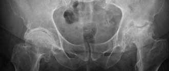
Diagnostics
The diagnosis usually occurs in the maternity hospital, in accordance with the signs of hip dysplasia in children, but can also be made at a later date. The issue of diagnosis can be dealt with by a pediatrician or highly specialized specialists, including a pediatric orthopedist.
If it is necessary to clarify the features, additional research methods are used. Let's look at them in more detail.
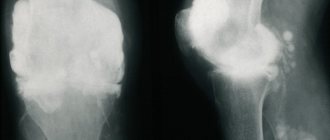
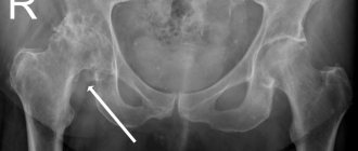
X-ray diagnostics
Children have certain structural features of the spine, which implies the absence of ossification of some components of the hip joint. In place of the femur and pelvic bone there is cartilaginous tissue, which is quite difficult to x-ray.
In order to assess the correct configuration of anatomical structures, special schemes are used, which include performing radiography in a direct projection with drawing auxiliary lines that have a name and are outlined in a certain place:
- midline – a vertical straight line passing through the middle of the sacrum;
- Hilgenreiner's line is a horizontal straight line passing through the upper outer edge of the acetabulum;
- Perkin's line is a vertical straight line passing through the upper outer edge of the acetabulum;
- Shenton's line is straight, continuing the edge of the obturator foramen of the pelvic joint.
The level of formation of the hip joint is determined by the acetabular angle, the normal indicators of which are considered to be:
- at birth – 25-29°;
- per year - for boys up to 18.5°, for girls up to 20°;
- at 5 years of age – 15°, regardless of gender.
Ultrasound diagnostics
Ultrasound diagnostics involves the use of ultrasonography and is actively used in the diagnosis of children under 1 year of age.
The key advantage of this technique is obtaining optimally accurate results, without causing harm to the child’s body. There are no contraindications to this type of procedure.
Indications for ultrasonography are considered to be:
- the presence of factors that include the patient at risk;
- determination of characteristic features.
During the diagnostic process, the doctor takes a picture, the results of which are in many ways similar to the results of an x-ray examination in the anteroposterior projection.
When forming a conclusion for a diagnostic procedure of the presented type, the following indicators are assessed:
- alpha angle – gives an assessment of the degree of formation and allows you to determine the inclination of the acetabulum (its bony part);
- beta angle - helps to assess the degree of formation and the angle of inclination of the acetabulum (its cartilaginous part).
Ultrasound is the preferred research method for suspected dysplasia in young children, due to the safety and informativeness of the results.
Treatment of hip dysplasia in children and adults
The disease has a number of features that can lead to serious consequences, and therefore it is recommended to begin treatment as early as possible.
Today, there are several areas of treatment for hip dysplasia, the combined use of which helps prevent complications and create conditions for successful treatment.
Physiotherapy
Physiotherapeutic treatment has a beneficial effect on the condition of the hip joint with dysplasia. The following may be prescribed as treatment and rehabilitation measures:
- electrophoresis using calcium, phosphorus and iodine;
- fresh baths;
- exposure to ultraviolet light.
The number and duration of procedures are determined by the attending physician, in accordance with the existing clinical picture.
Medicines
Diseases of the musculoskeletal system, in order to speed up treatment and achieve the fastest possible results, require taking a group of drugs of various types.
The determination of specific drugs, the duration of their use and, of course, the dosage is determined exclusively by the attending physician, taking into account the characteristics of the treatment and the age of the patient.
A special role in recovery from diseases of the musculoskeletal system is played by chondroproctors, one of the most effective of which is considered to be the drug “Artracam”. With their help, damaged tissues are regenerated faster, which helps prevent regression and improve the functionality of the affected joint.
Surgery
Some cases of dysplasia require surgery. Indications for the use of radical treatment are:
- late diagnosis of pathology (after the first year of life);
- presence of anatomical defects;
- pinching of cartilage tissue;
- severe displacement of the bones, without the possibility of their reduction using a closed method.
It is worth noting that surgical treatment can lead to a large number of complications, so it is important to identify the problem in a timely manner.
Therapeutic physical education (PT)
Therapeutic exercises are an integral part of the conservative treatment of the disease. The use of exercises is recommended not only during the treatment process, but also at the rehabilitation stage.
When treating dysplasia in patients under one year of age, exercise therapy is performed passively, as an element of a complex therapeutic massage.
Application of orthopedic structures
By orthopedic design, as a rule, we mean Pavlik Stirrups, which are the great development of a doctor from the Czech Republic since 1946. The design is soft enough to allow the child to move his limbs freely.
The structure of an orthopedic solution involves such structural elements as:
- chest band;
- a bandage attached to the lower leg;
strap connectors for bandages.
There are other types of orthopedic structures, including: Freik panties, Vilensky splint, CITO splint, etc.
Consequences
If there is no treatment, then at an early age this can threaten the child with serious troubles. Children develop a limp when walking; it can be either barely noticeable or pronounced. Also, the baby will not be able to move his leg to the side, or will do it with great difficulty. The child will be bothered by constant pain in the knees and pelvis with possible bone distortion. Depending on the severity of the symptoms of dysplasia, children experience muscle atrophy of varying severity.
Gradually, as the child grows, the consequences of untreated dysplasia will worsen and be expressed in the development of the so-called “duck gait,” when the baby rolls from one leg to the other, protruding the pelvis back. The motor activity of such a child will be limited, which will entail underdevelopment not only of other joints, but will also affect the functioning of all organs and overall physical development. In the future, the leg muscles can completely atrophy, and the person will begin to be haunted by constant, incessant pain. In adult patients, hyperlordosis of the spine in the lumbar region is observed. All organs located in the pelvic area are also affected.
All this can be avoided if you start treatment on time and follow preventive measures.
Treatment prognosis
Timely and high-quality treatment of pathological changes implies positive prognoses.
The absence or insufficiency of treatment, deviation from the course determined by the attending physician, or refusal of therapeutic procedures can lead to serious consequences, including the development of severe deforming arthrosis.
There are cases when patients with dysplasia live without knowing they have the disease. In a situation where pathology is detected by chance during an X-ray examination, it is important to ensure constant monitoring by an orthopedist (at least once a year).
In order to preserve the quality of life, it is recommended to conduct timely preventive examinations of all newborns, as well as to implement complete and timely treatment of the identified disease.
Possible complications
Without treatment, minor changes may go unnoticed quite unnoticeably, but not without consequences. In the period from 25 to 55 years, a patient with the diagnosis in question has a significantly increased risk of dysplastic type coxarthrosis, the primary symptoms of which begin to appear against the background of a significant decrease in motor activity.
The key features of such an unpleasant complication are considered to be the acute initial phase and the intensity of progression. The pathology manifests itself not only as unpleasant sensations localized in a certain part of the musculoskeletal system, but also as pain, as well as a significant limitation in the mobility of the limb.
Unreduced congenital dislocation of the hip over time can cause the formation of a new, defective joint, in addition to which shortening of the limb and disruption of the muscular frame can be observed.
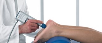
Physiotherapy
Many physiotherapeutic procedures are used that eliminate the inflammatory reaction, improve joint trophism and reduce joint pain. The most commonly used procedures are:
| Electrophoresis | Using this procedure, you can introduce anti-inflammatory and painkillers into the joint cavity. |
| Mud therapy | During this procedure, the blood vessels dilate, resulting in improved blood flow in the joints. |
| Ultrasound | This treatment also has an anti-inflammatory and resorption effect. |
How to prevent the disease?
As the main preventive measures to prevent the onset of the disease and its progress in general, it is recommended:
- early diagnosis aimed at identifying all kinds of pathologies of the musculoskeletal system (orthopedic examination in the maternity hospital, ultrasound, x-ray examination);
- using the loose swaddling technique - tight swaddling can provoke the development of the disease. It is best to adhere to the principles of maintaining the natural position of the limbs (bent at the hips, spread apart);
- using a sling to carry a child – carrying children on the parents’ side or back is considered to be a fairly physiological position;
- purchasing larger size diapers - the use of large size diapers will create a kind of orthopedic structure that contributes to the correct formation of the musculoskeletal system;
- attending massage courses and performing gymnastic exercises - courses of 10-15 sessions at an optimal interval of a month, together with daily general massage, will ensure strengthening of the muscular framework that limits mobility.
Simple recommendations for the prevention of musculoskeletal diseases will help create conditions for maintaining the highest possible level of quality of life.
Do you want your child to be healthy? Do not ignore the recommendations and make systematic visits to the doctor, which allows you not only to identify in time, but also to promptly eliminate pathology of the musculoskeletal system.