Cephalohematoma is a birth injury to the soft tissues of the skull, as a result of which blood accumulates between the periosteum and bone. The fetal vessels are very fragile, and any mechanical impact on them is accompanied by damage to the vascular wall and bleeding. The blood clotting of a newborn also has some features. It takes a child's body longer to form a blood clot than an adult's.
Cephalohematoma is a fairly common pathology. For every 1000 newborns, there are 2-5 children with this birth injury. The resulting hematoma tends to gradually increase, and reaches its maximum size 2-3 days after birth. This pathological formation has the appearance of a tumor, the boundaries of which coincide with the edges of the bone (most often the parietal and occipital bones).
The relevance of the problem of cephalohematoma in pediatric practice is explained by the close relationship between birth trauma and pathology of the nervous system. Traumatic brain damage in newborns with cephalohematoma occurs in 30% of cases.
General information
Cephalohematoma is a condition in which hemorrhage occurs, localized between the periosteum and the outer surface of the skull bones.
This tumor forms immediately after the birth of the baby, is limited to the edges of a certain bone of the skull and disappears, depending on the individual characteristics, after 3-8 weeks. This condition is referred to as birth injuries of the baby. Externally expressed by swelling in the head area. This happens, according to statistics, in 3-5 cases per 1000 babies. The disease code according to ICD-10 is P12.0. A cephalohematoma is formed during the birth of the baby's head through the birth canal due to the displacement of the skin with the periosteum. As a result, the vessels that are located between the skin and the periosteum rupture, and an area filled with blood is formed. This “pocket” can contain from 5 to 150 ml of blood, which remains in a liquid state for a long time, since the newborn does not have enough clotting factors to clot it.
Signs of ossified cephalohematoma
Cephalohematoma occurs within a few hours after the baby is born. Over the next 2-3 days, the formation increases in size, after which a period of regression begins. Complete resorption of the hematoma occurs by the end of 8 weeks.
Main symptoms:
- education has clear boundaries;
- on day 2-3, the growth of the hematoma stops;
- the color of the skin in the area of the cephalohematoma is unchanged;
- the hematoma is soft to the touch;
- the general condition of the baby is unchanged.
On our website Dobrobut.com you can make an appointment with a specialist and get an answer to any question. The doctor will talk about the main signs of ossified cephalohematoma and methods of treating the pathology.
Pathogenesis
Cephalohematoma is a consequence of soft tissue damage during childbirth . The essence of the mechanism of this injury is that as the child’s head passes through the birth canal, the bones of his skull are compressed, the soft tissues are displaced, and the vessels of the subosteum are ruptured. When the tendon helmet moves along with the skin, tension occurs on the fibrous fibers that connect it to the periosteum. Strong mechanical stress provokes damage to the small vessels of the periosteum, and blood from them pours into the subperiosteal space. As a result, a cephalohematoma is formed.
Until recently, experts believed that the main cause of this condition in infants was pathological childbirth. However, medical statistics record an increase in the number of cases of this diagnosis in recent years, and this also occurs in children born in physiological labor. Therefore, the development of cephalohematoma is currently associated with various factors, in particular with the incorrect position and presentation of the fetus, its large size, and diabetic fetopathy. Also, this pathology can be associated with various complications during pregnancy , the application of obstetric forceps during childbirth and the use of vacuum extraction.
Cephalohematoma of newborns in modern obstetrics
The problem of the occurrence of cephalohematomas in newborns exists at the intersection of two specialties: obstetrics and pediatrics. Cephalohematomas are a common manifestation of birth trauma. Currently, they occur not only during pathological, but also during physiological childbirth. However, the reasons for their occurrence have not yet been fully elucidated, which necessitates further detailed study of this issue. Our analysis of the literature devoted to this problem revealed that the etiology of cephalohematomas that arose during normal labor is not sufficiently illuminated.
Key words: cephalohematoma, birth trauma, pregnancy, childbirth.
The problem of infant kefalohematoma emergency existing between two specialties: obstetrics and pediatrics. Kefalohematoma is a frequent consequence of birth trauma. Nowadays it occurs not only in pathological parturition, but in physiological one too. But the reasons of its genesis still have not fully understood, what causes the need for further detailed research of this problem. The analysis of literature linked to this problem we made showed the shortage of information about emerged in normally occurring birth Kefalohematoma's etiology, what highlights the actuality of this pathology.
Key words: kfalohematoma, birth trauma, pregnancy, parturition.
Relevance
Despite the constant improvement of the technology for providing obstetric medical care, the frequency of birth injuries does not have a consistent downward trend and amounts to 2–7% of the total number of live births (T. K. Tikhonova, T. B. Shanina, 2011). A common consequence of birth trauma is the development of cephalohematomas. According to domestic authors, the frequency of their occurrence ranges from 0.9% to 3.2% of all cases of childbirth. According to the results of foreign researchers, this percentage ranges from 1% to 4% of cases [6]. However, the occurrence of cephalohematomas is not limited to a direct connection with birth trauma, since recently there has been a tendency to the formation of cephalohematomas during physiological childbirth.
The relevance of this problem is also emphasized by the connection between cephalohematomas in newborns and perinatal damage to the nervous system. One of the forms of perinatal damage to the NS is birth traumatic brain injury, which is often accompanied by both intracranial and subperiosteal hemorrhages-cephalohematomas [7]. Subperiosteal hemorrhage, as a consequence of perinatal brain damage of traumatic origin, occurs in 30% of cases, and in newborns with severe birth trauma to the head and neck - in 57% [8]. In some cases, subperiosteal hematomas can become a source of infection and lead to the development of inflammatory processes in the central nervous system or calvarial bones [9].
With all this, in practical healthcare there are no informative indicators that objectively reflect the severity of damage to the nervous system in newborns with birth traumatic brain injury complicated by cephalohematoma, which can determine the prognosis of the course of the disease. In this regard, the problem of cephalohematomas in obstetric and pediatric practice remains relevant and requires a thorough study of the causes of its occurrence.
Cephalohematoma is a subperiosteal hemorrhage that occurs due to compression of the bones of the skull and associated displacement of soft tissue. In this case, the movement of the tendon helmet together with the skin causes tension in the fibrous fibers connecting it to the periosteum. Under strong mechanical stress, small vessels of the periosteum cannot withstand stretching and compression and are damaged, followed by bleeding into the subperiosteal space [4]. Cephalohematoma can be focal in the form of a subperiosteal elevation of different sizes or widespread, involving one bone of the skull (usually the parietal) and not passing beyond the suture line. There are cases of combinations of several cephalohematomas in one child, for example, widespread on the right parietal bone and focal on the left parietal bone or on the occipital bone.
Cephalohematoma can be detected within the first day or delayed, with an increase in its volume within 2–3 days. This is due to a temporary deficiency of blood clotting factors in the newborn in the first days of life. On the other hand, the delayed appearance of cephalohematoma indicates systemic disorders in the child. Most often, cephalohematomas form on one or both parietal bones, less often on the occipital and frontal bones, and even less often on the temporal bone of the skull, but do not spread to the sutures and fontanelles [6].
Based on the size of the subperiosteal hemorrhage, three degrees of cephalohematoma are distinguished:
1st degree - hemorrhage diameter is 4 cm or less;
2nd degree - diameter of cephalohematoma 4.1–8 cm;
Grade 3 - the diameter of the hemorrhage is more than 8 cm (in the case of multiple cephalohematomas, the total area of hemorrhage is assessed).
Etiology. For a long time, there was an idea of cephalohematoma only as a result of pathological childbirth. However, today the idea of the etiology of cephalohematomas has changed somewhat: their frequent formation during normal labor has been noted [4]. Moreover, the reasons for the formation of cephalohematomas are multifactorial. On the fetal side, most often these are a large fetus, diabetic fetopathy, abnormal position and presentation of the fetus. And on the mother’s side - various complications of pregnancy, such as gestosis and extragenital pathology, and childbirth (prolonged or rapid labor, exostoses of the pelvic bones, a history of pelvic trauma, discrepancy between the size of the pelvis and the size of the fetus). In addition, cephalohematoma can occur due to fetal hypoxia.
Also, the reasons may be the application of obstetric forceps or vacuum extraction of the fetus (currently in the Russian Federation, surgical delivery in the form of the use of obstetric forceps occurs in 0.21% of cases, and vacuum extraction of the fetus in 0.066% (Akhvlediani A. N., Logutova L . S. 2009). At the same time, the frequency of cephalohematomas remains within 0.2–2.5% (Shabalov N.P. 2007, Barashnev Yu.I. 2005) [5].
The occurrence of cephalohematomas is more typical for vaginal delivery, although it also occurs during surgical intervention in the form of a cesarean section. According to the literature, the incidence of cephalohematomas during delivery by cesarean section is 0.3% [4].
The literature suggests that preventive routine prescription in antenatal clinics or self-administration of certain drugs by pregnant women can affect the coagulation system and contribute to the formation of cephalohematomas [4]. It has been proven that all hemotropic medications are capable of actively interfering with the metabolic processes of all tissues of the fetal body, including the vascular wall, changing their permeability and reducing the strength of the vascular wall. Ultimately, all these factors predispose to the formation of cephalohematomas [6].
Diagnostics. In the presence of an uncomplicated cephalohematoma, examination data come to the fore. Diagnosis of cephalohematoma in this case is not difficult. Examination of the head with characteristic signs in the vast majority of cases makes it possible to establish a diagnosis. In the case of ossification of cephalohematoma, CT data are of paramount importance. Also used for diagnosis are methods such as ultrasound, which allows you to determine its size, the presence or absence of a cranial bone fracture or cerebral hernia; craniography in two standard projections (direct and lateral); multislice CT.
Differential diagnosis is carried out with a birth tumor, hemorrhage under the aponeurosis (has a doughy consistency, is flat and crosses the sutures), cerebral hernia (protrusion of the meninges through the fontanel).
The clinical picture of cephalohematoma is nonspecific; symptoms of damage to the nervous system are more pronounced.
The study of the neurological status in the first week of life in newborns revealed a number of patterns in the clinical picture of the pathology being studied. In most newborns, the clinical picture is dominated by symptoms of depression of the nervous system. In other cases, there is an increase in neuro-reflex excitability; dominance of hypertensive-hydrocephalic syndrome; scattered focal symptoms without cerebral disorders.
A connection between the location of subperiosteal hemorrhage and neurological symptoms has been established and confirmed by statistical methods. Thus, with cephalohematomas of the occipital region, the most characteristic syndrome is depression of the nervous system; with subperiosteal hemorrhage of the right parietal region, a syndrome of increased neuro-reflex excitability was detected [3].
Externally, a cephalohematoma appears as a local bulge, most often in the parietal region with clear boundaries along the edge of the bone. At first it is dense, then fluctuation is determined and a ridge along the periphery of the PNH is palpated, which often creates a false impression of the presence of a depressed fracture in this area [1].
Outcomes and complications of cephalohematomas. The outcome of the development of cephalohematomas is favorable in most cases. About 80% of subperiosteal hematomas significantly decrease in the first week of a child’s life and usually resolve within 2–3 weeks.
But if this does not happen, the cephalohematoma begins to ossify. Usually, 4 weeks after the birth of the child, the cephalohematoma begins to be covered along the periphery with a dense capsule and is eventually completely encapsulated.
There are several options for the further development of ossified cephalohematomas:
1) gradual flattening and fusion of the resulting bone capsule of the hematoma with the underlying bone;
2) organization and ossification of the cephalohematoma cavity consisting of blood clots with the formation of local hyperostosis in its place;
3) gradual uneven flattening of the cephalohematoma, thinning and resorption with the formation of bone defects that can merge;
4) spontaneous resorption of the cephalohematoma with the appearance of a through defect of the skull bone formed under it [2].
In addition to local bone changes, subperiosteal hematoma can complicate the course of the early perinatal period, characterized by the following long-term consequences: the most common complication is the development of jaundice in newborns in the early neonatal period (blood that has permeated the surrounding tissues breaks down into bilirubin and enters the vascular bed); the formation of anemization due to the accumulation of a significant volume of blood in the cavity of the cephalohematoma; various forms of purulent-septic infection that developed due to infection of the cephalohematoma; lysis of the underlying bone.
It is also believed that cephalohematoma may be one of the indirect signs of birth injury to the central nervous system. Its presence is associated with hemodynamic disturbances not only in the skull, but also in the newborn’s brain, which cannot but affect his condition (the presence of a cephalohematoma is accompanied by impaired blood flow in the middle cerebral artery basin) (Safronova L. E., 2009). In this case, the development of such consequences as the child’s mental and speech development lag, cerebral palsy, the appearance of neurological symptoms, and general health problems is possible.
Treatment. Despite the high prevalence of cephalohematomas, there is no single idea and single scheme in determining the indications for surgical (puncture) treatment. However, puncture aspiration of the contents of a cephalohematoma has a therapeutic and, to a greater extent, preventive orientation, as it prevents ossification and further complications of the cephalohematoma [2].
Inpatient treatment of uncomplicated cephalohematoma lasts 7–10 days. Subsequently, the child is registered with a pediatric surgeon and neurologist for a year.
Literature:
- Obstetrics. National leadership / Ed. E. K. Ailamazyan, V. I. Kulakov, V. E. Radzinsky, G. M. Savelyeva. - M.: GEOTAR-Media, 2007. - P. 1160–1161.
- Bardeeva K. A. Multislice computed tomography is a new method for diagnosing the condition of cephalohematoma in newborns / Bardeeva K. A., Pisklakova A. V., Lukash A. A. // Modern problems of science and education - 2015 - No. 3, - p. 93–97.
- Georgieva O.A. Cephalohematomas in newborns with perinatal damage to the nervous system: clinical picture, diagnosis, treatment: abstract thesis. Ph.D. Georgieva O. A. - Saratov - 2015 - p. 18.
- Kerchelaeva S.B. Cephalohematoma as a result of complications of childbirth / Kerchelaeva S.B., Tyagunova A.V., Kuznetsova O.V. // Attending physician - 2015. - No. 10 - p. 88–92.
- Safronova L. E. Study of risk factors for the formation of cephalohematomas in newborns in modern conditions: abstract thesis. Ph.D. Safronova L. E. - Chelyabinsk - 2009 - p. 1–5.
- Tikhonova T.K. causes and mechanisms of occurrence of cephalohematomas in newborns / Tikhonova T.K., Shanina T.B.// Pediatrician -2011 –II -No. 4- p. 57–59.
- Merck Manuals. Birth Trauma / Merck Manuals //The Merck Manuals of Diagnosis and therapy. Section 19. Pediatrics. Chapter 260. Disturbances in newborns and infants. Jul 24 2003- p. 127–130.
- Mitsunori Tsuda MD Ph. D. Boys and large babies have more head and neck trauma at birth / MD Ph. D. TsudaMitsunori // Department of Medical informatics Mie University school of medicine. 1998. - May 12.P.100–105.
- Nirupama Laroina MD Birth Trauma / MD NirupamaLaroina // American Academy of Pediatrics. Section 4, topic 2836. May 13, 2003 –p.84–87.
Classification
Cephalohematomas are divided according to certain criteria.
According to the size of the subperiosteal hemorrhage:
- First degree – with a diameter of 4 cm or less.
- Second degree – with a diameter from 4.1 to 8 cm.
- Third degree – more than 8 cm.
If a baby is diagnosed with multiple cephalohematomas, the specialist evaluates the total area of hemorrhages.
In combination with other possible damage:
- with brain damage ( cerebral edema or cerebral hemorrhage, epidural hematoma);
- with a fracture of the skull bones;
- with neurological symptoms (focal and general brain symptoms).
According to the location of the cephalohematoma:
- parietal – occurs most often;
- frontal and occipital - less common;
- temporal bone - in very rare cases.
In terms of position:
- left-handed;
- right-handed;
- two-way.
By prevalence:
- focal - looks like a subperiosteal elevation, can have different sizes;
- widespread - spreads to one bone of the skull and does not extend beyond the suture line;
- mixed - one patient has several cephalohematomas.
Causes
The formation of cephalohematoma can be associated with both the fetus and the mother.
Causes related to the fetus:
- diabetic fetopathy;
- large fruit;
- presentation of the fetus, its incorrect position;
- developmental defects ( hydrocephalus );
- post-term fetus - the bones in this case become too hard and do not allow the head to transform during childbirth.
Reasons related to the mother:
- vacuum extraction of the fetus and application of obstetric forceps - such methods are used very rarely;
- discoordination of generic forces;
- protracted or rapid labor process;
- too narrow maternal pelvis and wide fetal head;
- previous pelvic injuries, exostoses of the pelvic bones;
- The age of the woman giving birth is over 35 years.
The hypoxic origin of cephalohematoma is also possible - it can occur as a result of entanglement or compression by the umbilical cord, a large amount of mucus in the child’s respiratory tract, or retraction of the tongue.
Symptoms
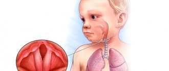
Cephalohematoma in newborns becomes noticeable on the second or third day, when the birth tumor subsides.
Photo of cephalohematoma in newborns
From the first day, the size of the hemorrhage increases, since newborns lack blood clotting factors, therefore, it remains liquid for a long time, and damaged vessels are not thrombosed by blood clots. As pediatrician Komarovsky notes about cephalohematoma on the head in newborns, its size depends on how severe the bleeding .
The symptoms of cephalohematoma are especially pronounced on the second or third day after the baby is born. Its distinctive feature is precisely the gradual increase these days. The child's head becomes asymmetrical due to the appearance of a formation.
If you try this formation by touch, it will be elastic, and with slight pressure on it you can feel the liquid moving. The skin over it is unchanged, elastic.
There can be from 5 to 150 ml of blood under the periosteum. Accordingly, if the cephalohematoma is small, then it decreases approximately a week after its appearance, and no intervention is required.
If the cephalohematoma is isolated, the child’s health remains normal. If it is combined with another pathology, then the baby experiences neurological symptoms. In most cases, there is depression of the function of the central nervous system, which is manifested by a sluggish reaction to the action of pathogens and poor expression of reflexes.
In some cases, it is possible to manifest strong central nervous system excitation similar to a hydrocephalic or hypertensive complex of symptoms.
If the size of the formation is large or the child has impaired blood clotting, and the cephalohematoma does not shrink on its own, anemia , ossification , resorption hyperbilirubinemia , and infection of the hematoma are likely.
With large cephalohematomas, rapid hemolysis of red blood cells , as a result of which a yellowish tint of the skin and mucous membranes is observed in the first days of the baby’s life. In this case, jaundice persists for 10 days or more.
Sometimes, where the formation is localized, a fracture (crack) of the bone is noted.
Tests and diagnostics
In the process of establishing a diagnosis, differential diagnosis is carried out with a birth tumor, cerebral hernia, hemorrhage under the aponeurosis . If we are talking about an uncomplicated cephalohematoma, the doctor is primarily guided by the examination data. In this case, it is not difficult to establish a diagnosis if you examine the head and detect the characteristic signs of the disease.
The clinical picture of cephalohematoma is nonspecific; symptoms of damage to the nervous system are more pronounced.
In addition, during the diagnostic process, if necessary, the following methods are used:
- computed tomography – used if there is suspicion of damage to brain tissue;
- ultrasound examination - makes it possible to find out whether there is a fracture of the cranial bone, a cerebral hernia, and also determine the size of the tumor;
- Craniography in direct and lateral projections – allows to exclude bone damage;
- multislice CT;
- neurosonography – allows you to determine the presence of damage in the brain.
Prevention
To reduce the risk of this pathology in a child, the expectant mother must follow the following rules:
- her lifestyle and diet should be as healthy as possible;
- she must undergo all necessary examinations during pregnancy and not miss visits to the doctor;
- it is important to identify all risk factors and follow your doctor’s advice;
- supervision should be provided by a professional specialist who can correctly conduct labor.
Consequences and complications
As a rule, this formation goes away on its own, and the consequences of cephalohematoma do not appear later. But sometimes, if a large cephalohematoma forms on the head of newborns, consequences in the future are still noted. True, this happens only in rare cases.
The consequences of a large cephalohematoma on the head in the future may be as follows:
- lagging child in speech and mental development;
- neurological symptoms;
- cerebral palsy;
- presence of other health problems.
However, it is important to understand that such consequences are observed infrequently.
Quite serious complications of this pathology can be the following manifestations:
- anemia as a result of blood loss;
- jaundice due to bilirubin the bloodstream;
- infection of the meninges;
- suppuration, compression of the optic (auditory) nerves;
- ossification of cephalohematoma and irreversible deformation of the skull bones.
Symptoms of pathology
The symptoms of subperiosteal hemorrhage in newborns on the head can be scanty, and the hemorrhage itself does not always have a significant impact on the general condition and well-being of the baby. If the hematoma is large, signs of trouble may appear already in the first hours of the baby’s life, in mild cases - 2-3 days after birth.
The general condition of the baby is determined by the absence or presence of other disorders - respiratory pathology, hemolytic jaundice, etc., while isolated hematomas do not affect the child’s condition in any way, representing only a cosmetic defect that may soon disappear completely.
Babies with cephalohematoma and without other pathologies behave the same as other babies, but they may be more restless and sensitive to changes in weather and the situation in the house. Perhaps this is due to the general difficult course of labor, great stress for the child, and hypoxia.
In some cases, in the first few days from the moment of hematoma formation, an increase in blood volume occurs due to a lack of coagulation factors, which is physiological for newborns, so it is not always possible to judge the final size of the cephalohematoma as soon as the doctor discovers it.
Externally, a generic cephalohematoma is a limited area of bulging skin with a subcutaneous layer, under which, when palpated, liquid contents are detected. Large hemorrhages give the skull an asymmetry, which decreases as the blood masses are absorbed. Palpation of the formation does not cause pain to the baby.
There can be several cephalohematomas, large hemorrhages can be multi-chambered, they are often located in those places that were most tightly pressed against the walls of the birth canal during childbirth or experienced maximum mechanical stress. If a vacuum extractor was used during the birth process, then hemorrhage is expected to form at the site where the device is applied to the head of the newborn baby.
Subperiosteal hemorrhages, regardless of volume, are limited to the edges of the corresponding bones and do not extend beyond them due to the presence of sutures. Their diameter reaches 5-7 cm. In severe and very rare cases, hematomas can form on several bones at once, but this is rather an exception to the rule and the result of a severe birth injury.
In the maternity hospital, a neonatologist or pediatrician examines the baby’s head daily, monitoring the size of the cephalohematoma and the dynamics of its regression. The hemorrhage has an elastic consistency, somewhat compacted at the edges; when palpated, the doctor notices fluctuations in the fluid inside in the absence of other pulsations.
A small cephalohematoma in newborns (up to 2-3 cm) is prone to spontaneous resorption during the first one to one and a half weeks, and after one and a half to two months there is no trace left of it. Large hemorrhages with a diameter of eight or more centimeters are not capable of regression, so they are eliminated by puncture.
Complications from cephalohematomas are not so common if the baby is under constant medical supervision, but the most likely among them are:
- Anemia due to the accumulation of a certain amount of blood under the periosteum;
- Infection and suppuration;
- Hemolytic jaundice due to the breakdown of red blood cells inside the hematoma and the release of bilirubin into the bloodstream (if the baby has signs of physiological jaundice, they may become more pronounced);
- Petrification is the deposition of calcium salts with calcification of the periosteum and soft tissues in the area of the former hematoma.
Signs of anemia in a newborn occur with significant blood loss or hemolysis, while the newborn has pale skin, increased heart rate and breathing.
When bacterial microflora (beta-hemolytic streptococcus, etc.) is attached, specific signs of the inflammatory process appear - fever, deterioration of the baby’s condition, redness and swelling of the hematoma, increased skin temperature over the site of inflammation, tachycardia, characteristic signs in a blood test, etc.
Petrification, or calcification, is one of the relatively harmless consequences of cephalohematoma, which consists of the deposition of calcium salts into the periosteum and soft tissue in the area of hemorrhage. In itself, it does not interfere with the child’s well-being and development, but it can constitute a significant cosmetic defect, leading to deformation of the skull and its asymmetry. At the request of the patient and parents, surgery to remove the ossified periosteum to correct the shape of the skull is possible.
Video: example of a large cephalohematoma
Forecast
As a rule, the prognosis for this disease is favorable.
Serious complications occur in a small number of children and are a consequence of displacement of brain structures, which are pressed by a hematoma and accumulation of blood.
Statistics show that in approximately 80% of cases, subperiosteal hematomas significantly decrease in the first week after their appearance, and completely resolve within 2-3 weeks. However, if this does not happen, then the cephalohematoma begins to ossify.
Symptoms and clinical picture of cephalohematoma
Manifestations of cephalohematoma are visualized several days after the birth of the child. The formation of a small lump without any changes on the skin is observed. The most common location of the tumor is the parietal region. When you feel the pathological swelling, you feel moving liquid contents.
Symptoms of cephalohematoma:
- the occurrence of tumor formation;
- head asymmetry;
- appearance after 2-3 weeks of icteric color.
List of sources
- Obstetrics. National leadership / Ed. E. K. Ailamazyan, V. I. Kulakov, V. E. Radzinsky, G. M. Savelyeva. - M.: GEOTAR-Media, 2007. - P. 1160-1161.
- Bardeeva K.A., Pisklakov A.V., Lukash A.A. A new look at the treatment of cephalohematomas in children // Modern problems of science and education. – 2015. – No. 5.;
- Ch. 6. Children's diseases // Directory of a paramedic / Professor L.A. Isaeva. - Moscow: “Medicine”, 1975. - P. 319. - 662 p. — 280,000 copies.
- Kerchelaeva S.B. Cephalohematoma as a result of complications of childbirth / Kerchelaeva S.B., Tyagunova A.V., Kuznetsova O.V. // Attending physician - 2015. - No. 10 - p. 88–92.
- Handbook of obstetrics, gynecology and perinatology / Ed. G. M. Savelyeva. - M.: MIA, 2006. - P. 344-348.
Treatment of cephalohematoma in a child
Hemorrhage is treated by a neonatologist or pediatric surgeon. For a small hematoma, there is no need for special treatment. The baby is prescribed calcium and vitamin K supplements for 5-7 days. The course of treatment for uncomplicated pathology is 7–10 days. Treatment of a cephalohematoma in a child that occurs with complications will require at least a month. A child with such a pathology must be registered with a surgeon and neurologist.
To speed up the healing of the formation, neonatologists advise using Troxerutin. The gel is applied to the hematoma area twice a day. The drug increases vascular tone and prevents further penetration of blood.
In case of extensive hemorrhage (more than 9 cm in volume), the doctor will prescribe surgery. Puncture of a cephalohematoma of the parietal bone is a procedure that is safe for the baby’s health and takes no more than 10 minutes. The child’s skin at the site of the hematoma is pierced with a special needle, the accumulated blood is sucked out, after which the puncture site is disinfected and a pressure bandage is applied. Surgical removal of cephalohematoma is indicated in the presence of purulent contents of the cavity, as well as in the III degree of development of the pathology.
Doctor's recommendations after puncture:
- parents need to strictly follow the specialist’s instructions;
- In no case should you self-medicate;
- protect the newborn’s head from damage;
- use a hat one size larger;
- carefully monitor the general condition of the baby.