Osteogenesis imperfecta (Lobstein-Frolik disease, congenital bone fragility, periosteal dystrophy) is a group of genetic pathologies characterized by impaired bone tissue formation. Then the child’s bone fragility increases, and as a result, pathological fractures occur. In addition, bones become deformed, muscles become thinner, joint hypermobility occurs, hearing is impaired, etc.
The congenital form of the disease is the most dangerous; it has a severe course and leads to death from numerous complications. The prognosis for the late form is more favorable. It is impossible to completely cure the pathology. Supportive treatment is carried out to help strengthen bone tissue and prevent fractures.
Description of the pathology
Lobstein-Frolik disease is a genetically determined disease that occurs as a result of impaired bone formation. It leads to a decrease in bone mass and increased fragility. The pathology develops as a result of a defect in type 1 collagen, which is an important protein in the bone structure. Then it is produced in insufficient quantities or the structure of the substance is disturbed. For this reason, bones become weak and brittle. Because of this, the pathology is called “crystal disease.”
Reference. According to statistics, in approximately 50% of cases, imperfect bone formation is provoked by spontaneous mutations. The disease is diagnosed in 1 child out of 10–20 thousand newborns.
Crystal disease is incurable, but with the right approach it can make a child’s life much easier.
Treatment of osteoporosis at the Echinacea Clinic
Treatment in our clinic will be structured as follows:
- Relieve pain. The pain can be caused by bone deformation, pinched nerves and can be quite severe and long-lasting. In this case, a neurologist is involved in the treatment. Pain can be relieved with the help of medications that block pain at the level of the nervous system, local administration of drugs, and relaxation of overstrained muscles.
- Stop bone destruction and, if possible, return bones to normal density and strength. Just giving the patient calcium is ineffective. Treatment is based on the results of the examination. In order for calcium to be absorbed, it is necessary to find and treat impaired metabolic processes .
We will offer you an individual osteoporosis treatment plan. Most often, we treat osteoporosis in several areas:
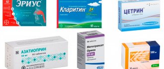
- The main treatment for osteoporosis (Miacalcic, calcium supplements, vitamin D3 and bisphosphonates).
- Gymnastics must be very careful, so we select it individually for each patient and teach it to be performed independently. Stress on bones and muscles improves bone strength and helps fight osteoporosis. The dosage of physical activity should be based on tests and osteodensitometry. We will definitely tell you how much and how to exercise. If the situation is critical, we advise you to refrain from any stress for a while.
- Nutrition. Useful foods containing calcium and vitamin D for the treatment of osteoporosis: dairy products, sea fish (except sturgeon), hazelnuts, cauliflower, seaweed. Vitamin D is also found in eggs, cocoa, and cream, but the consumption of these products will have to be reduced if there is excess cholesterol in the blood. Food products must also contain sufficient amounts of protein. There is a lot of protein in meat, fish, poultry, and legumes, but you should consult your doctor to see if there are any restrictions on these products due to concomitant diseases. It should be remembered that calcium is poorly absorbed together with foods containing oxalic acid - spinach, gooseberries, currants, rhubarb.
- Pain relief . Pain relief is almost always possible. We combine different groups of analgesics and drug blockades, sleeping pills, minor muscle relaxants, positional treatment, selection of pain relief is required. Treatment of osteoporosis itself, as a rule, over time allows you to abandon constant pain relief.
Symptoms
Symptoms depend on the type of pathology.
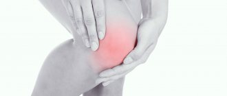
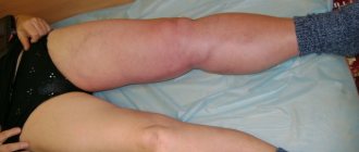
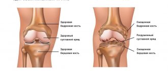
Osteogenesis imperfecta is manifested by pathological fractures, bone deformation
The early form of the disease is the most dangerous, as sometimes children die in the womb. Most newborns die in the first days or months of life. This is associated with intracranial birth injuries, severe respiratory disorders, and acute respiratory viral infections.
Osteogenesis imperfecta in children is manifested by the following symptoms:
- Thin, pale skin, thinning subcutaneous fat.
- General weakness, hypotension.
- Bone fractures (femur, lower leg, forearm, shoulders) with minimal impact.
Usually, in the early form of the pathology, the child dies within 2 years.
The late form is manifested by the following symptoms:
Paget's disease of bone
- Increased bone fragility.
- Blue discoloration of the whites of the eyes.
- Hearing impairment, up to complete deafness.
- Late overgrowth of the fontanel.
- Slowing down of the child's physical development.
- Excessive joint flexibility due to weak ligaments.
- Muscle thinning.
- Dislocations, fractures with minimal impact.
- Curvature or shortening of bones after their fusion.
- Deformation of the sternum or spine.
- Late teething (after 1.5 years), dental anomalies, caries, rapid abrasion and destruction of teeth, staining them yellow.
- Hearing impairment, deafness.
Crystalline disease may be accompanied by bulging of the wall of the mitral valve of the heart or its functional failure, kidney stones, inguinal hernias, nasal hemorrhages, etc.
Classification of crystalline disease
There are 2 known forms of pathology:
- Congenital. Fractures occur in the womb and immediately after birth.
- Late. Bones are injured when the child is already walking. This form of the disease has a milder course.
Types of crystal disease:
- Osteogenesis imperfecta type 1 - fractures occur after birth until adolescence, the spine is slightly curved, the ligaments and joints are weak, and muscle tone is reduced. The whites of the eyes become discolored, children lose their hearing early, and their eyes are slightly bulging.
- Type 2 – the development of the skeleton is disrupted, the bones are deformed or shortened, and protrusions remain at the sites of fractures after fusion of the bone tissue. Children develop slowly physically. This type of disease is considered the most severe. A child can die before the age of 1 year from functional lung failure or hemorrhage into the cranial cavity. The bones are severely deformed, the patient is short in stature.
- Type 3 – bones are injured after birth until puberty. Severe deformations of the bones, spine, chest, breathing problems, weak muscles, joints and ligaments are possible. The sclera becomes discolored, and hearing impairment quickly progresses.
- Type 4 – symptoms of bone development disorders are practically not noticeable, but patients develop premature osteoporosis (decreased bone density). Fractures are typical before adolescence, the curvature of the bones is of mild or moderate severity. The patient is short in stature and may lose hearing early.
- Type 5 – the course of the disease is the same as in type 4 pathology. The only difference is that the bone has a mesh structure.
- Type 6 – symptoms are the same as for type 4 disease, but the bone structure resembles fish scales.
- Type 7 – disorders associated with mutations in cartilage tissue.
- Type 8 – there is a strong change in the protein, which contains leucine and proline (amino acids). This type of pathology has a severe course and ends in death.
Reference. Depending on the type of inheritance, autosomal dominant and autosomal recessive osteogenesis imperfecta are distinguished. The first type is typical for types 1 – 5 of pathology, and the second – for types 7 – 8.
Causes of Lobstein-Frolik disease
The causes of osteogenesis imperfecta are associated with genetic pathologies. The gene for collagen A1 and A2 mutates, causing a lack of protein or its structure being disrupted. Then the fragility of bone tissue increases, especially the tubular bones (shoulders, forearms, thighs, legs) suffer. They have a porous structure, bone islands, a large number of sinuses, which are filled with loose tissue, the outer layer is thinned.
Doctors distinguish 2 types of inheritance of crystalline disease:
- Autosomal dominant - the disease is passed on to the child from one parent who also suffers from it. Then bones are more often injured after 1 year.
- Autosomal recessive - a mutated gene is passed on from both parents. The disease has a severe course, pathological fractures are possible in the womb or immediately after birth.
Reference. Osteogenesis imperfecta with an autosomal dominant type of inheritance is more often diagnosed.
III. Forecasts
Due to the enormous individual variability of the disease, it is impossible to make general statements about what treatment and rehabilitation methods will be correct in each individual case. Some predictions can be made based on statistics for each type of OI, but no guarantees can be made regarding the number of fractures, the height of a frail person, or his mobility.
Even among “fragile” people within the same family, differences can be quite significant, and in each specific case it is recommended to seek individual advice from a specialist.
In many cases, bone fragility decreases after puberty, but may increase again after age forty, so physical activity is important throughout the life of a person with OI.
Establishing a diagnosis
The congenital form of the pathology can be detected as early as 16 weeks of pregnancy using ultrasound. If necessary, a chorionic villus biopsy and gene diagnosis are performed to confirm the presence of a mutated gene.
In other cases, the diagnosis of osteogenesis imperfecta consists of the following methods:
- Collection of anamnesis, patient complaints. Signs of pathology: frequent fractures, abnormal bone shape, difficulty gait, short stature, bad teeth, hearing impairment.
- Visual inspection. The doctor evaluates height, body weight, hearing, condition of teeth, color of the whites of the eyes, and conducts neurological tests. The orthopedist is interested in the shape, length of the limbs, deformations, range of motion in the joints.
- Laboratory tests of blood and urine will help detect the level of proteins, glucose, urea, calcium, phosphorus, etc.
- An X-ray of the limbs, spine, skull will show that bone density has decreased, bone calluses after healing of pathological fractures, etc.
- A bone biopsy (examination of a fragment of bone tissue) is used to confirm a decrease in its density and thinning of the outer layer.
- A skin biopsy is performed to examine the collagen defect.
- Molecular genetic testing will help detect the mutated gene. To do this, the patient's blood or saliva is studied.
Reference. Differential diagnosis will help to distinguish crystalline disease from rickets, achondroplasia (a malformation of the cartilage-forming system of the fetus), desmogenesis imperfecta (hyperelasticity of the skin).
Are there any prospects?
We, the doctors at GMS Hospital Congenital Pathology Center, would really like to see Osteogenesis Imperfecta effectively treated using modern technology across the country. It is already obvious that in the near future we will have more patients than we can properly care for, so we will have to expand our staff again. We would like to train specialists from other cities and send some patients to them. But, unfortunately, today our center has no alternative in this direction. In other institutions that are trying to deal with this disease, there is a severe shortage of certain resources, and sending patients there means acting to their detriment. We constantly accept patients who previously received care in government institutions, and are faced with the fact that we have to correct other people’s mistakes and regret about lost time and missed opportunities.
If you know doctors who are interested in adopting our experience, studying from foreign literature, who are not indifferent to the global outcome of their treatment, who are ready to introduce new methods of therapy, surgery, and rehabilitation, feel free to give them our contact information. We want there to be more centers like ours. Patients fly to us from the Far East and Yakutia. It would be much better if, instead of wasting money, time and effort on flying across the country, they could contact a center, for example, in Novosibirsk or Yekaterinburg.
If you know people or organizations that are ready to pay for travel, technical equipment or accommodation for our patients in Moscow while they undergo a rehabilitation course in our center (funds pay for treatment, but not rehabilitation courses after fractures or operations), share information about us with them. We will tell them how to provide targeted help: we have lists of children who need their charity more than others.
If you know children with brittle bones whose parents may not even know that this disease can be successfully treated, tell them about us. We will be happy to invite them to a consultation and, if necessary, accept them into our treatment program.
We want awareness of osteogenesis imperfecta to grow, so that doctors stop being afraid of this diagnosis, and parents understand that the disease should not be an obstacle to a long and happy life.
Treatment methods
As already mentioned, osteogenesis imperfecta is incurable. Treatment is carried out to alleviate the patient’s condition and strengthen bone tissue. For this purpose, the following methods are used:
- Drug therapy. The patient takes drugs based on somatotropin (growth hormone) to stimulate collagen synthesis. In addition, antioxidants, medicines containing calcium, phosphorus, and vitamin D2 are indicated.
- Then the patient is prescribed drugs that accelerate the formation and mineralization of bone tissue, which contain extract of the thyroid glands of cattle and cholecalciferol. And bisphosphonates slow down the process of bone destruction; pamidronic acid, zoledronic acid, and residronate are used for this purpose.
- Physiotherapeutic procedures: electrophoresis with calcium chloride (penetration of a medicinal substance through the skin using an electric current), ultraviolet irradiation of blood, magnetic therapy, inductothermy, etc. Children are also prescribed massage, therapeutic exercises to strengthen muscles and ligaments.
Medicines will help strengthen bone tissue and alleviate the patient’s condition
In addition, the patient may need treatment from a psychologist. The use of orthopedic devices, such as shoes or corsets, is also recommended.
In case of severe bone deformation after fractures, a corrective osteotomy is performed. The surgery helps correct the shape and size of the limbs. During the procedure, the affected bone is dissected, the irregular shape is corrected, and bone fragments are fixed with special pins or bolts (osteosynthesis).
There are 2 types of osteosynthesis: bone and intramedullary. In the first case, the fixation structure is located in the patient's body, but outside the bone. The disadvantage of this treatment method is that the periosteum is damaged. In the second case, the fixator is placed inside the bone.
Attention. Surgery for osteogenesis imperfecta is contraindicated if the patient’s condition is severe, he suffers from functional failure of the heart, lungs, or it is impossible to fix the fixator due to lack of bone tissue.
Why do we recommend treating osteogenesis imperfecta in the central ventricle
Unique experience
The head of the Central Clinical Hospital, geneticist-endocrinologist Natalia Belova, has been treating children with OI for more than 30 years, orthopedist-traumatologist Vladimir Kotov for more than 25 years, pediatrician Fedor Katasonov for more than 5 years. Natalia Belova was the first to begin researching this pathology in the USSR, wrote articles about it for the encyclopedia, and compiled recommendations for the treatment of OI and a protocol for the treatment of children with OI for the Ministry of Health. Currently, she is the main freelance employee of the Ministry of Health of the Russian Federation on this disease. On the initiative of Natalia Belova, a patient organization for patients with OI was created. Thousands of children diagnosed with osteogenesis imperfecta have passed through Dr. Belova. Over the five-year history of the CVP, more than 350 patients with OI were observed here, and about 200 children from Russia, Kazakhstan, Ukraine, Belarus, Moldova, Kyrgyzstan and Tajikistan received systematic treatment. CVP is the largest center in Europe for the treatment of congenital bone fragility.
Complex treatment
Osteogenesis imperfecta affects all areas of human life. The key to successful treatment lies exclusively in a multidisciplinary approach, in which the efforts of many specialists are combined. If at least one direction of treatment is excluded, the chances of success are significantly reduced.
- The center's pediatricians provide drug treatment, prescribing drugs from the group of bisphosphonates that strengthen bone tissue to patients. The responsibilities of these specialists include coordination of all areas of treatment and continuous pediatric monitoring.
- Orthopedists and traumatologists provide adequate treatment of fractures and timely surgical correction. The center performs almost all types of surgeries required for OI, with the exception of spinal surgeries.
- Rehabilitation specialists engage in physical activation of patients. It is known that improving the quality of life and reducing the number of fractures is possible only with certain physical activity. Experts select special exercises and teach parents how to practice with their children at home.
- The psychologist’s tasks are to help the patient accept his diagnosis without internal conflict, relieve anxiety (fear of fractures can often be the only obstacle slowing down treatment), motivate physical activity and solve many other psychological problems.
- Specialists in the selection of technical equipment help to choose the necessary aids (crutches, walkers, chairs), most suitable for the level of physical activity and size, and also to receive financial compensation from the state for their purchase.
- Related health problems are solved by qualified doctors: ophthalmologists, ENT specialists, neurologists, etc. The studies necessary for patients, even those not included in the standard of treatment for OI, are carried out at GMS Hospital or partner clinics.
This comprehensive approach makes the CVP unique.
Natalia Belova considers the criteria for selecting specialists to work in the center to be their high professionalism, personal involvement in the process, ability to work collectively, as well as a special medical culture, aimed primarily at serving the interests of the patient and providing assistance to him. Five years ago, the CVP team was lucky enough to find a private GMS clinic, which took responsibility for promoting this socially important, although at that time questionable from a financial point of view, direction. Today, as a result of this fruitful collaboration, the clinic can simultaneously treat at least twice as many small patients with OI. Western standards of treatment (evidence-based medicine)
Continuous staff development
Regular interaction with leading Russian and foreign medical institutions
Modern medical equipment and advanced diagnostic and treatment methods
Unified standard of service
We work around the clock 24/7/365
Make an appointment We will be happy to answer any questions Coordinator Oksana
Correct goal setting
The goal of our treatment is not to reduce the number of fractures or increase bone density (this happens naturally during the work), but to improve the patient’s quality of life. We work to raise every fragile child to be a full-fledged member of society, minimally limited by their illness. We strive to ensure that our patients receive a good education, start a family and become happy people.
Modern approach
The Center for Congenital Pathology GMS Hospital is the only organization in the territory of the former USSR that provides the most up-to-date approach to the treatment of OI. Many doctors and organizations trying to treat OI are still guided by the recommendations compiled by Natalia Belova more than 20 years ago, which today have lost their relevance.
- Modern diagnostics became possible as a result of cooperation with the Genomed laboratory, the only one in Russia that performs a full-fledged study on OI. Genetic research allows you to clarify the type of disease and select accurate and effective recommendations for each specific case. In addition, information about the gene mutation in parents gives them the opportunity for further family planning. If there is a risk of having a child with a pathology, clients can resort to the latest reproductive technologies at the GMSEKO clinic.
- Modern drug treatment includes the prescription of drugs from the group of bisphosphonates (pamidronate, zoledronate), and the treatment regimen (doses, frequency of administration, duration of treatment) is constantly adjusted taking into account the latest developments. In the near future, CVP plans to be the first in our country to begin trial treatment with the drug denosumab, which is more effective in some rare types of OI. The center’s doctors do not use outdated and harmful treatment methods based on calcium, collagen, osteogenone, somatotropin, etc., as is done in other medical institutions.
- Modern surgical treatment involves the use exclusively of intraosseous structures to fix bones after correction of deformities or fractures. Bony structures (plates, bolts), acceptable for healthy people, but destroying the bones of our patients, are used in our country in medical institutions working the old fashioned way, although they have long been abandoned all over the world. In addition, the TsVP became the first center in Russia where they began to install telescopic rods that lengthen as the child grows. This global “gold standard” of surgery for OI was unavailable in our country for a long time, so we used to send patients with this prescription for treatment abroad.
- The modern approach to physical therapy involves intense exercise and an active lifestyle. Unfortunately, the level of rehabilitation in our country is quite low, and the words “therapeutic physical education” mean meaningless activities that do not really load any muscle group. Dr. Andreas Stromberger, who studied and practiced in Germany for many years, is responsible for physical therapy at the Center. The goal of rehabilitation is not to develop muscles, but to develop skills. At the center, children learn to crawl, walk and run, maximizing their everyday capabilities. The rehabilitation room at GMS Hospital is equipped with sports equipment, exercise equipment and a new effective means of rehabilitation - the Galileo vibration platform.
- A modern approach to hospitalization means the most comfortable stay in the GMS Hospital. The patient is in the room with one of the close adults (mom, dad, grandmother, sister). The wards, equipped at the level of European clinics, are designed for one or two families. Polite, highly qualified nurses, many of whom studied abroad, have extensive experience working with OI, in which, for example, due to the characteristics of the vessels, it can be difficult to place a venous catheter. The wards have TVs and air conditioning, and meals are served by maids. The hospital has free access for visitors around the clock, and patients themselves are free to walk around the city when they do not have procedures scheduled.
Continuous learning
International conferences on osteogenesis imperfecta, which take place every three years, are the main specialized event, which brings together all the leading specialists in fragile bones: geneticists, surgeons, pediatricians, rehabilitation specialists, representatives of patient organizations, etc. This is where you can learn about the latest technologies and trends in the development of research on OI. TsVP specialists are the only permanent conference participants from Russia. The last conference in Oslo in 2021, which became our fourth, was attended by Natalia Belova, Fedor Katasonov, orthopedic surgeon Alexey Rykunov and geneticist of the Genomed laboratory Fedor Konovalov, together with whom the TsVP staff prepared three poster presentations. In addition to ongoing training, participation in conferences allows CVP doctors to maintain relationships with the global medical community, thanks to which, in particular, our patients were previously able to be sent for treatment abroad.
Concern
We stay in touch with our patients at all times – even after they return home between hospitalizations or outgrow childhood. We know how difficult it is to get adequate medical care in the community when any problem is written off as a rare diagnosis and they don’t want to deal with it, or they offer inadequate treatment methods and a protective lifestyle. We also understand the need for normal education, promote the enrollment of our patients in schools and kindergartens and fight discrimination against them, sometimes even with the involvement of journalists and lawyers. We do not ignore any aspect of children's lives, be it difficult circumstances in the family or difficulties in obtaining an education, and we do everything in our power to help.
Availability
Treatment at the Center for Congenital Pathology is free for patients (they only need to pay for travel). This became possible thanks to the contribution of charitable foundations under a program organized by Natalia Belova. Funds (primarily Rusfond) pay for course treatment and operations, including abroad, and we also, if possible, look for funds for special equipment (walkers, chairs) and even for legal assistance. Expensive genetic research is also carried out at the expense of philanthropists.
You have questions? We will be happy to answer any questions Coordinator Tatyana
The most important
Thus, the most dangerous form of pathology is considered to be the early one, in which most children die within the first months or years. This occurs due to multiple injuries and infections (pneumonia, sepsis). The late form of crystalline disease has a more favorable prognosis, although the quality of life is reduced. Maintenance drug therapy will help get rid of the symptoms of the pathology, strengthen bone tissue, and improve the general condition of the patient. In case of severe bone deformation due to fractures, a corrective osteotomy is performed. Treatment is complemented by physiotherapy, exercise therapy, and massage. Doctors strongly recommend medical genetic counseling for expectant mothers whose families have patients with osteogenesis imperfecta.