One day hospital 3rd KO
Dadyev
Islam Arturovich
11 years of experience
Oncologist (OMC), surgical oncologist, candidate of medical sciences, member of the Russian Association of Oncologists, member of the Russian society of chemotherapists RUSSCO
Make an appointment
Esophageal cancer is a malignant pathology that involves the degeneration of cells in the mucous membrane of the esophagus and the subsequent formation of a tumor. Increasing in size, the neoplasm partially or completely blocks the lumen of the esophagus, grows through its wall and spreads to nearby organs. Without treatment, death is inevitable. The disease affects men four times more often than women. The main risk group is elderly people: the average age of male patients is 64 years, female patients – 70 years.
Kinds
According to histological characteristics, esophageal cancer is divided into:
- squamous - developing from epithelial squamous cells, in turn, can be superficial and deeply invasive;
- adenocarcinoma – developing from cells that produce protective mucus.
Tumors of the upper (10%), middle (35%) and lower (55%) parts of the esophagus are distinguished by location.
Squamous cell tumors in the superficial form respond well to treatment. An invasive tumor is prone to growing deep into tissue and forming metastases to nearby organs. Adenocarcinoma is much less common and has a severe course of the disease.
Chemotherapy
Chemotherapy, that is, taking drugs that have an inhibitory and destructive effect on neoplasm cells, can be adjuvant, neoadjuvant, or independent.
Preoperative chemotherapy is especially effective for adenocarcinoma, for which 2-3 courses of treatment are prescribed. Postoperative chemotherapy is usually indicated in conjunction with neoadjuvant chemotherapy and is not recommended without it for adenocarcinoma and squamous cell carcinoma. Completely independent chemotherapy is recommended only for inoperable patients with contraindications to radiological treatment methods.
Symptoms
As a rule, the first symptoms of esophageal cancer appear too late, when the tumor has already acquired significant size, or accidentally, during examination for another disease. Typically the disease manifests itself through:
- difficulty swallowing food, especially solid food, due to which the patient is forced to reduce portions and give preference to liquid dishes;
- feeling of a constant lump in the throat;
- increased secretion of saliva, often even in the absence of food;
- a feeling of discomfort or even pain in the chest;
- weight loss due to eating problems;
- hiccups, cough, voice changes are rarer symptoms;
- bleeding in the esophagus, due to which the stool becomes dark, almost black;
- anemia, pale skin, constant weakness and drowsiness.
If these signs appear, you should consult a doctor as soon as possible for a thorough examination.
Clinical picture of the disease
Clinical symptoms depend on the extent of the disease
The clinical picture directly depends on the degree of damage to the esophagus. Cancer of the upper thoracic and upper esophagus is especially painful. Patients often complain of symptoms such as the sensation of a foreign body in the esophagus, scratching while eating, etc.
A little later, signs of pharyngeal insufficiency also appear - frequent regurgitation, dysphagia, and sometimes asphyxia.
If the tumor has affected the mid-thoracic esophagus, then first of all the patient will experience dysphagia and chest pain. Once the tumor begins to grow into neighboring tissues and organs, the pain will intensify. A tumor of the lower thoracic esophagus is characterized by dysphagia on the left side of the chest, as well as progressive angina.
Causes and risk factors
The mechanism of occurrence and development of esophageal cancer has not yet been thoroughly studied. However, factors have been identified that increase the risk of tumor development:
- smoking and drinking strong alcoholic drinks that burn the esophageal mucosa;
- old age: at least 85% of patients are people over 55 lei;
- overweight, obesity;
- habit of drinking very hot food and drinks;
- the presence of “Barrett's esophagus” - pathological areas of the intestinal epithelium in the lower part of the esophagus, resulting from chronic burns from gastric juice;
- inflammatory diseases of the esophagus;
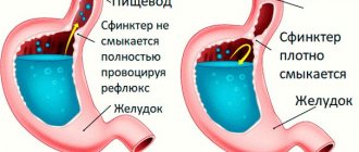
- decreased locking function of the sphincter between the esophagus and stomach;
- Plummer-Wilson syndrome (simultaneous dysfunction of swallowing, narrowing of the lumen of the esophagus and iron deficiency anemia);
- constant contact with fumes of carcinogenic substances, acids and other aggressive reagents;
- papillomavirus infection.
The increased incidence of men compared to women is explained by the fact that they are much more often exposed to the listed negative factors.
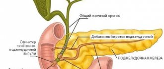
Anatomy of the esophagus
The esophagus is a hollow muscular tube that connects the pharynx and oral cavity to the stomach. The wall of the esophagus is built from the mucous membrane, submucosa, muscular and adventitial membranes. The muscular lining of the esophagus consists of two layers: the outer longitudinal and the inner circular. In the upper part of the esophagus, the muscular layer is formed by transverse muscle fibers. At approximately the level of one third of the esophagus (counting from the top), striated muscle fibers are gradually replaced by smooth muscle fibers. In the lower part, the muscular layer consists only of smooth muscle tissue.
The mucous membrane is covered with stratified squamous epithelium; the submucosa contains mucous glands that open into the lumen of the organ.
In the esophagus there is a skin-type mucous membrane. The epithelium is multilayered squamous, non-keratinizing, lying on fine-fibrous connective tissue - its own layer of the mucous membrane, consisting of thin bundles of collagen fibers; also contains reticulin fibers and connective tissue cells. The own layer of the mucous membrane protrudes into the epithelium in the form of papillae.
A special feature of the esophagus is the absence of a serous membrane; the outer layer is represented by adventitia, loose connective tissue. Only in the lower third of the esophagus, more precisely its abdominal section, is covered with serosa.
Stages
Oncologists distinguish the following stages of esophageal cancer.
- Zero. The neoplasm is located on the surface of the epithelial layer, without even growing deep into the epithelium, and is a cancer “in situ”, i.e. "on the spot." It can only be detected by chance during an endoscopic examination of the esophagus from the inside.
- First. Malignant cells spread to the entire epithelium, but do not yet affect muscle tissue. The lymph nodes are not affected, there are no metastases.
- Second. The tumor either grows through the entire wall of the esophagus, including muscle and connective tissue, but without affecting the lymph nodes, or spreads only to the muscle layer and metastasizes to several nearby lymph nodes.
- Third. The size of the tumor creates obstacles to swallowing. Malignant cells affect nearby organs and regional lymph nodes.
- Fourth. Regardless of the size of the tumor and the degree of its spread to neighboring tissues, metastases of esophageal cancer are detected in distant organs - liver, lungs, bone structures.
Differential diagnosis
At the stage of determining esophageal cancer, it is extremely important to exclude all other types of diseases that are characterized by dysphagia: scar narrowing after chemical burns, stenosis in patients with esophagitis, tuberculosis, benign neoplasms. It is also necessary to exclude external displacement of the esophagus by tumors of the posterior mediastinum, intrathoracic goiter, and enlarged lymph nodes.
Displacement of the esophagus is the first sign of compression, which is determined by x-ray. Even a significant increase in neighboring organs cannot cause dysphagia, because the likelihood of displacement of the esophagus is very high.
If the esophagus is compressed and patency is impaired, this may also be due to fibrous mediastinitis, which can develop after inflammatory processes in the lungs and lymph nodes. Endoscopy can also be used in combination with a biopsy, which increases diagnostic accuracy to 90%.
Based on everything described above, it is worth noting that the procedure for examining a patient with suspected esophageal cancer should include an X-ray examination, esophagoscopy, endoscopy, ultrasound, multiple biopsy, and computed tomography of the lungs.
Diagnostics
If the patient's complaints are similar to those of esophageal cancer, the oncologist will prescribe a series of diagnostic tests to confirm or refute this suspicion.
- X-ray of the esophagus. The test is performed with a contrast agent, which the patient drinks to make the esophagus more visible on the image. The image shows a narrowing of the lumen, allows you to determine the thickness of the walls, and indicates the presence of an ulcer.
- Esophagoscopy. An endoscopic examination is carried out using a flexible probe with a video camera, which broadcasts an image of the internal walls on a screen. The endoscope allows not only to detect pathologies, but also to take samples of altered tissue for analysis.
- Ultrasound of the esophagus and abdominal cavity. It is carried out to determine the size of the tumor and to identify metastases in the lymph nodes and peritoneal organs.
- MSCT of the chest cavity. The procedure allows not only to identify even a small tumor, but also to determine the extent of its spread to neighboring tissues, to identify metastases in the lymph nodes and other organs, including those developing in bone tissue.
- Laparoscopy. A probe is inserted into the abdominal cavity through a small puncture to examine the organs to detect secondary tumors and biopsy the affected tissue.
- Histological analysis of the biopsy specimen. Tissue samples taken are dissected and examined under a microscope to determine the type of tumor cells. This is one of the most important methods for diagnosing esophageal cancer.
Attention!
You can receive free medical care at JSC “Medicine” (clinic of Academician Roitberg) under the program of State guarantees of compulsory medical insurance (Compulsory health insurance) and high-tech medical care.
To find out more, please call +7, or you can read more details here...
Tomotherapy
Ionizing radiation leads to changes in the DNA of cancer cells, which prevents the transfer of genetic information to daughter tumor cells. This leads to a slowdown in their growth and division. Under the influence of radiation, damage to neoplasm cells develops with further necrosis and replacement by healthy tissues. These processes do not occur instantly, and the therapeutic effect of radiation therapy can be assessed after some time.
There are several options for using radiation therapy:
- Adjuvant, carried out after surgery, in order to “finish off” the surviving tumor cells;
- Neoadjuvant, on the contrary, is used before surgery to reduce the size of the tumor and make it operable;
- Radiation therapy not associated with surgical treatment.
Standard radiation alone can be used if the tumor is relatively small in size, there are no distant metastases, and the patient’s general condition is relatively satisfactory. At the same time, irradiation of the primary tumor in esophageal cancer without the use of other treatment methods, as a rule, has low effectiveness, which is partly explained by the inability to deliver a significant dose of radiation to the tumor without harming surrounding organs.
A fundamentally different approach involves the use of an innovative treatment method using the Tomotherapy device. This device is an irradiator combined with a tomograph, which allows you to contour the tumor as accurately as possible and not irradiate the surrounding tissue. The duration of treatment on the device is up to one and a half months, and the total delivered dose of radiation is 40-50 Gray.
Treatment
To ensure that the treatment of esophageal cancer is as successful as possible, oncologists use different methods and directions, building a therapeutic strategy in accordance with the degree of spread of the process, the patient’s condition, the stage of the disease, etc.
- Surgery. Surgery to remove pathological tissue is most effective when the tumor is localized in the middle or lower esophagus. In some cases, complete removal of the esophagus and lymph nodes affected by metastases is performed, followed by reconstruction of the organ from a segment of the small intestine. In the final stages, the intervention is performed to open the lumen of the esophagus in order to return the patient to the ability to eat in the usual way.
- Endoscopy. If cancer is detected at the zero or first stage, gentle endoscopic surgery is possible. A probe with a camera, surgical loop or laser emitter is inserted through the mouth and the diseased tissue is removed. In 70% of cases, such an operation returns the patient to his usual lifestyle.
- Radiation therapy. Radiation is often given before surgery to shrink the tumor. Postoperative therapy is carried out to reduce the risks of relapse and destroy residual malignant cells. For inoperable esophageal cancer, treatment can reduce the growth rate of malignant tissue and prolong the patient's life.
- Chemotherapy. Modern methods of combining cytostatic drugs with radiation therapy make it possible to achieve a significant reduction in tumors and reduce the risk of metastasis.
Statistics
This is one of the most aggressive malignant diseases. Esophageal cancer is the 8th leading cause of death worldwide. According to the International Agency for Research on Cancer, in 2018 the incidence was 7.49 cases per 100,000 people per year, and the mortality rate was 6.62. Calculations by Rosstat of the Russian Ministry of Health say that the incidence is 5.6 cases per 100,000 people. Among men – 9.43 per 100,000, among women – 2.29 per 100,000. The disease is most often diagnosed in the so-called “Asian belt”, that is, from the northern part of Iran, through Central Asia and to the central regions of Japan and China, also capturing Siberia. This is largely due to the peculiarities of the diet of people living in these areas.
Most often (up to 80% of cases), the neoplasm is located in the lower and middle thoracic sections of the esophagus. With a frequency of 10-15% of cases, cancer of the cervical esophagus is diagnosed.
Rehabilitation
In the first few days after surgery, the patient is usually prescribed intravenous nutrition, or nutritional mixtures are introduced into the stomach through a thin tube. Routine pain relief is performed to facilitate the recovery process. An important element of clinical recommendations after esophageal cancer is therapeutic and breathing exercises, which are first performed in bed, and then in a sitting and upright position. A bed with a raised headboard prevents the development of reflux. As the patient recovers, he is allowed to eat normally while following a special diet.
Consequences of therapy
Surgical removal of the tumor is associated with the usual postoperative risks, as well as the possibility of infection of surrounding tissues due to leakage of digestive tract contents in the suture area. Therefore, after an esophagectomy, you may be advised to have a feeding tube to prevent infection and promote better healing.
Side effects from radiation therapy are rare and usually include transient redness of the skin and painful or difficult swallowing.
The consequences of chemotherapy are determined by the characteristics of action and the doses of chemotherapy drugs received.
Diagnosis and treatment of esophageal cancer in Moscow
If signs of esophageal cancer appear, contact the Medicine clinic and undergo diagnostics using modern, highly informative medical equipment. Consultations and subsequent treatment are carried out by oncologists and gastroenterologists of the highest category with many years of experience in the treatment of diseases of the esophagus, including oncopathologies. The clinic's laboratory performs all types of tests that are necessary during the treatment process. The inpatient department has all the conditions for a comfortable stay during the treatment and rehabilitation period.
Classification
To correctly assess the extent of the process, select a treatment method and reliably assess the long-term results of treatment, the classification of esophageal cancer by stages is used.
- Stage I - a small tumor affecting the mucous membrane and submucosa of the esophageal wall, without invading its muscular layer. There is no narrowing of the lumen of the esophagus. There are no metastases.
- Stage II - the tumor also affects the muscular lining of the esophagus, but does not extend beyond the wall of the organ. There is a narrowing of the lumen of the esophagus. Single metastases are detected in regional lymph nodes.
- Stage III - the tumor grows through all layers of the esophageal wall, passing through the peri-esophageal tissue or serosa. There is no germination into neighboring organs. Narrowing of the esophagus. Multiple metastases in regional lymph nodes.
- Stage IV - the tumor grows through all layers of the esophageal wall, periesophageal tissue, and spreads to adjacent organs. There may be an esophageal-tracheal or esophageal-bronchial fistula, multiple metastases to regional and distant lymph nodes (classification adopted by the USSR Ministry of Health in 1956).
Questions and answers
How long do people live with esophageal cancer?
In the absence of medical care, the patient’s life expectancy is no more than 5-9 months from the moment the tumor is detected. Even in the most advanced cases, life expectancy as a result of competent treatment increases to several years, and with early detection of the disease, the chances of a complete cure range from 85 to 100%.
What does esophageal cancer look like?
With esophageal cancer, there are no obvious changes in the patient’s appearance even at a late stage. It is impossible to independently determine this disease, as special laboratory tests and diagnostic studies are required. If you suspect the presence of cancer, you must contact an oncologist as soon as possible for examination and qualified diagnosis.
Is there a cure for esophageal cancer?
The earlier a tumor is detected, the higher the chances of a complete cure, so if you have the slightest ailment, you should visit a gastroenterologist or oncologist. This is especially important for people at risk. To prevent the disease from progressing to an inoperable stage, you should undergo an annual screening examination.
Attention! You can cure this disease for free and receive medical care at JSC "Medicine" (clinic of Academician Roitberg) under the State Guarantees program of Compulsory Medical Insurance (Compulsory Medical Insurance) and High-Tech Medical Care. To find out more, please call +7(495) 775-73-60, or on the VMP page for compulsory medical insurance
Observation
Active surveillance is indicated for patients for early detection of relapse in the esophagus for the purpose of surgical intervention or relapse in the mediastinum for the purpose of chemoradiotherapy. The scope of the examination depends on the stage of the disease and previous treatment:
Stage I (after endoscopic mucosal/submucosal resections) and stages II–III (after chemoradiotherapy, with prospect of esophagectomy in case of recurrence):
- EGDS – every 3–4 months. during the first two years, every 6 months. – during the third year, then annually until the total observation period is 5 years.
- CT scan of the chest and abdominal organs – every 6 months. during the first 2 years, then annually until a total observation period of 5 years;
Stage I–III (after surgical treatment):
- CT scan of the chest and abdominal organs – every 6 months. during the first 2 years, then annually until a total duration of 5 years.
Other examination methods are recommended for other categories of patients if clinically indicated. Performing PET/CT and determination of serum markers for monitoring patients is not recommended.
Reasons for tumor development
A connection has been proven between malignant processes in the mucous membrane of the esophagus and the constant irritating effect of hot food or toxic products, for example, chewing drugs - nas or betel. Smoking and alcohol affect esophageal health; under their influence, chronic inflammation of the mucous membrane is maintained with increased proliferation and the development of dysplasia.
There is a high probability of a malignant neoplasm with GERD, which is common today due to a hiatal hernia. the symptom of which is the reflux of stomach contents into the esophagus. Insufficiency of closure of the esophagogastric muscle sphincter leads to the constant reflux of gastric contents that are caustic due to hydrochloric acid and chronic inflammation - esophagitis with a transition through the stage of dysplasia to cancer.
The likelihood of cancer increases 30 times when the stomach lining moves into the lower esophagus, which is called Barrett's esophagus and is considered a precancerous process.