There are contraindications. Specialist consultation is required.
Neoplasms, tumors on the skin: causes Types of neoplasms on the skin Symptoms and diagnosis Treatment methods Prevention of skin tumors
The term skin neoplasms refers to areas of pathological growth of the epidermis and dermis with an increase in the size or number of cells. These altered cells form tumor-like formations of limited size.
Usually the number of dying cells in the body is equal to the number of newly formed ones. But if the body is affected by unfavorable external or internal factors, uncontrolled cell proliferation is possible. Moreover, they can divide before they have fully matured, so they cannot fully perform the functions assigned to them. When it comes to malignant skin tumors, it is often extremely difficult to determine in which layer of the dermis the tumor originally originated.
Neoplasms, tumors on the skin: causes
There are quite a lot of various factors that can trigger the uncontrolled division of cellular elements of the dermis and lead to skin neoplasms. But among the most key reasons are frequent injuries, due to which cells must regenerate more often. As a result, uncontrolled proliferation of epidermal cells may begin. Various types of radiation (ultraviolet, X-ray, ionizing) can also act as provocateurs.
Hereditary predisposition, skin phototype, the presence of pigmentation, moles are important in provoking a neoplasm on the skin of the face or body. Usually these elements are benign, but some can undergo malignancy, which leads to their transformation into cancer.
How to avoid getting skin cancer? What to avoid?
Sunlight. The most proven cause of both types of skin cancer, as well as melanoma, is exposure to sunlight. If you like to travel to hot countries, have fair hair and skin, or your work involves prolonged exposure to the sun, you should seriously consider UV protection.
Precancerous skin diseases are the next factor that may precede the development of the squamous cell form: actinic (solar) keratoses and cheilitis, leukoplakia, human papillomavirus infection of the mucous membranes and genitals. This type of tumor can also develop against the background of scar changes after burns or radiation therapy.
Contact with carcinogens
Various chemicals can lead to the development of skin cancer: arsenic and petroleum products.
Weakened immune system. People taking immunosuppressive drugs after an organ transplant or people living with HIV have an increased risk of developing squamous cell skin cancer.
Types of skin tumors
Any tumors or neoplasms localized on the skin can be classified into three large groups:
- benign elements;
- precancerous formations;
- malignant tumors.
Although benign skin tumors themselves do not threaten the patient’s life, if they are located in an inconvenient location and grow to large sizes, their presence leads to dysfunction of organs; they put pressure on nerve trunks, causing pain, and disrupt blood circulation, pinching arteries or veins.
If skin tumors classified as benign are constantly influenced by various external aggressive factors - infectious agents (bacteria, fungi or viruses), injuries, chemical irritants, radiation, a transition to precancerous and malignant lesions is possible. However, hereditary predisposition to such diseases is also important.
Types of benign skin tumors
Although cells in benign elements on the skin have lost control of division, they can be easily identified and partially retain their functional activity. Such tumors of the skin of the face or body tend to grow very slowly, pressing on the surrounding tissues, moving them, but without penetrating into the surrounding organs.
Various types of neoplasms on human skin are distinguished, depending on the predominant cells and localization of the process, the possibility of transition to more serious forms of the disease.
Lesions of adipose tissue can be in the form of lipoma and atheroma . The first has the appearance of a round formation from the fatty layer and does not have a tendency to develop into cancer. Atheroma is an epithelial cyst in origin; in rare cases it can transform into liposarcoma.
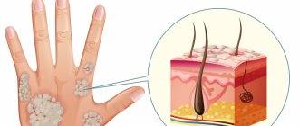
Viral elements - warts, papillomas, condylomas - look like bulges, growths on a stalk or a broad base. Some forms may become malignant due to frequent trauma, or initially have rapid growth and large size (Buschke's condyloma).
Dermatofibromas come from connective tissue, are more common in young women, grow slowly and have few symptoms. In extremely rare cases, they can become malignant. Externally they look like a nodule deep in the tissue with a convex hemisphere, smooth or slightly flaky surface.
Nevi , moles are pigmented areas of the skin formed by clusters of pigment cells. Typically caused by excess sun exposure. Although they themselves are not dangerous, about half of all melanomas (skin cancer) form in the area of birthmarks or nevi.
Types of precancerous skin tumors
With increased congenital sensitivity of the skin to the sun and other types of irradiation, xeroderma pigmentosum may occur. It begins as a large number of freckles, which develop into wart-like growths. With frequent UV irradiation, the elements turn into different forms of cancer (squamous cell or basal cell).
Bowen's disease is a neoplasm in the form of spots and nodules, plaques or bumps that tend to merge and form surfaces that are covered with growths. It happens especially often in older people. Often develop into undifferentiated forms of skin cancer.
Keyer's disease is bright red nodules that have a velvety appearance, are prone to ulceration and become covered with papillomas. It rarely turns into cancer, but often gets injured, hurts and bleeds.
Senile keratomas are limited nodules in which the skin intensively divides, they turn into plaques and become covered with dense crusts.
Cutaneous horn - occurs in areas where the skin is injured. A cone-shaped skin compaction up to several millimeters long is formed. Prone to developing into cancer.
Malignant neoplasms of the skin
They account for up to 10% of all patrols. They occur more often in elderly patients; they differ from benign elements in that the formation cells are difficult to differentiate already in the initial stage of growth. Cells lose their functions, grow into neighboring organs and tissues, and metastasize through blood capillaries and lymphatic vessels. This group includes melanoma , as well as non-melanoma cancers (squamous cell, basal cell), epithelioma , Kaposi's sarcoma .
ABCD principle
In dermatology, a simple rule is used to determine the signs of malignancy called ABCD. Each letter of the word corresponds to a specific characteristic of a mole:
A - asymmetry:
symmetrical (good) asymmetrical (bad).
B - random coloring:
uniform coloring (good) uneven coloring (bad).
B - height:
uniform height (good) uneven height (bad).
G - boundaries:
clear boundaries (good) unclear boundaries (bad).
D - diameter:
not growing (good) growing (bad).
Important: any changes in moles or their trauma is a reason to pay attention to them! Carefully examine all your formations; if you notice any suspicious signs, contact a specialist immediately!
Symptoms and diagnosis
Typically, benign and malignant skin tumors begin as a small area of skin change: the appearance of growths, crusts, and round formations. In addition, any moles or nevi that change their size, shape, or color are dangerous. In these cases, if there are any changes in the epithelium and underlying tissues, targeted diagnosis of skin tumors is needed. It is performed by a dermatologist, and if there are signs of melanoma or another type of cancer, by a dermato-oncologist. The doctor will ask you in detail about the complaints, evaluate what the skin growths look like, clarify growth dynamics, color changes, additional complaints (itching, pain, ulcers, bleeding).
If a tumor is suspected, the doctor will perform a biopsy of the suspicious element, which will accurately determine the nature of the tumor.
Do benign formations hide the danger?
Benign neoplasms are unpredictable structures that can manifest themselves at any time or not at all. The process of their transformation into malignant ones has not been fully studied. There is no clear answer to the question of what exactly activates this process. It is believed that mechanical trauma, excess ultraviolet radiation, metabolic disorders and other factors contribute to degeneration. One way or another, if you have a benign skin lesion, you should not experiment and rely on chance. Moreover, today removal does not cause difficulties.
Treatment methods
The main method of therapy is radical removal of the affected area with a small inclusion of healthy tissue. Benign elements are removed using the Surgitron apparatus, which avoids relapses, since the removed tissues are cauterized and there is no spread of tumor cells. Electrocoagulation of the affected areas is also used. Many clinics recommend radio wave therapy when removing skin tumors.
If it is cancer that cannot be removed, chemotherapy and radiation are used. With malignant processes, the prognosis is unfavorable, tumors can metastasize, so it is important to consult a doctor immediately at the first sign of skin problems. Modern removal techniques make it possible to prevent cancer and remove formations without scarring.
Dangerous degeneration of moles into melanoma!
Every year, doctors diagnose 200,000 patients with melanoma, and a third of cases are fatal. On average, after positive treatment results, patients with melanoma live about 5 years. These scary statistics are presented here so that you think about your health and pay attention to it.
How to spot melanoma?
Melanoma, in simple words, is a former mole. The harbingers are situations when a mole changes color, redness appears around the mole, grows unevenly, hurts, and bleeding appears. It is possible that new moles will appear during life, which look unusual and differ from those that previously existed on the body. Any changes or appearance of “unusual” moles require special attention and are a reason to consult a dermatologist.
Gentle removal of tumors
New growths can regularly appear on human skin, which, as we indicated above, must be treated with caution and monitor for changes. The most effective treatment for skin lesions is laser techniques, which not only provide an excellent cosmetic effect, but also reduce the number of complications and relapses.
Therefore, if skin formations bother you and you want to get rid of them, there is an opportunity not to put off taking care of yourself and quickly and delicately remove them using the Deka SmartXide CO2 laser. In what cases should you apply for removal? If the neoplasm:
- was traumatized;
- changed shape or size;
- causes physical discomfort;
- aesthetically you don't like it.
Using a specific CO2 attachment, the laser precisely affects the tumor and does not affect healthy tissue. This gentle removal promotes rapid healing.The Deka SmartXide laser is an ablative CO2 laser that includes a fractional scanner and a dermatology attachment. The fractional scanner is used for laser resurfacing, but the dermatological attachment is designed to remove various tumors.
The SmartXide system is considered the “Gold Standard” in laser surgery due to its wavelength absorption, technology, ergonomics and ease of use.
Please note that when removing skin lesions, the laser affects only the pathological tissue to which the wave is directed, and it minimally affects the healthy skin around it. The specialist controls the depth of exposure and has the ability to remove the formation layer by layer, while virtually eliminating the possibility of scarring.
Why is it worth removing tumors with a laser?
Deka SmartXide laser technology has several advantages:
- painlessness of the procedure (anesthesia is either not used, or application or infiltration anesthesia is used);
- the risks of inflammation are minimized because the laser beam is sterile;
- gentle removal of the pathological focus;
- lack of blood, because only coagulation of blood vessels is carried out;
- no repeat sessions are required.
Indications for histological examination
Histological examination differentiates a benign process from a malignant one. For histological analysis, fragments of a tumor or mole taken during removal are used. Histological analysis consists of examining thin sections of pathological tissue using an electron microscope. If histological results show signs of a malignant process, the skin in the area of the removed tumor is excised more widely.
All tumors removed surgically or excised by other methods can be subjected to histological examination. With laser destruction, tumor tissue is destroyed, and histological analysis becomes impossible.