There are contraindications. Specialist consultation is required.
Types and characteristics of neoplasms Signs of breast cysts Causes of occurrence Diagnostics Methods of surgical treatment Possible complications Prevention
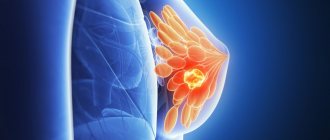
A breast cyst is a fluid-filled capsule that forms in the breast ducts. The liquid is non-inflammatory. Usually the disease begins against the background of other diseases, such as fibrocystic mastopathy. A benign neoplasm can appear in one gland (unilateral pathology) or in both at once (bilateral). There are both single lesions and multiple ones, spreading throughout the organ. At risk are women from 30 to 55 years old, especially those without children.
A woman can feel the tumor by palpating her breast during a self-examination. It will be oval, round or irregular in shape. The size can reach 5 cm in difficult cases. A typical formation has smooth walls, while an atypical one has growth directed inside the cavity.
Multiple cysts of different sizes are called "polycystic". Over time, they unite into multi-chamber conglomerates. Modified cystic tissue can occupy more than half of the breast.
This disease is not a threat to life, but can reduce its quality. It rarely degenerates into oncology, but its nodular forms can become a trigger for the development of cancer.
Cysts: types, diagnosis, surgical removal
What is a breast cyst
Cysts are benign, fluid-filled sacs that develop in the breast. They can be single or multiple. Cysts are usually round with regular edges and resemble a water balloon in appearance.
These types of growths are usually benign and do not require any special treatment until they become painful. In this case, there is a need for aspiration (surgical removal). Excision is also performed in that case. If the tumor increases in size.
Cysts are symptoms of nodular mastopathy. They are formed due to the expansion of one of the ducts of the mammary gland; first, a capsule is formed from the connective tissue, limiting the cavity, and then non-inflammatory fluid accumulates in the cavity. The shape of the formations can be round, oval or irregular.
The size of the cyst ranges from a few millimeters to 3-5 centimeters. The practice of mammology confirms that formations that are especially large in diameter can lead to deformation and change in the shape of the breast or to a complete replacement of its tissue itself.
Surgical removal of cysts
When cysts grow and the pills are no longer effective, surgical intervention becomes necessary. There are several methods for removing tumors:
- Puncture - performed in cases where the cyst consists of vesicles connected to each other by connective tissue and filled with liquid inside. During the manipulation, the cyst is punctured and all the secretion is removed from it.
- Laser removal is a modern and safe, but not a cheap method. Excision of a cyst with a laser is carried out quickly and painlessly, without scarring. LED exposure destroys only atypical cells. Over the next two months, they are completely replaced by healthy glandular cells.
- Surgical intervention is necessary for large cysts or high-density neoplasms. The operation is carried out with high precision; upon completion, the tissue at the incision site is stitched together in layers. A sick leave after surgery is issued by the doctor for the entire recovery period.
How to identify a cyst in the mammary gland: symptoms
Clinical signs depend on the location and size of the formation, as well as on the emotional state of the patient. The breast has a smooth, round lump that moves easily and has clear edges.
Small cysts usually do not cause discomfort. Often they can only be detected during a routine examination by a gynecologist.
If the cyst grows to a significant size or compresses the ducts and nerve endings, then the woman may experience the following complaints:
- aching pain in the area of education;
- breast irregularities and changes in its shape;
- the skin in this area may turn red and even blue;
- upon palpation of the chest, it is defined as a smooth round formation, painless and not welded to the surrounding tissues;
- may rarely be accompanied by nipple discharge;
- with inflammation and suppuration, many other symptoms are associated.
Pain syndrome has a relationship with cyclic changes. When
lumps in the chest, you need to visit a doctor for a comprehensive examination.
Symptoms and complications
Small breast cysts are often discovered accidentally during an ultrasound examination. Large cystic formations can be palpated by a woman during self-examination. A breast cyst is manifested by the following symptoms:
- Painful sensations, a feeling of discomfort, burning and tingling in the chest shortly before menstruation;
- Chest pain during menstruation, which goes away at the end of menstruation;
- Discharge of various types from the nipple;
- The presence of a round or oval formation of soft elastic consistency with a smooth surface, which is not fused with the surrounding tissues.
Can cysts in the mammary glands resolve? Small cystic formations can undergo reverse development if you follow a diet, a healthy lifestyle and systematic use of medications. We should not forget that some cysts have a tendency to become malignant. For this reason, if a cystic formation is detected, you should not self-medicate or use “traditional medicine”. If you detect a lump in the mammary gland, contact the Yusupov Hospital.
Breast cysts are dangerous due to the following complications:
- Inflammation;
- Suppuration;
- Breast deformation;
- Degeneration into a cancerous tumor.
Inflammation of the cyst occurs in the event of excessive heat exposure to the breast, weakened immunity, injury to the mammary gland, or infection caused by sore throat, flu, and caries entering the breast tissue. Symptoms of inflammation of the cyst are expressed in increased body temperature, fever, chest pain, redness of the skin over the formation. With inadequate anti-inflammatory therapy or severe immunodeficiency, the cyst suppurates. Body temperature rises to 41-41°C, regional lymph nodes enlarge, and “softening” appears in the center of the cyst. During puncture, pus is visible in the syringe.
Large cysts can deform the mammary gland, which causes psychological discomfort in a woman. When it degenerates into a malignant neoplasm, the cyst becomes dense, the skin above it takes on the appearance of a “lemon peel”, and the nipple is retracted into the breast. It may ooze blood or brown fluid. In order to prevent this formidable complication, you do not need to wait until the breast cyst “resolves” on its own. At the first symptoms of the disease, make an appointment with mammologists at the Yusupov Hospital.
What types of cysts are there in the mammary glands?
Depending on the number of cysts, a single formation (single cyst) and multiple formations (polycystic mammary glands) are distinguished. Also, depending on the number of cavities, the cyst can be single-chamber or multi-chamber. If the capsule of the cyst has growths from the inside, then they speak of an atypical cyst.
There are two types of cysts based on size:
- Microcysts
. Quite small formations that cannot be detected by palpation. They can be visualized using ultrasound and mammography. - Macrokists
. Determined by palpation. The size varies from 2.5 to 5 cm. Large formations compress the nerve endings in the chest, causing discomfort and pain.
Depending on the type of breast cyst, there are:
- Simple
. They consist of a single (solitary) formation, which is an elastic neoplasm of a round shape. The nodes most often contain a fibrous component. - Complex
. The formation consists of simple cysts fused together. May contain fibrous tissue growths. These also include atypical cysts.
Our services
The administration of CELT JSC regularly updates the price list posted on the clinic’s website. However, in order to avoid possible misunderstandings, we ask you to clarify the cost of services by phone: +7
| Service name | Price in rubles |
| Needle biopsy (without the cost of histological examination) | 2 500 |
| Ultrasound of the mammary glands | 3 000 |
| Mammography | 3 000 |
All services
Make an appointment through the application or by calling +7 +7 We work every day:
- Monday—Friday: 8.00—20.00
- Saturday: 8.00–18.00
- Sunday is a day off
The nearest metro and MCC stations to the clinic:
- Highway of Enthusiasts or Perovo
- Partisan
- Enthusiast Highway
Driving directions
Reasons for development
In most cases, a cyst develops under the influence of hormonal changes in the body, for example, during pregnancy or prolonged fasting.
The neoplasm appears against the background of the following factors:
- metabolic disorders in the body (diabetes mellitus, etc.);
- psycho-emotional shocks (stress, depression, etc.);
- long absence of sexual intercourse;
- birth of a child at the age of 30-35 years;
- diseases of the female reproductive system (adnexitis, endometritis, etc.);
- breast injuries and wearing uncomfortable underwear;
- inflammatory processes in the breast (mastitis, etc.).
If a woman has already been diagnosed with cysts, for example in the paranasal sinuses, gallbladder, this is not a predisposing factor for the development of cysts in the breast. This is due to various reasons for the appearance of neoplasms.
Survey
After conducting an independent minimal examination of the breast, the woman herself is able to confirm the suspicion of a neoplasm in the mammary gland. But only a mammologist makes an accurate diagnosis, determining the type of cyst or possible other formation. For a complete diagnosis, mammography alone is not enough. A comprehensive examination is required. Ultrasound examination and pneumocystography will give an unambiguous result in determining the diagnosis. Sometimes the liquid inside the formation becomes very cloudy and thickened due to the pus formed in it. This makes it difficult to establish an accurate diagnosis. With such manifestations, it is necessary to make sure that there are no malignant formations and conduct an examination of the walls. Such an examination cannot be postponed, since in 10-17% of all cases the cyst may degenerate into breast cancer.
Complications of breast cysts
Common problems associated with breast cysts include inflammation and suppuration. If treatment measures are not taken in time, this can lead to the development of cavities filled with pus. Both a small cyst and a large formation can fester.
Most often, complications develop when a woman wears tight underwear or has intense breast massage.
NB!
The danger of such breast cysts is the possibility of their transformation into cancer. This happens in 1-3% of cases. A cyst, large or small, should be under the supervision of a specialist. To find out about its presence, it is enough to undergo routine examinations on time, and when confirming the diagnosis, follow the recommendations of your doctor.
Rules for disease prevention
- Annual visit to a mammologist, regular mammography or ultrasound of the mammary glands.
- Regular self-examination.
- Taking care of the mammary gland.
- Take COCs only as prescribed and under the supervision of a physician.
- Rejection of bad habits.
- Selecting and wearing the right bra.
CELT clinic specialists will help you choose the appropriate treatment and maintain your health. The latest equipment, advanced techniques, professional doctors - CELT offers complete patient support, from diagnosis to recovery.
Diagnosis of breast cyst
As soon as a breast tumor is detected, the attending physician will immediately prescribe screening tests. The specialist will also ask about the duration of the pain and perform a physical examination.
Mammography
A screening x-ray method that is widely used to examine patients aged 40 years and older. The diagnostic procedure is based on the ability of adipose tissue to transmit x-rays to varying degrees. By obtaining different images on a mammogram, the doctor can visualize the problem to make a diagnosis and prescribe effective treatment.
Ultrasonography
An ultrasound is performed to find out whether the cyst is filled with fluid or is a solid growth. Depending on the type of non-cancerous growth, a biopsy may be performed to rule out cancer.
Fine needle aspiration is by far the most preferred test for cyst confirmation. During this procedure, a needle is inserted into the cyst cavity, the fluid is aspirated under local anesthesia and sent to the laboratory for analysis.
This study completely confirms the diagnosis. If the clear fluid is aspirated and the lump disappears, no treatment or further testing is required.
What you need to tell at an appointment with an oncologist-mammologist
At the appointment, the specialist makes a first assessment of the patient’s condition, based on the collected anamnesis and medical history. It is for this reason that you need to inform your doctor about your symptoms and their relationship with the menstrual cycle.
At the appointment, the mammologist should tell you about the presence of:
- symptoms that caused you to visit the clinic;
- the stresses, experiences and changes that have occurred in life;
- drug and vitamin therapy, use of dietary supplements.
You should prepare in advance a list of questions that will be asked to the specialist. First of all, you should ask about the risk of malignant degeneration and the examinations that need to be completed. It is also recommended to clarify the principle of treatment and inquire about the availability of additional individual recommendations.
At an appointment with a mammologist
Consultation and diagnosis of women begins with a survey. The mammologist must find out the patient’s complaints and identify unfavorable factors that can affect the normal functioning of the mammary glands. Next, a careful examination of the breast is necessary - palpation of all areas of the mammary gland, lymph nodes, zones of lymphatic drainage, nipples and areolas.
Only comprehensive diagnostics can guarantee accurate diagnosis. It includes a clinical examination of the patient with palpation of the mammary glands, ultrasound, mammography (X-rays taken on special devices with a low dose of radiation, mammographs). If formations are detected, puncture and biopsy are performed. If there is a suspicion of a connection between breast disease and pathology of other organs and systems of the body, the woman is prescribed appropriate tests. The mammologist will also indicate that consultations with a gynecologist, endocrinologist and other specialists necessary for your case are mandatory.
Visits at the clinic are conducted by doctors of the highest category - mammologist, mammologist-oncologist. Experienced surgeons at our clinic provide surgical treatment of the mammary glands: removal of intraductal papilloma, removal of skin lesions, sectoral resection, removal of benign lesions, as well as conservative treatment of all breast diseases. Equipping the clinic with the most modern medical equipment allows us to diagnose and treat mammological diseases using the most modern minimally invasive techniques.
In our clinic you will be greeted by experienced, highly qualified specialists. Consultations and explanations from the clinic’s doctors will not leave you with any questions. Our clients are surrounded by the care and attention of the best mammologists in the capital. We will always help you and bring back the joy of life.
Treatment of breast cyst
If the cyst is very small and only visible on x-rays, no treatment is required. In this case, a wait-and-see approach is recommended. This is especially true if the woman has not reached menopause. The most preferred treatment for breast cysts is aspiration.
If the cyst is quite large and needle aspiration does not resolve it, surgical excision is recommended. Cysts are quite common and tend to recur frequently and may require further intervention.
Drug therapy
Regulating your menstrual cycle can help reduce relapses. This can be done with the help of hormones, but the treatment must be under the strict supervision of a gynecologist, as there are many side effects and complications associated with the use of hormonal medications.
Hormonal therapy is carried out using oral contraceptives (birth control pills). Aimed at restoring menstrual cycles and reducing the likelihood of relapse. Hormones are prescribed only in the presence of severe symptoms, which is associated with the risk of adverse reactions.
Iodine preparations and sedatives are also prescribed. For severe pain, your doctor may recommend taking diuretics and anti-inflammatory drugs.
Physiotherapy
Physiotherapeutic intervention is necessary when medications do not give the desired result. The doctor prescribes procedures that increase the effectiveness of medications.
Physiotherapy has an analgesic, absorbable and anti-inflammatory effect. A good result is observed after electrophoresis and magnetic therapy.
Surgical intervention
The decision about surgery is made by the doctor, who is based on the patient’s well-being. Painless small cysts (up to 1 cm) are left for dynamic observation. But for most women, neoplasms themselves cause psychological discomfort. In this case, the issue of removing the tumor is also resolved.
Removal is necessary for recurrent formations and in the presence of discharge from the nipple. Surgical intervention is not necessary if the cyst contains bloody fluid or the tumor tends to grow.
Cyst removal is carried out in various ways:
- Puncture and removal of contents
. This operation to remove a formation in the mammary gland is quite safe, but often causes relapses, because its walls remain and the process is repeated after some time. It is performed using fine needle aspiration under the control of an ultrasound probe. - Laparoscopy
. Allows you to remove the formation and maintain an aesthetically attractive breast without rough postoperative scars. - Sectoral resection and mastectomy
. Removal of part or all of the mammary gland is rarely performed, in advanced cases, with its gigantic size or abscesses that cannot be treated conservatively.
Many online sources offer recipes for treating cysts with medicinal herbs and infusions that contain phytoestrogens and affect hormonal balance. Such therapy should be carried out under the supervision of a specialist. Otherwise, the patient's condition will only worsen.
Over-the-counter pain relievers and NSAIDs (such as ibuprofen) can be used to reduce pain and discomfort due to breast cysts.
Non-drug treatment of cysts
Physiotherapy
A breast cyst can be treated with physical therapy, but such a program should be approached with great caution. The main condition is the absence of malignant neoplasms, pain and swelling.
For treatment, two methods can be used:
- Electrophoresis - used in low doses and under the strict supervision of the attending physician. During the procedures, medications are applied to the skin, which, under the influence of weak electrical impulses, are absorbed into the parenchyma of the gland. In 10-15 sessions, swelling and inflammation are relieved, immunity is increased, and with full conservative therapy, cysts resolve.
- Radon baths - before using these procedures, a woman should definitely visit a doctor; if there is a risk of tumor formation, treatment is excluded. In other cases, such physiotherapy will be beneficial - it will relieve pastiness, activate blood flow, and create conditions for the resorption of compactions.
Other physical procedures are strictly prohibited. If necessary, they can be replaced with therapeutic exercises.
Exercise therapy
If the disease is in remission, physical therapy can be included in the program. Exercises should be light and not overload a weakened body.
There are two types of exercises you can do at home:
- Wall push-ups are done measuredly and accurately, with a straight back and straight legs. It is optimal to do 2-3 approaches 5-8 times.
- Raising arms fastened into a lock - perform with moderate effort for 1-3 minutes.
These exercises are aimed at training the pectoral muscles and activating blood circulation.
Prevention
Proper prevention implies timely detection and treatment of diseases of the female genital organs, digestive and nervous systems, and metabolic and endocrine disorders. It is recommended to take measures to prevent the development of metabolic syndrome (limit fatty and sweet foods, increase physical activity). It is necessary to exclude abortions and extend breastfeeding to at least 6 months.
The following measures can help prevent breast lumps:
- Wearing a bra that provides good breast support.
- Conducting a self-examination.
- Regular examinations by a mammologist - once a year.
Reducing caffeine intake is important in reducing cyst symptoms. Salt restriction has also been shown to be effective in relieving symptoms of the disease.
Women's theme
The mammary gland (breast gland) is a paired glandular organ. It functions only in women, producing milk after childbirth. The mammary gland is an organ of increased vulnerability, as it is under fire from many hormones. During the menstrual cycle, pregnancy, and lactation, the mammary glands are influenced by 15 hormones. The breast is susceptible to changes when at least one of them is produced in the wrong quantity. This is where women's problems begin.
Every woman over 17 years of age should undergo regular preventive examination by a mammologist. The frequency of visiting a mammologist depends on various factors: age, heredity, the presence of gynecological diseases, the presence of complaints. Women over 30 who have no complaints need to visit a mammologist every 1-1.5 years. Women over the age of forty are recommended to have a mammogram. Timely diagnosis and examination make it possible to detect various mammological diseases in the early stages. And this, in turn, will prevent breast cancer.
Question answer
Question: Can a cyst in the mammary gland resolve on its own/disappear?
In very rare cases, the cyst goes away on its own, so you should not let the disease take its course. Even small formations do not resolve on their own, but they can be treated conservatively. If the cyst reaches 1.5 cm, a puncture is made to take out the contents and introduce air into the cavity.
Any traditional methods of treatment (herbs, compresses), in the hope that the formation will resolve itself, are unacceptable. Only an experienced mammologist can prescribe effective treatment, carefully monitoring the dynamics of cysts. It is unacceptable to carry out drug therapy without confirming the diagnosis. Experimenting with health in the hope that the lump will resolve on its own is unacceptable.
Question: Can a cyst in the mammary gland hurt?
The causes of severe discomfort are often not in the cyst itself, but in swelling or plethora, which are characteristic of hormonal changes. If pain occurs in the projection of the cyst, then this indicates inflammation or tension. A puncture will bring relief.
Question: Can a cyst in the mammary gland burst?
In medical practice, cases of spontaneous opening and disappearance of a tumor in the breast have been described. But this is extremely rare.
Question: Can a breast cyst become inflamed?
If treatment measures are not taken in time, suppuration will develop. This condition is accompanied by severe pain and fever. In this case, treatment is carried out by external drainage (opening) under general or local anesthesia.
Question: Can a breast cyst be malignant?
Only macrocysts can transform into a malignant cyst tumor. It all depends on the morphological properties of the neoplasm. The risk of malignancy ranges from 1 to 30%.
There are three degrees of histological severity of proliferation:
- I – absent, so the likelihood of degeneration into cancer is minimal.
- II – epithelial proliferation is present, but without the risk of atypia of cellular structures. The risk of malignant transformation is moderate.
- III – the proliferative process occurs in conjunction with cell atypia. The risk of malignant transformation is high.
Proliferating forms of breast cysts occur in 0.3-1.4% of cases. They are classified as precancerous conditions. With pronounced proliferation of the epithelium, the risk increases to 25-30%.
Question: Can a breast cyst get bigger?
Non-tumor neoplasms become larger before menstruation. During this period, there is also an increase in pain.
Question: Can a breast cyst shrink?
A decrease in cyst size is observed after menstruation every month.
Types of neoplasms
Breast cysts can be atypical, fibrous, fatty, solitary, multi-chamber, ductal, developing against the background of diffuse mastopathy - let's look at their features and symptoms.
Atypical
They grow inside the cavity due to their lack of walls, form in an enlarged duct, can become inflamed, and are prone to relapses. There may be papillomomatous neoplasms in the cavity, both benign and malignant.
Fibrous
Their distinctive feature is the proliferation of connective (fibrous) tissue, leading to the formation of cavities that accumulate fluid. Over time, this fluid comes out of the nipples. These neoplasms create the background for the occurrence of cancer.
Fatty
The reason for its appearance is blockage of the sebaceous gland in the skin and overflow with secretion (milk). A benign formation has smooth walls and appears during pregnancy or breastfeeding.
Solitary
They may contain liquid of different colors and are benign. They form only in one breast and resemble a dense capsule.
Multi-chamber
Small breast cysts that eventually merge into one.
Ductal
They appear after 50 years and are benign in nature, but in medicine they are considered a precancerous condition.
Against the background of diffuse mastopathy
Diffuse mastopathy is characterized by a change in the structure of the breast tissue, in which the correct ratio of connective tissue and epithelial structures is disrupted. With diffuse changes in tissues, connective tissue structures grow unevenly, forming cords and nodules. This leads to the fact that the structure of the breast lobules and its ducts is disrupted, and very small cysts appear. Under certain conditions, this form of the disease can develop into nodular mastopathy, which is a risk factor for cancer.
conclusions
A breast cyst can be one or more fluid-filled cavities in the breast tissue. The contents may be pus or blood plasma. For a long time, the cyst may not give any symptoms, perhaps only a slight burning sensation and soreness in the mammary gland, which intensifies before the onset of menstruation.
The main cause of breast cysts is hormonal imbalances in the ratio of estrogen and progesterone. The cyst can become inflamed and inflamed, which is a dangerous and extremely unpleasant complication. Also, some types of tumors can develop into cancer. To exclude such an outcome, you should consult a doctor in time.
Sign up at the Medscan clinic to see a mammologist with extensive practical experience. The medical center is equipped with modern equipment, which allows you to make an accurate diagnosis in the shortest possible time.
Methods of surgical treatment
Surgery is needed when a breast cyst grows rapidly and its contents become infected. The classic technique is sectoral resection, in which the fibrous membrane itself and part of the tissue surrounding it are excised. In general, the intervention technique depends on the location of the tumor, its type, quantity and cause of appearance. Indications for surgery:
- lack of effect from conservative treatment;
- multilocular tumors;
- frequent relapses;
- inflammation of the capsule contents;
- neoplasms larger than 1.5 cm.
Modern techniques include sclerotherapy, in which a puncture of the mammary gland is first performed to pump out the contents of the cyst, and then a special drug for sclerosis is injected into the remaining capsule. As a result, the walls of the neoplasm stick together and fluid no longer accumulates. The pumped-out contents are sent for laboratory testing to rule out oncology.
Also at SM-Clinic it is proposed to treat breast cysts using laparoscopy. This is a low-invasive intervention in which both the contents of the capsule and the capsule itself are removed.
Another method is laser resection. The cystic tumor is destroyed by a directed laser beam. This is also a minimally invasive technique, which minimizes the risk of complications, blood loss and wound infection.
You can see the cost of removing a breast cyst on our website in the price list, as well as the prices for preoperative tests.
Literature
- Mumladze R.B., Dzukaeva N.T. New possibilities for treating patients with mammary gland cysts [Electronic resource] // Journal “Annals of Surgery”, 2015.
- Odintsov V.A., Dzidzaeva I.I., Bakhovadinova Sh.B. Modern ideas about risk factors for fibrocystic breast disease [Electronic resource] // Journal “Modern problems of science of education” - No. 5, 2021.
- Travina M.L., Popov A.G., Popov S.A. Cystic changes in the structure of the mammary gland in different age periods [Electronic resource] // Journal “Women's Health” No. 1, 2021.
Kinds
There are the following types of breast cysts:
- Shape: oval, round, irregular;
- By size – small, medium and large;
- By quantity – single or multiple;
- By the number of cameras - single-chamber or multi-chamber;
- Based on the presence of signs of inflammation - cysts with or without inflammation.
Typical cysts have smooth internal walls, while atypical cysts have growths on the walls. There is an atypical, fibrous, ductal, solitary, multilocular cyst of the mammary glands. If a complex breast cyst contains pinpoint tissue inclusions, doctors conduct in-depth diagnostics.
Atypical breast cyst
An atypical breast cyst is an accumulation of fluid in the cavity, which was formed when the gland duct expanded. It is round or oval in shape, can be of different sizes, and has a fibrous capsule. An atypical cyst is a benign neoplasm with growths that protrude into the cavity.
- An atypical cyst is formed for the following reasons:
- Long period of existence of cystic formation;
- Often recurrent breast cysts:
- Inflammatory processes that occur in the cyst;
- The presence of papillomatous formations in the cystic cavity.
If the cavity of an atypical cyst is small, it does not pose a problem for the patient and does not reveal its presence for a long time. After the cavity enlarges, the first signs of pathology appear - nagging pain in the mammary glands, which intensifies during menstruation. If the neoplasm is large, characteristic bumps are visible on the chest. They can be easily felt with your fingers. A large cyst in the mammary gland deforms a woman's breasts.
Fibrous cyst of the breast
Fibrous cyst of the mammary gland occupies a central place in mammology and is a background disease in the development of breast cancer. There are non-proliferative and proliferative forms of fibrous cyst. Fibrous cysts of the mammary glands develop under the influence of the following provoking factors:
- Hormonal imbalance;
- Weakened immunity;
- Disorders of internal organs (liver, thyroid gland).
The disease manifests itself with characteristic symptoms:
- Increased pain on the eve of menstruation;
- The presence of cluster-shaped nodular seals in the mammary gland;
- Discharge of fluid from the nipples.
A fibrous cyst with dense mammary contents is predominantly expelled by papillomas.
Solitary cyst of the mammary gland
A solitary cyst of the mammary gland is a benign dysplasia that does not degenerate into a malignant tumor. The neoplasm has a round shape and elastic consistency. It is a cavity filled with liquid of various colors and surrounded by a capsule. Brown fluid in a breast cyst may be evidence of atypia or vascular microtrauma. The longer the cyst exists, the denser its capsule. A solitary cyst is located in one breast.
The following reasons for the development of a solitary cyst are distinguished:
- Burdened heredity;
- Neuroendocrine disorders;
- Multiple abortions;
- Age after 35 years;
- Overweight;
- Late birth.
The cause of the disease can be stress, nervous strain, early puberty, complete refusal of breastfeeding, mastitis and trauma to the mammary gland.
A solitary cyst is manifested by pulling, pressing or aching pain in the mammary gland, which intensifies in the second phase of the menstrual cycle. The pain may radiate to the shoulder blade, neck or shoulder. At the end of menstruation, the pain stops. A woman can identify a large solitary cyst on her own by palpation.
Ductal cysts of the mammary glands develop at any age, but are most often detected after 48 years. This disease is diagnosed in 1% of cases among all breast tumors. Ductal cysts are small growths inside the breast. This is a benign tumor that is a precancerous condition. The risk of developing a malignant breast tumor against this background is high.
Ductal cysts develop under the influence of the following provoking factors:
- Hormonal disorders;
- Ovarian dysfunction;
- Abortion;
- Inflammation of the uterine appendages;
- Obesity;
- Diabetes mellitus and other endocrine diseases.
Clinically, this pathology is manifested by discharge from the nipples of a clear, brown, bloody or greenish fluid. A ductal cyst can be palpated if it is present in the area of the milk ducts. It is palpated as a dense, painless formation. The disease is most often discovered by chance, during a routine examination.
Multilocular breast cyst
A multi-chamber cyst develops with an unhealthy lifestyle, alcohol abuse, nicotine and drug use, and poor diet. The cause of the disease may be poor nutrition, metabolic disorders, stress, anxiety, and hormonal imbalances. First, one cystic cavity develops, then new ones are born, after which they merge. This is how a multi-chamber cyst is formed.
A multi-chamber cyst of the mammary glands is a dangerous disease that can develop into a malignant tumor. When felt, cysts resemble soft balloons, but sometimes they are hard. May be round, oval or irregular in shape.
Mastopathy
Mastopathy
This is a tissue pathology that occurs as a result of a change in the ratio of epithelial and connective tissue in the structure of the mammary gland. Such changes, in turn, lead to the appearance of cystic and fibrous neoplasms. Mastopathy is common in women of reproductive age and is most often associated with hormonal disorders. By type it is divided into diffuse, fibrocystic and nodular.
Risk factors that can trigger the occurrence of mastopathy:
- heredity;
- diseases caused by disruption of the endocrine system - diabetes, obesity, hypothyroidism, metabolic syndrome, polycystic ovary syndrome;
- inflammatory diseases of the reproductive system
- chronic liver diseases;
- psycho-emotional overload and stress;
- reproductive factors: late pregnancy, no pregnancy, a large number of abortions, early menarche, late menopause;
- lack of sexual life, late onset of sexual life (after 30 years), decreased sexual desire.
Signs and symptoms of mastopathy:
- pain in the mammary glands;
- a feeling of heaviness and fullness in the mammary glands;
- the appearance of hardened dense neoplasms-nodules;
- cracks in the nipples or changes in their skin, discharge from the nipples;
- enlarged axillary lymph nodes.
How to diagnose mastopathy
Mastopathy appears already during a visual examination by a mammologist and is confirmed using mammography in two projections or during ultrasound of the mammary glands. Additional diagnostic procedures may be prescribed: pneumocystography, tissue biopsy with cytological examination, hormone analysis. Early diagnosis of mastopathy is important because it is also a prevention of breast cancer.
Treatment of mastopathy
As in the case of treatment of cysts, mastopathy has a medicinal or surgical method of treatment. Drug therapy is primarily aimed at correcting endocrine problems. Patients are prescribed hormonal medications to eliminate hormonal imbalances, as well as non-hormonal therapy - diet, vitamins, immunomodulators, non-steroidal anti-inflammatory drugs, sedatives. Surgical treatment - lumpectomy (minimally invasive removal of breast tumors) or breast resection.
Diagnostics
Mammologists at the Yusupov Hospital make a diagnosis of “breast cyst” based on clinical, instrumental and laboratory examination of patients. After clarifying the complaints and collecting anamnesis, the doctor prescribes the following examination:
- Mammography (breast x-ray), which allows you to determine the presence, size, shape and number of cysts;
- Ultrasound (ultrasound) – helps to study the pathology in detail;
- Computer and magnetic resonance imaging;
- Radiothermometry;
- Fine-needle aspiration biopsy under the control of an ultrasound probe;
- Pneumocystography;
- Cytological examination.
Having our own clinical diagnostic laboratory allows mammologists at the Yusupov Hospital to promptly receive test results, including blood tests for cancer markers and hormones. Complex tests can be performed at partner clinics.
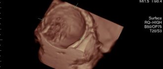
During ultrasound examination, breast cysts look like round formations with clear contours that are completely black in color (in medicine this phenomenon is called anechoic). Blood flow in cysts is not recorded during Doppler examination. The size of detected cysts can vary from a few millimeters to several centimeters. Sonologists often identify multiple breast cysts, as well as complex cysts communicating with each other. The vascular locus in the mammary gland in a complex cyst is detected using color Doppler mapping. Dotted tissue inclusions on ultrasound indicate the presence of a breast cyst. If a clot is found in a breast cyst, the cause of its appearance is determined.
Computed tomography of the mammary glands is prescribed as an additional examination method if a malignant cyst is suspected. Magnetic resonance imaging of the mammary glands does not allow clear differentiation between a normal gland and a cyst. MRI is always performed after a traditional radiation examination, and the data obtained are assessed comprehensively.
A cytological examination is carried out in the presence of bloody or profuse serous discharge from one duct. The material for cytology is biological material obtained by puncture with a thin needle, as well as impression smears from surfaces or discharge from the mammary gland.
Cytological examination of punctate allows one to reveal the malignant nature of the pathological process by identifying cancer cells or to diagnose benign changes by determining the presence and degree of proliferation and dysplasia of epithelial cells of the gland. The accuracy of cytological examination reaches 40-90%.
Only a biopsy can determine the nature of the cyst and establish whether it is a malignant or benign tumor. Methods for puncture of the mammary gland are non-operative and operative.
Mammologists perform a breast biopsy in the following cases:
- The patient or attending physician discovered a lump in the mammary gland;
- A mammogram or ultrasound reveals suspicious areas;
- Crusts, peeling, ulcers, or bloody discharge from the breast have appeared in the nipple area.
Fine-needle aspiration puncture of the mammary gland is the simplest way to obtain material for cytological examination on an outpatient basis and does not require anesthesia. Doctors use a regular injection syringe with a volume of 20 ml3. When performing a puncture of a cyst, a needle is inserted into the cavity and the contents of the cyst are aspirated. During the procedure, the doctor avoids unnecessary movements of the needle so as not to injure the cyst wall. If the contents are bloody, cytology of the breast cyst is performed and pneumocystography is performed.
In the presence of breast cysts, fine needle aspiration is not only a diagnostic, but also a therapeutic measure. After aspiration of the contents, the cyst cavity is filled with an ozone-oxygen mixture, after which the walls of the formation “stick together” and the cavity is empty.
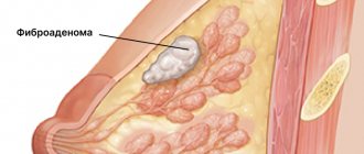
Fine needle aspiration biopsy is a quick and minimally invasive diagnostic procedure that can differentiate a fibroadenoma from a breast cyst. Depending on the results of the cytological examination, mammologists at the Yusupov Hospital individually decide on treatment tactics in each specific case.
Stereotactic needle biopsy is designed to precisely guide a needle into the area of suspected lesion using X-ray mammography or ultrasound. The procedure is used in cases where the cystic formation cannot be felt and is located deep in the tissues.
The use of a special needle allows one to obtain the required amount of tissue for a histological conclusion of the nature of the pathological process, including the difference between invasive tumors and cancer in situ, the presence of estrogen and progesterone receptors in tumors, and the degree of tumor differentiation. This method is performed in a clinic under local anesthesia. For tumors that are not palpable or the presence of microcalcifications, the needle is performed under ultrasound or mammography control (stereotactic biopsy). In the case of a biopsy of a tissue area with microcalcifications, an image of the resulting tissue sections is taken to confirm the presence of microcalcifications in them.
To perform a breast biopsy, a biopsy gun and disposable needles are used. This needle consists of two parts, a knife and a special tube. The gun fires a knife at high speed, which “cuts” a thin column of fabric. The procedure allows you to obtain not just a few cells, but a full-fledged tissue of formation.
To determine the differences between a cyst and malignant neoplasms of the breast, mammologists at the Yusupov Hospital perform biopsies under imaging control. This is a needle biopsy using ultrasound and magnetic resonance imaging. The method of taking material for research is the same as when performing a stereotactic needle biopsy. The main differences between the above methods of performing a biopsy are the imaging methods. In one case, this is high-frequency sound (ultrasound); in the other, it is a 3-D reconstruction, which is created by images of the breast in many planes.
If differential diagnosis of a breast cyst with a malignant tumor is necessary, an incisional biopsy is performed. It involves excision of a small piece of tumor tissue. This research method is similar to surgery. Incisional biopsy is performed when the results of aspiration biopsy are not sufficiently reliable. An excisional biopsy is a mini-surgery during which the surgeon removes part or all of the tumor. If the tumor is large, only part of it is removed.