Early recognition makes it possible to completely remove the lesion.
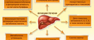
Thus, alveococcosis of the liver remains a surgically dependent disease. Radical liver resection for alveococcosis can completely cure most patients and gives good long-term results. When using modern research methods, there is no problem of making a correct diagnosis, but the lack of a screening system leads to the detection of the disease in late stages. As a consequence, at the time of diagnosis, in most cases, the possibility of radical surgery is excluded. Methods of local destruction and liver transplantation are used in limited observations due to the specificity of the parasitic disease.
Publications in the media
Regarding the treatment of this disease, you can contact the Department of X-ray surgical methods of diagnosis and treatment of the Clinic of Faculty Surgery named after. N.N. Burdenko
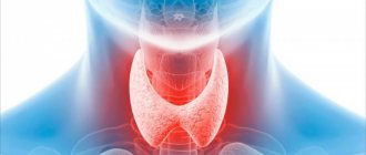
Alveococcosis is a zoonotic helminthic invasion that occurs as a volumetric process in the liver. In some cases, infiltrating growth and development of metastases in the lungs, brain, heart and bones are possible.
Etiology. The causative agent is the flatworm (cestode) Echinococcus multilocularis, type Platyhelminthes.
Epidemiology. The natural reservoir and definitive hosts are dogs (the main definitive host in Russia), arctic foxes, foxes, wolves, coyotes, and cats. Humans become infected by eating the liver of infected intermediate hosts. The eggs come out with feces, and active crawling of the segment is possible (they can crawl on the ground, leaving eggs behind), which leads to contamination of the fur, the environment (soil, water) and objects. Intermediate hosts (muskrats, voles) swallow segments or oncospheres (eggs in a special shell), which penetrate through the bloodstream into the liver, where the hatched larvae form a parasitic node. Most often, a person becomes infected when cutting carcasses or skins of infected animals, or by not following the rules of personal hygiene and keeping animals (especially dogs). Less common cases of infection are observed when eating wild berries and herbs contaminated with animal feces. In the Russian Federation, alveococcosis is widespread mainly in the Republic of Sakha (Yakutia), Krasnoyarsk, Altai and Khabarovsk territories, in Tomsk, Omsk and other regions. The disease is also reported sporadically in Tatarstan and Bashkortostan. Clinical picture. Compared to echinococcosis, it is more malignant. Characteristic is a gradual increase in symptoms. The main target organ is the liver. An enlarged and hardened liver (“iron” liver), pain and a feeling of heaviness in the right hypochondrium, decreased appetite, weight loss, jaundice and (rarely) ascites are observed.
Diagnostics • OAC: pronounced eosinophilia, increased ESR • Serological and immunological methods (RNGA, ELISA, RSK, latex agglutination reaction with Ag from the fluid of echinococcal blisters) give positive results in 60–90% of cases. A skin allergy test (Cazzoni reaction) is also used; it is most informative for liver echinococcosis • X-ray methods: cysts in the liver (against the background of pneumoperitoneum) or in the lungs look like rounded shadows with clear contours; rings of calcification are often found around cysts in the liver • Ultrasound, computed tomography, angiography.
Treatment. Surgical removal of parasitic nodes. Before and after surgery, mebendazole is prescribed in courses of 30 days in increasing daily doses from 200 to 600 mg or more.
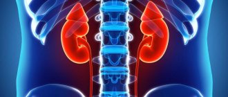
Complications • Portal hypertension • Budd–Chiari syndrome • Perihepatitis • Spreading and metastasis to internal organs (gallbladder, anterior abdominal wall, diaphragm, pericardium, lungs, brain).
Synonyms • Alveolar echinococcosis • Multilocular echinococcosis.
ICD-10 • B67.5 Liver invasion caused by Echinococcus multilocularis • B67.6 Invasion of other sites and multiple echinococcosis caused by Echinococcus multilocularis • B67.7 Invasion caused by Echinococcus multilocularis, unspecified
Consequences
The most common complication of alveococcosis is obstructive jaundice, which occurs due to compression of the bile ducts. Others include:
- aspiration pneumonia;
- chronic grosserulonephritis;
- renal failure;
- dilation of the blood vessels of the stomach and intestines with bleeding;
- fevers accompanied by severe chills and heavy sweats;
- breakthroughs of larvocysts into the cavity with the occurrence of peritonitis;
- abscesses due to bacterial infections;
- fistulas in the liver, lungs and pleura;
- germination of the parasitic node through the omentum and diaphragm into the mediastinum, lungs and heart.
Malignant alveococcosis with metastasis to the brain is particularly severe.
Forecast
The prognosis for alveococcosis is always serious. Without appropriate treatment, about 90% of patients die within 10 years. Lead to death:
- distant metastasis to the brain;
- tumor infiltration into neighboring organs with disruption of their functions;
- profuse bleeding;
- liver failure;
- purulent complications.
Surgical intervention leads to complete recovery, with early diagnosis and timely treatment, but the risk of incomplete removal of nodes and their further development cannot be excluded.
How can you get infected?
People become infected by ingesting parasite eggs or failing to comply with hygiene rules. There is a high risk of infection among hunters and anyone who has anything to do with animals. In rare cases, you can become infected from domestic animals, but when cutting up a fox carcass you need to be especially careful.
The disease does not spread from person to person because the parasite inside cannot reach maturity and cannot produce eggs. People do not infect each other with alveococcosis; this is impossible.
It is possible to become infected by eating unwashed berries and herbs that have been contaminated with the feces of various wild animals. In rare cases, eggs enter the human body through inhalation of dust.
Pathogen
The larva of the alveococcus tapeworm Echinococcus multilocularis is the cause of a parasitic disease; this parasite is from the family Echinococcus. Experts classify it as a species of Alveococcus, which is why it is called alveococcus. This parasite is popularly called the fox tapeworm, due to the fact that foxes are often its final hosts.
- The parasite in the larval stage looks like a small bubble with a diameter of 1-20 mm; this nodule causes the pathogen alveococcosis in both humans and animals. These larvae are dangerous because they reproduce vegetatively; they can bud and, with a prolonged illness, a conglomerate of very small bubbles (vesicles) is formed in the patient’s body. As a result of reproduction, they adhere tightly to each other, and sometimes grow together. The bubbles are filled with a viscous liquid of a yellowish or dark color; they contain the heads of parasites (scolex).
- In the normal life cycle of the parasite, such small bubbles are preferred by the organisms of small infected animals, these are rodents, which most often feed on foxes and other canines. Next, the larvae turn into adult worms, living in the small intestine, they reproduce. For the development of alveococcus, humans are a dead end because the larva will not be swallowed by the definitive host.
As an adult, the parasite reaches a length of up to 4 mm. This helminth is a species of flatworm, class cestodes (tapeworms), order Tapeworm (Cyclophyllidea). But it differs from its well-known counterparts (bovine or pork tapeworm) in that it is content with only a maximum of five segments, and does not have thousands, like others.
Alveococcosis in the liver
Stages of the disease
During alveococcosis there are several stages:
| Asymptomatic | Can last up to 10 years. The disease is discovered as an incidental diagnostic finding during the examination of a patient for another reason. |
| Uncomplicated | The pathological process is localized in the liver, that is, the location of the primary tumor. Patients complain of digestive disorders. |
| Complicated | It is characterized by the presence of metastatic tumors and significant dysfunction of a number of internal organs. |
Diagnostics
When examining patients with suspected alveococcosis, an epidemiological history is ascertained (residing in endemic areas, hunting, collecting wild berries, processing skins and carcasses of wild animals, occupational risks, etc.). The early stages are characterized by positive allergic tests (eosinophilia, Casoni reaction with echinococcal antigen).
Specific methods for laboratory diagnosis of alveococcosis include immunological reactions (RIGA, RLA, ELISA), PCR. To identify alveolar echinococcosis of the liver, the size and location of the parasitic node, plain radiography of the abdominal cavity, ultrasound and Dopplerography of the liver are used. A non-invasive alternative to arteriography and splenoportography is computed tomography. In difficult situations, liver scintigraphy and diagnostic laparoscopy are used.
If alveococcosis is suspected, other focal liver lesions are excluded: tumors, hemangiomas, polycystic disease, cirrhosis, echinococcosis. To identify metastases, chest X-ray, MRI of the brain, ultrasound of the kidneys and adrenal glands, etc. are performed.
Echinococcosis of the liver and lungs
The localization of echinococcosis is noteworthy: liver damage in 36 patients, 10 patients suffer from lung damage, and one patient has a generalized process.
Echinococcosis is represented in humans by two types of invasions: echinococcosis and alveococcosis.
Echinococcosis –
zoonotic biohelminthiasis caused by the larval stage of the tapeworm Echinococcus granulosus, characterized by a chronic course with the development mainly in the liver, less often in the lungs and other organs of solitary or multiple cystic formations prone to expansive growth.
Alveococcosis
– zoonotic biohelminthiasis caused by the larval stage of Echinococcus multilocalis, vogeli, oligarthrum and is characterized by a chronic progressive course with the development of multiple cystic formations in the liver or other organs, capable of infiltrative growth and metastasis. The life cycle and epidemiology of these parasitic diseases is similar.
Up
Symptoms of alveococcosis
For a long time (sometimes for many years), the symptoms of alveolar echinococcosis may not appear; the long latent period is explained by the slow growth of the cyst (larvocyst). The first signs of the disease are usually associated with dysfunction of the liver: it may be enlarged in size and lumpy to the touch. Pain in the right hypochondrium may appear, fatigue may increase, general weakness may develop, and appetite may decrease.
The growth of larvocyst causes disruption of blood flow in organs; toxins released by the parasite lead to tissue necrosis and acute allergic reactions. With the development of alveococcosis of the liver, symptoms such as:
- Increased pain in the liver (become permanent), liver dystrophy develops, and cirrhosis appears;
- The functioning of the gastrointestinal tract is disrupted (a feeling of heaviness after eating, diarrhea or, conversely, constipation);
- The appearance of skin itching (caused by both liver dysfunction and acute allergic reactions to worm toxins);
- Edema, varicose veins of the esophagus, accumulation of fluid in the abdominal cavity (if larvocysts have damaged large vessels);
- Disruption of the functioning of organs and tissues affected by metastases: if these are the kidneys, then problems with urination begin, if the tumor grows in the brain, symptoms of schizophrenia may appear, and so on.
In the last stages of alveococcosis of the liver (with the formation of a large number of metastases), the patient sharply loses weight, experiences severe pain, the functioning of almost all organ systems is disrupted - clinically the disease manifests itself as cancer. Alveococcosis causes serious immunity disorders and often causes complications such as abscesses of internal organs, purulent cholangitis, and amyloidosis.
Feeding at the expense of the host organism, alveococcus absorbs vital microelements that are not reproduced in the human body, but come from the external environment. Their deficiency provokes the development of severe endocrine disorders and has a detrimental effect on the functioning of the nervous system and the entire body as a whole.
Treatment of alveococcosis at the EUGENE clinic.
Our clinic offers you an innovative drug treatment method for alveococcosis. Our chief physician and part-time director of the clinic, Evgeniy Li, developed this technique in order to find a way to effectively get rid of parasites without the use of surgery.
And for ten years now we have been successfully treating patients with these problems. During this time, we have cured a lot of people who now live full lives and are periodically observed in our clinic for the purpose of prevention.
Get rid of alveococcosis with the EUGENE clinic and breathe deeply again!