According to the World Health Organization, in most developed countries, deep vein thrombosis and pulmonary embolism have become one of the leading causes of mortality. Millions of people around the world die every year from acute blockage (embolism) of a blood vessel by a thrombus. The danger of thrombosis is that a clot can reach a size comparable to the diameter of a venous vessel and completely block the blood flow in it. Or, if it ruptures, the clot can travel with the blood into the vessels of the lungs and lead to pulmonary thromboembolism. Often with fatal outcome.
Related article Catch it if you can. How to detect and destroy a blood clot
“A blood clot has broken off,” they sometimes say in this case about the cause of death. Even fairly young people often die from detached blood clots. According to specialists from the Moscow State Medical and Dental University named after A. I. Evdokimov, more than 90% of deaths from pulmonary embolism occur in patients who were not diagnosed with embolism and who did not receive treatment.
What causes blood clots?
Thrombi, in the form of blood clots, form in blood vessels in response to damage to the vascular wall, slowing of blood flow and changes in blood composition. “Thrombosis does not occur out of nowhere; there must be some prerequisites. This is facilitated by the presence of varicose veins in the lower extremities. If it is, you should pay attention to compactions that may appear in the veins,” says phlebologist, surgeon Fedor Shpachenko .
According to the phlebologist, compactions may indicate that blood clots have already formed in the veins. “Often, these blood clots may not occur in the superficial veins that we see, but in deep veins, and it is impossible to visualize them without special research methods,” warns Shpachenko.
Causes of blood clot separation
The thrombus can be parietal, or it can be floating. If a blood clot is located near a cholesterol plaque, it is less likely to come off than a floating blood clot. The latter “sits” on a thin leg, which has a weak attachment to the vascular wall. It is floating blood clots that most often lead to the development of pulmonary embolism and strokes.
Also, wandering blood clots may be present in the body, which pose a serious threat to life.
A blood clot can break off as a result of the following influences:
- The blood flow accelerates greatly.
- A thrombus has formed in a wide vessel.
- The stem of the thrombus is weak.
After breaking off, the thrombus begins traveling with the bloodstream along the vascular bed. At this time, it often breaks up into small fractions. Once in a vessel that does not exceed its size, the thrombus clogs it. This entails stopping blood flow and disrupting the nutrition of the limb or organ. This pathological condition is called occlusion.
Who is at risk?
Those who are most likely to develop thrombosis are people who are obese and have varicose veins, women who take oral contraceptives, and those who take many long flights. But a high risk of blood clots occurs when all these factors are combined.
“Patients with obesity and varicose veins are at risk. As for medications, in particular oral contraceptives, now most of them are well balanced and we rarely see the development of thrombosis and thromboembolism in women taking these drugs. People with a sedentary lifestyle, impaired hemostasis, namely blood clotting disorders, also fall into this group. If the patient has all these provoking factors, then the risk of developing thrombosis increases. Age also makes it worse. Over the years, the vessels lose elasticity and become brittle, plus a number of concomitant diseases cause swelling of the lower extremities,” says Shpachenko.
How do blood clots occur?
If a blood clot has formed on the wall of an artery, its appearance can be described in the following stages:
- Some process damages the artery wall.
- The body notices the violation and begins to build protection against blood loss, forming a large number of special blood cells - platelets, which, attaching to the damaged area, form a kind of patch.
- In case of coagulation disorders or changes in the hematopoietic system, platelet formation does not stop on time and continues longer than expected. This causes too much growth to form on the wall. Or platelets, which are in small quantities in the blood, floating past in the bloodstream, stick to the resulting accumulation.
The causes of damage to the walls of blood vessels can be:
- mechanical disruption of the structure due to injury;
- infectious lesion;
- high levels of glucose molecules in the blood;
- immune system dysfunction.
If there are no factors that contribute to the formation of blood clots, any injury or other damage will not lead to a large accumulation of blood cells. Under the layer of platelets, the artery wall will heal and recover, and the crust will resolve over time.
There are several stages of thrombus formation:
- disruption of the structure of the inner surface of the artery;
- activation of blood clotting factors;
- platelet adhesion at the site of injury;
- the appearance of substances that trigger a chain of reactions that form fibrin threads, which contribute to thrombus formation;
- a kind of network of fibrin threads is formed, into which blood cells enter, creating a large clot;
- Over time, the clot thickens, forming a thrombus.
When a blood clot breaks off under the influence of any factors, it begins to move through the bloodstream. Once it hits the nearest bottleneck, the blood flow will be blocked. If a similar situation happens outside a medical facility, it is impossible to save the person.
How to diagnose thrombosis at an early stage?
Thrombosis at an early stage can be detected with timely diagnosis of the veins of the lower extremities. To confirm or refute the diagnosis, phlebologists often recommend laboratory and ultrasound duplex scanning (USD) of the veins. This diagnostic method allows you to see the walls and lumen of the veins, the presence of a blood clot in them, its size, and even roughly judge how long ago the process is. You can also do angiography: a contrast study of blood vessels. And the predisposition to the appearance of blood clots can be identified using a coagulogram: a comprehensive analysis of blood clotting indicators.
If a blood clot comes off, what should you do?
If a person has a blood clot, then you cannot hesitate to call an ambulance. Moreover, even a doctor will not be able to make an accurate diagnosis without special equipment. Therefore, the victim must be urgently hospitalized.
There can be two treatment regimens for thrombosis: either the patient is prescribed medication correction or he is referred for surgery.
Drug therapy includes:
- Prescription of anticoagulants. These drugs help thin the blood (Heparin, Warfarin).
- Course treatment with statins.
- Prescription of fibrinolytics.
- Prescribing thrombolytics, which are necessary to dissolve the blood clot.
If the problem cannot be solved with the help of medications, or the person develops complications of thrombosis, he is indicated for an operation called thrombectomy. It is also possible to install special vena cava filters that will “catch” blood clots, preventing them from clogging vital vessels.
How to determine deep vein thrombosis?
Thrombosis often occurs asymptomatically or with mild symptoms. The nature of patient complaints may vary depending on the location of the blood clot, the duration of the disease and the nature of the lesion. The main symptom of deep vein thrombosis is pain in the leg, namely in the calf muscle. Swelling may also be a concern. When a blood clot forms in the deep veins of the lower extremities, the skin may take on a reddish or blue tint.
Article on the topic
Dissolve the clot. What foods improve blood quality
“Signs of thrombosis can manifest themselves as swelling and pain in the lower extremities, and the location of this pain can be very diverse. Sometimes you can’t guess what the pain is due to: either osteochondrosis, or blood clots, or other diseases. Therefore, if your legs swell, or there are varicose veins in your legs, then you should pay attention to the possible development of blood clots,” says Shpachenko. An ultrasound duplex scan or angiography will help you understand the exact picture.
Call your doctor or nurse if you have:
- pain has intensified, swelling has increased, or both of these symptoms are observed on the arm or leg where the blood clot has formed;
- there is pain, swelling, or both of these symptoms on the other arm or leg;
- breathing problems;
- severe or persistent headache;
- nosebleeds;
- bleeding gums;
- blood in your urine, stool (feces), vomit, or sputum you cough up through your mouth;
- heavier menstrual bleeding than usual;
- continuous bleeding;
- a bruise that does not go away;
- you fell or were injured in any way;
- you are planning a dental procedure or surgery;
- you stop taking your blood thinner for any reason;
- unexpected, unexplained side effects occurred;
- have any questions or concerns.
to come back to the beginning
How is pulmonary embolism diagnosed?
As a result of pulmonary embolism, cardiac function, pulmonary blood flow and gas exchange are disrupted. This condition is accompanied by characteristic symptoms, primarily a rapid heartbeat. There may also be a stabbing pain in the chest that gets worse while breathing. Patients may experience severe shortness of breath, and the respiratory rate may increase to 30-40 per minute. In addition, cyanosis or pallor of the skin, decreased blood pressure, cough, and hemoptysis may be observed.
“In the case of pulmonary embolism, difficulty breathing and chest pain may occur. In people with a disease of the vascular-cardiac system, for example, atrial fibrillation, pulmonary embolism can occur quite often,” says Shpachenko.
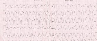
If thromboembolism is suspected, urgent hospitalization is required. To confirm the diagnosis, the hospital will order electrocardiography, chest x-ray and echocardiography. A radionuclide scan of the lungs, probing of the right side of the heart, and computed tomography with contrast of the pulmonary arteries can also be performed.
What is thrombosis?
Thrombosis can be venous or arterial. Venous is considered an acute disease; it appears due to disruption of the structure of the venous wall during injury, surgery, radiation or chemotherapy, due to increased blood clotting, due to a slowdown in the speed of blood flow.
Arterial thrombosis is a pathological condition. Patients have atherosclerotic plaques - cholesterol deposits that narrow the lumen of the artery and provoke circulatory deficiency. Over time, the plaques become clotted.
There is also pulmonary embolism (PE), a fatal disease when the lumen of the pulmonary artery is completely or partially blocked by a blood clot. According to statistics, almost 50% of patients with massive pulmonary embolism die within 30 minutes after its occurrence.
Treatment measures
These may be conservative therapy measures, such as medications and other therapeutic procedures or various surgical methods.
If it is determined that the clot is floating, first of all, the actions of doctors should be aimed at preventing its further movement.
The set of therapeutic measures to eliminate the danger to the life and health of the patient is as follows:
- The patient is kept in bed around the clock, during which the limb affected by thrombosis must be fixed in an elevated position.
- The leg should be wrapped with an elastic bandage.
- Thrombosis should be treated in a medical facility, and patients are prescribed anticoagulants - blood thinning drugs. Such products, according to the instructions, have multiple side effects and contraindications. Heparin-based medications are considered the best. Heparin is injected into a vein or via a drip through a catheter. Subsequently, the patient is transferred to tablet-type anticoagulants, for example Warfarin, and often such therapy can be lifelong.
- In addition to anticoagulants, the treatment regimen includes taking anti-inflammatory drugs to prevent the risk of thrombophlebitis - inflammation of the venous wall.
- During the entire period of hospitalization, doctors monitor the rate of blood clotting by prescribing various tests: CBC, coagulogram, thrombocrit.
Thrombosis of the lower extremities - symptoms
An additional danger of thrombosis is that the disease may not manifest itself for a long time. Symptoms appear when the blood clot reaches a large size and spreads up the vessel. Then the patient begins to complain of pain, which decreases at rest and increases with physical activity. Then, due to the deterioration of blood outflow, swelling is added to the pain. The skin on the leg turns pale (to the point of cyanosis) and “tightens”, acquiring a glossy shine.
To diagnose the localization of a blood clot, an ultrasound of the veins is performed, and in particularly difficult cases, angiography, an X-ray examination with the introduction of a contrast agent into the vessels.
Causes and risk factors
Blood clot on the arm: symptoms, first signs, photos
Among the causes are primary, associated with hereditary blood clotting disorders, and secondary, arising under the influence of external conditions.
For the first time, the process of thrombus formation was studied by R. Virchow, who identified several causes of this phenomenon: irritation of the vessel, coagulation disorders and blood flow disorders (Virchow’s triad).
The following risk groups for DVT and PE are distinguished:
- Low risk: patients under 40 years of age and those undergoing minor operations;
- Moderate risk: 40-60 years, minor or major surgery;
- High risk: after 40 years, major operations;
- Very high risk: after 60 years of age, major leg surgeries, hip fractures and other complicated injuries.
About ¼ of all cases of the disease occur after fractures of the long tubular bones of the legs, major operations on the joints and peritoneum. Thrombosis threatens a person both in the early and late postoperative periods.
Hip and knee surgery increases the risk of DVT to 30-50%, while minor procedures lasting up to 30 minutes have a risk of less than 10%.
Main risk factors:
- Prolonged immobility: condition after a stroke, bed rest, physical inactivity, air travel;
Obesity accompanied by changes in fibrinolytic activity;
General anesthesia with the use of muscle relaxants, when the muscle pump is turned off;
Pregnancy and the postpartum period, when changes in hemostasis and compression of blood vessels by the growing fetus occur;
Taking certain medications (thalidomide, heparin) and birth control pills with estrogen;
Oncological diseases: the combination of a tumor process and DVT is known as Trousseau's syndrome; elderly patients with DVT must be examined for oncological pathology;
Hemostasis disorders - thrombophilia;
Coronary heart disease along with chronic heart failure;
Viral infections;
Age-related changes: weakening of blood circulation, weakness of the venous wall, increased blood viscosity;
A history of DVT increases the risk of recurrence after injury and surgery.
Hemostasis disorders that increase the likelihood of thrombosis are combined under the term thrombophilia. There are congenital genetic defects that determine the tendency to form thrombosis: Leiden mutation, deficiency of antithrombin III, proteins S and C. Thrombophilia can be suspected if thrombosis occurs at a young age.
Size matters
Which blood clot is the most dangerous? The larger the vein and the larger the size of the clot, the higher the chance of thromboembolism. The most dangerous are proximal (located close to the center of the body) blood clots - in the veins of the thigh, pelvis and retroperitoneum. The incidence of embolism in this location is above 50%.
A small peripheral blood clot (for example, in the lower leg) can also become hypothetically dangerous. It also migrates to the pulmonary arteries and disrupts blood flow. Minor embolisms are asymptomatic in 75% of cases, but they still pose a threat to life, albeit a delayed one. Sooner or later, if left untreated, they can lead to circulatory problems and, in some cases, death. Therefore, any identified blood clot in the veins requires active treatment.
Why does thrombophlebitis develop? Causes of thrombophlebitis
As already mentioned, a favorable condition for the development of thrombophlebitis is varicose veins of the legs. In this case, there is stagnation of blood in the veins, weakness of the venous walls, venous insufficiency, increased venous pressure and deformation of the venous walls. All of these factors are enough for any infection to penetrate the vessel wall and cause inflammation.
Reduced immunity, thickening of the blood caused by illness or medication also facilitate the establishment of infectious pathogens on the vascular wall.
Thrombophlebitis can be triggered by injury to the venous wall, a medical procedure (intravenous injection, catheterization of a vein, vascular surgery, etc.), an infectious disease, for example, influenza, ARVI, pneumonia, tonsillitis, tuberculosis and even caries.
Treatment of the disease
Gastrointestinal bleeding: first aid, causes, symptoms, signs, treatment, consequences
Once the disease is diagnosed, the patient with a blood clot is treated as an inpatient. Treatment methods in this case can be conservative and surgical. When a clot is detected that floats, doctors first of all set the goal of preventing it from breaking away from the vessel wall.
Treatment of a floating thrombus is not easy. Treatment procedures include:
- Bed rest for the patient. The patient should rest as often as possible, while his lower limb with a blood clot should be in an elevated state.
- The lower limb with a pathological formation is fixed with an elastic bandage.
- If necessary, the patient is prescribed medications that help dissolve the clot. Such treatment must be carried out in a hospital, since the patient requires constant monitoring. Drugs that dissolve the clot have many contraindications and side effects. Medicines are administered by a vascular surgeon through a special catheter in a vein.
- During treatment, the patient is given a coagulogram, which is used to determine blood clotting.
- A modern method of treating a floating thrombus is to install a vena cava filter into the vein. This device looks like an umbrella. It is installed for a while and removed from the vein when the clot gets into this kind of filter.
- As a conservative treatment, the patient is prescribed anti-inflammatory medications.
- Hirudotherapy is sometimes used as an alternative treatment.
- Surgical method: thrombectomy or bypass surgery.
Possible complications and prognosis
Possible complications of thrombophlebitis and thrombosis of the thigh veins include:
- chronic venous insufficiency;
- TELA;
- formation of trophic ulcers;
- lipodermatosclerosis.
Without treatment, the risk of complications is high. In the absence of taking anticoagulant drugs, approximately 30% of patients experience a relapse of the pathology within 3 months, and after a year - in 70%. After treatment of thrombosis, the probability of relapse after 1 year is about 4-5%.
Thrombosis and thrombophlebitis are dangerous pathologies that can cause serious complications. To avoid this, it is necessary to carry out timely diagnostic procedures, as well as the correct choice of treatment tactics.