The foramen ovale is the communication between the two upper chambers of the heart, the atria.
Every unborn baby has an oval window; it performs an important function - while the fetus’s lungs are not breathing, thanks to the presence of a special opening (oval window), most of the blood “bypasses” them. At the same time, oxygenated blood flows directly from the right atrium to the left and then to the actively developing organism.
While the baby is in the womb, the task of the “lungs” – to saturate the blood with oxygen – is taken over by the mother’s body, and specifically the blood vessels of the placenta. This type of blood circulation is called fetal.
What is this defect and what is its physiology?
An open foramen ovale in the heart of a newborn is a normal variant. What is it for? Blood circulation in the fetus starts at the beginning of the second trimester of pregnancy. Like an adult, a baby’s heart has four chambers:
- In the third week of intrauterine development, there is already a wall separating the atria - the primary interatrial septum (PAS) with a wide opening.
- In the fifth week of pregnancy, it overgrows, but a new one appears - a secondary one (opposite which an oval window is formed in the child’s heart), and another wall is formed to the right of the primordial septum.
- By the end of the seventh week, the primary MPP turns into a valve covering the oval window.
The fetus does not breathe, and its “point of gas exchange” is the placenta, therefore, the body does not need a pulmonary circulation. Instead it has:
- ductus arteriosus - a vessel connecting the aorta to the pulmonary artery;
- Foramen ovale is the opening between the ventricles.
The unborn baby receives oxygenated blood from the mother's placenta, from where it flows through the two umbilical arteries into the inferior vena cava. Venous blood from the baby’s internal organs also enters there. This mixed blood (arterial for the mother and venous for the fetus) flows into the right half of the heart.
The window between the atria is necessary for maximum blood flow into the left chambers of the heart, and then into the aorta. The remaining blood in the right atrium is sufficient to oxygenate the lung tissue.
How the pulmonary circulation turns on
With the first cry, when the mother's oxygenated arterial blood stops flowing, the newborn's carbon dioxide concentration increases. Vascular receptors “signal” about hypoxemia to the respiratory center of the brain. From there, the impulse goes to the skeletal muscles of the ribs, under the influence of which it expands. The pressure inside the chest cavity decreases, and a stream of air rushes into the lungs, thereby straightening them.
At the moment of the first breath:
- The pressure inside the left atrium increases significantly.
- The valve is pressed against the oval window.
- The communication between the atria is blocked.
- The flows of arterial (from the lungs) and venous (from the internal organs) blood no longer intersect.
Two circles of blood circulation begin to function. Subsequently, the septum secundum is fused to the wall of the heart. In most people, a fossa ovale forms at the site of the oval window.
Why doesn't the hole close?
If the closure of the interatrial communication is formed during the period of intrauterine development, such babies are born with serious dysfunctions of the cardiac and respiratory systems or die even before birth. In fact, an open foramen ovale in a child’s heart is an adaptive mechanism, and not a congenital anomaly.
In 50% of people, the valve fuses with the heart wall by the end of the twelfth month of life. If they do not heal before the age of two, cardiologists classify the condition as minor anomalies of the heart (MACD).
Distinctive features of MARS:
- there is no significant hemodynamic disturbance;
- symptoms are practically absent, and if present, they are not characterized by specificity.
25–30% of the adult population lives with an open foramen ovale. For some, the valve dimensions are insufficient to completely close the hole. The primary MPP may overlap the communication, but not fuse with the interatrial wall. If the pressure in the right side of the heart increases, the valve will open and some of the venous blood will flow through the ventricle into the aorta.
Experts believe that the main cause of MARS is genetic predisposition. The presence of connective tissue dysplasia in the mother significantly increases the likelihood of their occurrence. Risk factors for having a baby with a patent foramen ovale:
- smoking of the expectant mother;
- use of certain medications by a pregnant woman;
- adverse environmental influences.
Pediatricians note that in premature infants, an open foramen ovale is observed much more often than in babies born after the 36th week of gestation. In adults, the valve covering the opening between the atria may open:
- after excessive physical exertion, for example in athletes;
- if there is a history of decompression sickness;
- in people with thrombophlebitis after episodes of pulmonary embolism.
Sometimes the defect between the atria is combined with other, more serious heart anomalies.
Prevention
There is no special prevention for an open oval window. To prevent pathology from occurring in newborns, during pregnancy a woman should lead a healthy lifestyle, give up alcohol and smoking. She also needs to avoid contact with:
- chemicals (paints, varnish vapors, some medications);
- ionizing radiation (thermonuclear reactions, X-ray machine);
- patients with infectious diseases (if infected with rubella in the first trimester, the risk of heart disease in a child increases significantly).
This article is posted for educational purposes only and does not constitute scientific material or professional medical advice.
Child's symptoms and behavior
In most cases, an unclosed foramen ovale in a child’s heart is not characterized by clinical manifestations. Parents may not even be aware of the existing feature. Further, in adolescence, the following are noted:
- decreased tolerance to physical activity;
- tendency to faint;
- breathing disorders.
Your doctor may suspect a patent foramen ovale in your baby if:
- pale skin;
- murmur on auscultation of the heart;
- periodically occurring cyanosis (cyanosis) of the nasolabial triangle in a child during outdoor games, screaming, coughing;
- if respiratory diseases “pursue” you much more often than other children;
- when your son or daughter is behind in growth compared to their peers;
- if the child complains of fatigue, dizziness, headaches, shortness of breath during exercise.
The discovery of varicose veins and thrombophlebitis in people under 30 years of age may also lead the doctor to think about a possible unclosed foramen ovale. A young child is not always able to accurately describe his condition, and a teenager, trying to appear older and more independent, sometimes tends to hide painful symptoms. Try not to delay visiting your doctor if there are even minor changes in your health.
Diagnostics
Ultrasound of the heart is of primary clinical significance. The doctor clearly sees a small hole in the projection of the left atrium, as well as the direction of blood flow.
When listening to a heart murmur, the pediatrician will definitely refer your baby for this type of examination.
According to new standards, at 1 month all newborns must undergo ultrasound screening, including the heart.
As a rule, there are no pathological changes on the ECG with LLC.
Diagnosis criteria
The International Classification of Diseases, 10th revision, recommends that atrial septal defect and patent foramen ovale be coded in a card using one code - Q 21.1. But in practice, doctors separate these two conditions (table below).
| Differences | Atrial septal defect | Patent foramen ovale |
| By pathogenesis | The hole may be caused by a violation of the formation of both primary and secondary septa | Rupture or insufficient size of the oval window valve |
| By localization | Can be located in any area | At the bottom of the fossa ovale, on the inside of the right atrium |
| According to anatomical features | Defect in the form of a hole | Has a valve structure |
| For hemodynamic disorders | Blood can flow from one atrium to another in both directions | Blood is shunted more often from right to left |
Ultrasound data
Cardiologists call echocardiography the “gold standard” for determining congenital heart anomalies. An ultrasound doctor judges the presence of an open foramen ovale using the following criteria:
- two-dimensional echocardiography: interruption of the signal from the septum at the location of the fossa ovale, gradual wedge-shaped thinning towards the edges of the defect, absence of volume overload of the right half of the heart, paradoxical movements of the barrier between the atria;
- Doppler echocardiography: turbulent blood flows at the location of the oval window, blood flow indicators in the right ventricle and pulmonary artery are normal.
Transesophageal echocardiographic examination is considered the most informative. But this type of diagnosis is only possible for adults or adolescents.
Degrees
Patent foramen ovale is classified in different ways. Some sources divide it by hole size:
- small - less than 2 mm;
- average - from 2 to 4 mm;
- large - more than 4 mm (up to 18 mm).
Other authors believe: everything that is in the middle part of the septum is less than 5 mm in size and has periodic discharge from the right atrium to the left or vice versa is recognized as a patent foramen ovale. Anything larger than 5 mm with constant bleeding is a heart defect, atrial septal defect. The latter option manifests itself with more severe symptoms and requires treatment.
Additional examination methods
To determine patient management tactics, it is important to know how a hole in the interatrial septum affects heart function. Doctors do electrocardiography at rest and after exercise. This will help eliminate arrhythmia and conduction disturbances.
For adults, x-rays of the chest cavity are recommended, which will make it possible to judge the degree of hypertrophy of the right atrium and reveal congestion in the lungs.
Children with minor anomalies of heart development need consultation with a pulmonologist and immunologist. The presence of chronic foci of infections can aggravate the child’s condition, so you need to go to the otolaryngologist’s office or visit a dentist.
Possible complications
Most parents and pediatricians rightly believe that the presence of an open oval window does not threaten the child’s health. This opinion is supported by the statement of a popular pediatrician, Evgeniy Olegovich Komarovsky: this condition is not a malformation of the heart. Indeed, with a small hole, a person can play sports, work and study, and young men are even subject to conscription for military service.
But under certain conditions, a patent foramen ovale can cause the following unfavorable conditions in adults:
- Paradoxical venous embolism is the entry of a microthromb, a bubble of fat or air from the venous system into the left atrium, and then its penetration into the systemic circulation. This condition can provoke a transient ischemic attack or stroke.
- Migraine with a patent foramen ovale often occurs with an aura. It usually occurs in older women when blood is shunted from right to left.
- Platypnea-orthodeoxia is a syndrome characterized by shortness of breath that occurs when standing and subsides when lying down. It is usually observed when a patent foramen ovale is combined with deformation of the IV.
- Obstructive sleep apnea syndrome.
Some people with a patent foramen ovale develop transient global amnesia after a migraine or ischemic attack - a syndrome of memory impairment and inability to perceive new information.
Complications and prognosis
The most serious and only complication is paradoxical embolism. In this case, a blood clot (usually with thrombosis of the deep, superficial veins of the lower extremities, pelvis) or air bubbles (during diving, less often during fractures) from the venous system enter the arteries of the systemic circulation through the open oval window. Thus, ischemic stroke, myocardial infarction, intestinal ischemia, infarction of the spleen and other organs develop.
There are no clear statistics in Russia. According to studies in France and the USA, ischemic strokes account for up to 30% of cryptogenic (unexplained) strokes. Of these, about a little less than half of the cases are associated with an open foramen ovale detected in patients.
Forecast
The prognosis depends on the size of the open oval window. It is favorable for sizes up to 4 mm, low blood discharge and no increase in it over time.
In the case of the development of large blood shunts, which increase the blood supply to the right chambers of the heart and increase the degree of pulmonary hypertension, the prognosis sharply worsens.
Usually, before the development of severe pulmonary hypertension, the patient has time to undergo surgery - to close the functioning oval window with an occluder. The operation dramatically improves the prognosis for the patient.
Treatment
Holes with a diameter of 4 - 5 mm have a high chance of closing on their own. Approaches to the treatment of an open foramen ovale are individual and depend not only on the size of the defect, but also on the presence of increased pressure in the pulmonary trunk, the degree of myocardial hyperplasia, and the risk of complications.
For children with no clinical manifestations and stable defect sizes, drug therapy is not indicated.
If your child is diagnosed with a patent foramen ovale, he or she needs periodic examinations by a cardiologist.
What can happen to the oval window during life:
- closure;
- maintaining constant dimensions;
- expansion of the defect as they grow older.
Some parents give their children vitamin complexes and dietary supplements and resort to herbal treatment, but the effectiveness of such therapy has not been proven.
Indications for surgical correction of communication between the atria:
- signs of overload of the right atrium and ventricle;
- the occurrence of heart failure;
- presence of complications.
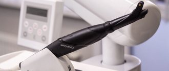
Transcatheter closure of the hole in the interatrial septum is performed. An occluder is passed to the human heart through large vessels using a catheter (see photo below), which plays the role of a patch. The manipulation is performed under X-ray and ultrasound control.
For preventive purposes, the operation is not indicated even for people of certain professions, although the available data from the medical literature confirm that performing transcatheter occlusion can help prevent decompression sickness in divers.
It is worth mentioning the possible adverse consequences of the intervention:
- embolization with occluder;
- infectious complications;
- erosion of the aorta or pericardium;
- atrial fibrillation;
- occluder thrombosis;
- the emergence of a new MPP defect.
Late complications of the transcatheter procedure include the occurrence of a residual shunt (residual discharge of blood into the left atrium) and the formation of a blood clot on the occluder. You should carefully discuss with your doctor all the pros and cons of surgical correction of the defect. Closing the oval window significantly improved the condition of patients with migraine, platypnea-orthodeoxia syndrome, and helped prevent recurrent ischemic strokes in adults. At the same time, pediatric cardiologists do not recommend surgical elimination of the IV defect in infants, considering the optimal age for radical correction to be from 2 to 5 years.
Patent foramen ovale in an adult
There is a high likelihood of ischemic stroke in young women with a patent foramen ovale who suffer from migraines.
In adults, after examination and ultrasound, cardiologists note the following risk factors for complications:
- dimensions of the oval window – more than 4 mm;
- tension of the interatrial septum during the Valsava maneuver;
- shunting even a small amount of blood to the left;
Sometimes doctors prescribe anticoagulants and antiplatelet agents to prevent the development of stroke and other thromboembolic complications.
Causes and risk factors
The main cause of patent oval window is a genetic factor. Most often, the anomaly occurs in people with a predisposition to connective tissue dysplasia, which is inherited. Therefore, in addition to the oval window, people often experience mitral valve prolapse.
There are other reasons and risk factors for the development of an open oval window:
- taking non-steroidal anti-inflammatory drugs (diclofenac, ibuprofen, nimesulide, etc.) during pregnancy, especially in late gestation (pregnancy);
- alcohol and nicotine consumption during pregnancy;
- premature birth.