Stereotypes and differences from radiography
When undergoing an examination, it is important to pay attention to the direction issued by the attending physician. If it says that you need to undergo fluorography, you do not need to change the order yourself, relying on a classic X-ray to better show the condition of your lungs. Such ignoring of specialist advice will only lead to unreasonable radiation exposure to the body. In medicine, radiation dose limits (1 mSv/year) are established only for healthy people when conducting preventive studies (for example, when applying for a job). Limits on radiation doses required for diagnostic purposes (in sick people) are not established by law. It only states that the doctor should strive for a minimum level of radiation, but not at the expense of the quality of diagnosis. In general, a reasonable postulate, but how is it implemented in practice? But in practice in Russia the average “medical” radiation dose turns out to be several times higher than in the UK, USA and France. At the same time, in some of our regions the average “medical” radiation dose exceeded 3 mSv. Fluorography provides less information than radiography. But the World Health Organization does not recommend the use of traditional film fluorography, even in underdeveloped countries. The solution to the problem was the transition to digital fluorography, which, with high information content, reduces the radiation dose several times. Here are the approximate radiation doses (mSv) that a person receives when examining the chest using various methods:
- radiography (in two projections) – 0.25;
- fluorography (in one projection) – 0.6–1.6;
- fluoroscopy – 3.0;
- digital fluorography – 0.01–0.06;
- computed tomography – 3.5–5.0.
So, if nothing bothers you, and you are referred for an X-ray for prevention, then it is better to undergo digital fluorography or radiography (“image”) instead of conventional fluorography. You should definitely refuse to perform fluoroscopy (when the doctor looks at the patient behind a screen): not only does the patient receive a large dose, but his image will not remain, and the conclusion will be solely on the conscience of the doctor who conducted the study.
Fluorography is one of the most important methods of modern diagnostics
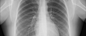
Fluorography is an x-ray examination in which an x-ray image of an object is photographed from a fluorescent screen onto photographic film or electronic media. The basic principles of fluorography were developed by Italian scientists A. Battelli and A. Carbasso and the American scientist D. M. Bleyer immediately after the discovery of X-rays.
A fluorogram appears on a digital camera immediately after the photo is taken. This allows you to reduce the appearance of uninformative images to zero and avoid the need for repeated fluorography. It is very important to know that the radiation dose during the study is reduced by 4-5 times! Digital devices give a radiation dose comparable to only part of the dose load for a person in one day from natural radiation sources! And no matter how irritated we are at times by the inexplicable demands to undergo fluorography at work, when visiting a clinic, undergoing various medical examinations or anywhere else, we should not refuse it.
Fluorography is one of the most popular services and one of the most important methods of modern diagnostics. This is an effective X-ray method for mass screening of the population. The main advantages of fluorography are cost-effectiveness and high availability. In 2007, a digital fluorograph was installed in the clinic of the Priargun Central District Hospital. An image of the lungs appears on the monitor screen after 6 seconds and the patient is immediately informed of the examination result. The main purpose of fluorography is to conduct preventive examinations of the population (adolescents and adults) for the early detection of diseases such as pneumonia, tuberculosis, pleurisy, and oncological formations at the earliest stage, which is the key to timely successful treatment.
Thanks to this method, in the Argunsky district in 2021, 9 new cases of tuberculosis and 7 new cases of lung cancer were detected. In the first quarter of 2021, 4 cases of tuberculosis and 2 cases of lung cancer have already been identified.
Very often, a person has to go to a medical institution not only due to illness, but also for a number of other reasons. This includes passing a medical examination when applying for a job, when preparing documents for the right to use weapons, for the right to drive a vehicle, before business and tourist trips abroad, to prepare documents for guardianship, adoption, and much more. In all these situations, acquaintance with a medical institution begins with a chest fluorography. Naturally, many visitors have questions about its feasibility. We will try to answer them. – What diseases can be detected using fluorography? – First of all, this is tuberculosis of the respiratory system, since its clinical manifestations, especially in the initial period, are extremely scarce or even absent. In addition, tuberculosis can occur with symptoms that mask the true cause of the disease “mask of tuberculosis”: acute respiratory viral infections, influenza, pneumonia, functional circulatory disorders, dyspepsia. In addition to tuberculosis, other diseases can be identified: oncological diseases of the chest organs, congenital anomalies of the visible skeleton and lungs, sarcoidosis, occupational lung diseases, pathologies of the heart, pleura, diaphragm, collarbones, ribs, etc. Therefore, considering that you do not have tuberculosis , you cannot refuse to undergo a preventive examination. – What is the difference between fluorography and radiography? – Fluorography is used as a preventive method of mass examination of the population, which is facilitated by a number of its advantages: high throughput, low economic and labor costs. Radiography is used as a diagnostic (more expensive) method when the doctor suspects a disease or injury. – Who is contraindicated for prophylactic fluorography? – Children under 15 years of age and pregnant women. – Where can I get fluorography? – Fluorography can be done at a clinic at your place of residence or place of work if you have a valid medical insurance policy. Paid and without insurance - in any medical institution. – How often do you need to undergo fluorography? – There are groups of people who are subject to preventive examinations 2 times a year :
- Persons who have had tuberculosis - during the first 3 years, once every 6 months.
- Persons in close contact with sources of tuberculosis infection.
- HIV-infected.
- Persons released from correctional institutions during the first 2 years.
Persons over 15 years of age who are at risk should undergo fluorography annually
- patients with chronic nonspecific lung diseases, diseases of the genitourinary system, peptic ulcers of the stomach and duodenum, diabetes mellitus;
- persons receiving corticosteroid, radiation and cytostatic therapy;
- persons without a fixed place of residence (homeless);
- migrants, refugees, internally displaced persons;
- persons living in inpatient institutions and social assistance institutions for persons without a fixed place of residence and occupation;
- employees of social service institutions for children and adolescents;
- workers of medical institutions, sanatoriums, educational and sports institutions for children and adolescents.
extraordinary preventive examinations :
- persons who sought medical help with suspected tuberculosis;
- persons living with pregnant women and newborns;
- citizens conscripted for military service or entering military service under a contract;
- persons diagnosed with HIV infection for the first time.
The entire population over 15 years of age must undergo fluorography once every 2 years. However, due to the unfavorable situation regarding tuberculosis, it is necessary for all citizens to undergo annual fluorography.
– How long are images stored?
– Fluorograms are stored in the archive for 5 years.
– Can I take them with me?
– Fluorograms and radiographs are issued only at the request of the attending physician or military registration and enlistment office, and the patient is given the result with a date stamp for this study and its result.
– What should people do who cannot come to the clinic and have fluorography done?
– For non-transportable patients and disabled people, the local nurse takes a sputum test at home to detect Mycobacterium tuberculosis.
Annual fluorography can protect you from deadly diseases. Since tuberculosis and lung cancer detected in time are sometimes the only chance of survival from these diseases.
Every resident of the district can undergo fluorographic examination at the clinic of the Priargun Central District Hospital. To do this, you must contact the reception desk during business hours and have a compulsory health insurance policy and an outpatient card with you. The fluorography room is open from Monday to Friday from 8.00 to 15.00. We invite everyone!
Radiologist of the State Healthcare Institution Priargunsky Central District Hospital Mironov K.O.
Classification of fluorography modes
A preventive examination of the lungs should be done once every 2 years, and for certain categories (for example, people with chronic diseases or members of other risk groups) - once a year or even 2 times a year.
Patients who are already registered at a tuberculosis dispensary are prescribed a test much more often: at the beginning of a course of therapy, the interval between procedures is about three to four months, depending on the intensity of treatment, the characteristics of the development of the disease, and the individual properties of the body.
With positive dynamics of treatment, the regimen changes. At first, monitoring is carried out approximately once every six months while maintenance therapy continues, and after complete recovery and deregistration, the patient needs to be examined on a general basis - annually.
Using a digital camera ensures that the image is quickly captured when the image is reconstructed using high-tech software. The result is displayed on a computer monitor, which you can:
- put on digital media;
- print on paper.
The examination results are stored in a database, which ensures their availability for the patient and doctor during the entire observation period. When you enter your last name along with your residential address, the program will display all previously recorded survey results. All that remains is to compare the updated and past information to track the dynamics.
If fluorography is bad, what could it be?
The transcript of the study is ready the next day, within 5 minutes after the study. If the result is poor, the person is referred to a TB specialist. It is too early to make a final diagnosis; a comprehensive diagnosis is carried out. Doctors may recommend repeat FLU. For the most part, the transformation of lung tissue is caused by the development of connective tissue in the lungs, which grows and layers.
Depending on the location and shape of the pathology focus, the doctor diagnoses fibrosis, sclerosis, adhesions, radiance, heaviness, shadows, and scar transformations. Based on the results, it is possible to diagnose oncology and emphysema, cysts, abscesses, calcifications, and other neoplasms and cavities.
Diagnostics can be done in every city, both in a private medical center and in a city clinic. The result obtained cannot be trusted 100%; an integrated approach to the health problem is needed.
Main indications
The main objective of the diagnostic method is to identify diseases of the lungs or bronchi. It is not for nothing that the technique is considered a preventive approach that prevents serious complications for diseases of the respiratory system.
As a rule, a local therapist or pulmonologist gives a referral for testing when a patient comes to them with characteristic symptoms:
- prolonged persistent cough;
- dyspnea;
- pain syndrome in the chest area.
When the above signs are detected, the chance of detecting a pulmonary or bronchial deviation increases significantly. Often the study allows you to establish:
- inflammatory process even at the initial stage of development;
- pneumothorax;
- neoplasms of malignant and benign nature;
- pleural damage;
- emphysema.
But most often, even during a preventive examination, tuberculosis is detected, so annual mandatory examinations are recommended, which help prevent the disease, which currently leads in a high percentage of deaths.
In addition to standard indications, there are two more categories for examination: family members living with pregnant women or newborns in order to minimize the risk of illness in infants and persons of military age.
The second category will include unscheduled fluorography if there is suspicion of:
- foreign body in the lungs;
- inflammation;
- tuberculosis;
- tumors of the mediastinal organs;
- diseases of the heart, large vessels.
Such precautions make it possible to recognize concomitant diseases such as fibrosis or sclerosis. Also, using a small image, it is possible to determine the localization:
- cysts;
- cavern;
- abscesses;
- infiltrates;
- accumulations of gases.
Despite the productivity of the technique, fluorography can hardly be called the main argument for diagnosing pulmonary pathologies. It is often necessary to use auxiliary tests, including sputum analysis, radiography, and MRI with contrast.
Why is fluorography necessary?
The main goal of diagnosis is to identify the pathological process at an early stage and begin treatment before specific symptoms appear. Here are the diseases that fluorography diagnoses:
- Pneumonia;
- Pleurisy;
- Tuberculosis;
- Malignant tumors of the lungs, mediastinum;
- Emphysema.
If a health problem is detected early, the chances of a favorable clinical outcome increase. This is what can be seen on fluorography:
- Lungs;
- Heart;
- Less often - bones.
This screening examination shows the inflammatory process, foreign bodies, tumor formations, accumulation of infiltrate, cavities of a non-physiological nature (various cysts). Fluorography even shows if the patient smokes. The result is not the main criterion for making a diagnosis; it allows a more accurate assessment of a specific clinical case.
Pregnancy and other contraindications
Contraindications to manipulation are pregnancy and childhood. It is believed that the minimum threshold is 15 years, since before this time it will be difficult for children to tolerate increased radiation exposure.
Among the relative contraindications, which are sometimes ignored when the benefit exceeds the possible harm, are:
- claustrophobia;
- dyspnea;
- general serious condition.
But sometimes even pregnancy, which is a contraindication for almost all types of diagnostics, does not become an obstacle to the procedure. It should be done only after preliminary consultation with your doctor. Control must be carried out by a specialist throughout the entire manipulation.
An important factor in the success of diagnosis and safety for the fetus is the appointment of an examination after 25 weeks. During this period, it had already formed its basic systems.
The reasons for “photographing” the respiratory system of pregnant women and women during the breastfeeding period should be as compelling as possible. Preventive X-ray examinations are not carried out for pregnant women and children under 15 years of age.
What results does chest fluorography show?
Chest fluorography is a method of primary diagnosis of lung diseases. That is, based on its result, the doctor can assume that the patient has some kind of pathology, but cannot make an accurate diagnosis. This is explained by the fact that fluorograms are small in size and have lower image quality when compared with classic x-rays. Therefore, the information obtained may not be enough to confirm the diagnosis.
On a fluorogram, the doctor can see:
- change in the structure of the pulmonary roots (they can expand, become compacted, stringy);
- change in the vascular pattern of the lungs (strengthening, deformation);
- focal shadows;
- fibrosis zones;
- calcifications;
- expansion or displacement of the mediastinal shadow.
Diseases that can be detected (suspected) using fluorography:
- tuberculosis;
- large tumors (small tumors may go unnoticed);
- pneumothorax;
- pleurisy;
- late stages of pneumonia;
- foreign bodies in the respiratory tract;
- heart diseases.
People whose radiologist has identified any abnormalities in the image require a more detailed examination, including either a chest x-ray or a computed tomography scan of the area. You may also need to consult with specialized specialists - a phthisiatrician, a pulmonologist, an oncologist, a cardiologist.
Fluorography
The study involves the use of x-rays, their effect on the body is negligible. Only organs located in the chest are examined, namely the lungs, large bronchi, and the heart. As a result, an image is obtained (often digital), but its size is quite small, it is 11 by 11 cm. Today, the devices with which the study is carried out are divided into 2 types:
- film devices;
- digital.
The technique is used for prevention purposes and is distinguished by the presence of contraindications, including: age under 14 years, pregnancy.
X-ray
Allows you to explore the internal structures of the body. The photo is usually life-size. The method can be survey or targeted, when a specific area of our body is studied. As a result, even small pathological changes can be seen. It is used only to clarify or make a correct diagnosis. It is not used for prophylactic purposes.
Examination helps to detect tuberculosis, pleurisy, cancer, fractures and other conditions. The image will allow you to form a clear picture regarding the size and contours of organs, the presence of cavities and foci of inflammation.
How does a CT scan differ from an X-ray of the lungs?
The fluorography method was patented in the 1940s. The first images were printed on film containing silver. The research was expensive and was not as widespread and widespread as it is today. However, in the middle of the 20th century, fluorography of the chest organs proved to be excellent in the diagnosis of tuberculosis, oncological tumors, and bone fractures. When images began to be displayed on a screen instead of film and digital fluorography appeared, the method became more accessible, images became more convenient to store, and it became possible to enlarge them and adjust the quality.
CT scanning was not invented and patented until the 1970s, but it revolutionized modern medical diagnostics and scientists Godfrey Hounsfield and Allan Cormack were awarded the Nobel Prize for their discovery. The novelty consisted of a fundamentally important and new opportunity to study internal organs, bones and the vascular bed “in volume”, that is, on slice images in three projections. Computer processing made it possible to combine multiple images obtained as a result of scanning the body with dynamically rotating sensitive sensors into a detailed and detailed 3D model.
Computed tomography (CT) differs from x-rays in a number of ways:
1. Result of lung examination. A fluorogram is a planar two-dimensional image in a frontal projection. Digitization of the image allows you to enlarge the image, but the information content of lung fluorography is lower due to an incomplete two-dimensional view and possible artifacts (distortions). Inaccuracies are possible due to the overlapping of shadows of organs on some areas of tissue. The result of a CT scan of the lungs is recorded on a DVD, which stores many cross-sectional scans (up to 1 mm thick) of the chest in three projections: horizontal, frontal, sagittal, as well as a 3D model of the respiratory tract, skeleton and vascular bed (if the study was carried out with contrast).
2. Technique. Both fluorography and CT of the lungs are performed quickly (1-3 minutes) and do not require preliminary preparation. An exception is a CT scan of the lungs with contrast - such an examination takes 10-15 minutes; there are recommendations that should be followed before the examination. Modern multislice tomographs (in particular, our Siemens somatom go.now device) scan the chest in dynamics - the patient moves on the tomograph table to the gantry (ring-shaped frame), and sensitive sensors located in several rows move in a spiral and take pictures. To obtain a fluorographic image, the patient needs to press his chest against the screen and hold his breath.
3. Image clarity. The clarity of chest CT images is improved. Make the picture more contrast, better examine the vessels and soft tissues, possibly by intravenous administration of an iodine-containing drug (CT scan with contrast). Thanks to the special technique of the X-Ray procedure, the rays obtained during tomography have a higher penetrating power.
4. Radiation dose. Radiation exposure during fluorography is minimal - 0.05–0.5 mSv. CT scan of the lungs shows 2–2.9 mSv. This is a harmless radiation exposure. It is believed that it is safe to do up to 5 CT scans within a year.
5. Availability. The cost of computed tomography is higher, and not all medical institutions have special equipment. Meanwhile, fluorography is suitable for annual preventive screening (if there are no special indications for CT) and various medical commissions.
How the procedure is performed for women
There are no differences in the procedure for performing the examination based on the patient’s gender characteristics. Let's look at the algorithm of how to do fluorography correctly:
- The patient takes off his clothes, exposing the chest. If there is no metal in the bra, you can leave it on, or you may be asked to remove it.
- To take a photo, women are asked to stand with their chest firmly pressed against the CCD matrix. In this case, you need to spread your arms to the sides. Brushes can be placed on the belt.
- The patient is required to hold his breath for a few seconds. At this moment, an X-ray tube (X-ray tube) transmits rays through the chest and captures the image on the CCD matrix.
Is it possible to do fluorography for girls during menstruation?
Doctors remind us that it is important for every girl of reproductive age to know the answer to this question. On critical days, the procedure is performed if there are compelling reasons.
Diagnosis for preventive purposes in the period from 1 to 8 days of the menstrual cycle is not recommended by doctors. These precautions will prevent accidental exposure of the egg if it is fertilized.
What is an X-ray?
X-ray (radiography) is a method of studying the internal structure of human organs. The main task is to identify whether there are pathologies in them or whether the organs are normal. Also, competently assess the dynamics of treatment. A digital X-ray machine is a device through which such diagnostics are carried out. The idea is that a beam of X-rays first penetrates the internal tissues of the human body, and then is projected onto special paper/film.
What diseases are digital radiography used to diagnose?
- respiratory organs and various chest injuries;
- the patient's cardiovascular system;
- human musculoskeletal system.