Quadriceps femoris (quadriceps)
belongs to the anterior muscle group of the thigh, the extensor muscle, which is responsible for straightening the leg at the knee joint. The quadriceps consists of four muscle bundles (heads): the rectus femoris, vastus lateralis (external), vastus medialis (internal), and vastus intermedius. The longest muscle is the rectus femoris, which originates on the pelvic bone. Together, these muscle heads form a single tendon that attaches to the patella. The tendon is a fairly strong and elastic formation. More vulnerable to damage are the areas where the tendon passes into the muscle or where the tendon attaches to the bone.
Types of injuries and damages.
The most common tendon injuries include:
- Tendonitis is a disease in which inflammation and degeneration of tendon tissue occurs. The disease most often affects those tendons that regularly and frequently experience constant physical stress.
- Sprains are the most common injury in which tendons and muscle fibers are damaged due to physical impact, but their integrity is not compromised.
- Partial rupture of the tendon, the integrity of the soft tissues is partially preserved
- Complete tendon rupture
Publications in the media
Tendonitis is an inflammation of tendon tissue, usually observed at the point of attachment to the bone or in the area of the musculotendinous junction; usually combined with inflammation of the tendon bursa or tendon sheath.
Etiology • Increased motor activity and microtraumatization • Diseases of a rheumatic nature •• Rheumatoid arthritis •• Gout •• Reactive arthritis.
Risk groups • Athletes • Manual workers.
Pathomorphology . Degenerative changes in tendons: presence of fibrinoid, mucoid or hyaline degeneration of connective tissue. Clinical picture
• Pain •• With active movements made with the participation of the affected tendon, while similar passive movements are painless •• With palpation along the affected tendon.
• Hyperemia, hyperthermia over the area of the affected tendon.
• Crepitus when the tendon moves, audible at a distance or only through a phonendoscope.
• Most common location •• Rotator cuff tendinitis, biceps tendonitis (see Periarthrosis humeroscapular) • • Lateral epicondylitis (tennis elbow) - tendinitis of the wrist extensor muscles (brachioradialis, extensor carpi radialis longus and brevis) ••• Pain when palpating the area of the lateral epicondyle of the humerus ••• Thomsen's test: when the patient tries to hold the hand clenched into a fist in the position of dorsiflexion, it lowers, moving to the position of palmar flexion ••• Belsh's test: the patient is given the command to simultaneously extend and supinate both forearms located at the level of the chin in a position of flexion and pronation, with the affected side lagging behind the healthy side • • Medial epicondylitis (“golfer’s elbow”) - tendinitis of the flexor and pronator muscles of the forearm (pronator teres, flexor carpi radialis and ulnaris, palmaris longus) ••• Pain on palpation of the medial epicondyle of the humerus ••• Pain with flexion and pronation of the forearm, radiating along its inner edge ••• Concomitant ulnar nerve neuropathy (25–50% of patients) • • Stenosing tenosynovitis of the extensor brevis and abductor longus muscles thumb (de Quervain's disease), accompanied by narrowing of the first canal of the dorsal ligament of the wrist ••• Pain when extending and abducting the thumb ••• Pain when palpating the styloid process of the radius ••• Elkin's test: pain when bringing the tip of the thumb together tips of the index finger and little finger • • Stenosing tenosynovitis of the extensor ulnaris (ulnar styloiditis) is accompanied by a narrowing of the VI canal of the dorsal ligament of the wrist ••• Pain in the area of the styloid process of the ulna ••• Swelling in the same area • • Tendonitis of the patellar ligament proper ••• Pain in the area of the tibial tuberosity when walking, running, going down stairs ••• Swelling in the area of the tibial tuberosity • • Tendinitis of the Achilles tendon and plantar tendons (talalgia) ••• Pain when stepping on the heel and when flexing the plantar ••• Local swelling - with concomitant achillobursitis and subcalcaneal bursitis.
• Children and teenagers. The most common form is patellar tendinitis associated with inflammation of the tibial apophysis (Osgood-Schlatter disease).
Research methods • Laboratory studies: changes are observed only with concomitant rheumatic pathology • X-ray examination •• Possible calcium deposits in the tendons •• Heel spurs - with tendonitis and tendobursitis of the Achilles tendon or plantaris tendon •• With tendonitis of the patellar ligament, signs of aseptic necrosis of the tuberosity are possible tibia (Osgood-Schlatter disease) • Special studies •• Echography of the tendon - contraction of the tendon, changes in its structure. It is necessary to ensure that the ultrasound wave does not cross the tendon along the oblique diameter. •• CT/MRI are informative for identifying tendon ruptures, but are not very informative in diagnosing stenosing tenosynovitis.
Differential diagnosis • Tendon avulsion • Bursitis (often combined with tendonitis should be remembered) • Infectious tenosynovitis (usually on the arm; pain on palpation and swelling are located along the tendon sheath, and not at the point of attachment to the bone).
Treatment • Management •• In the acute phase - rest, immobilization ••• Shoulder sling or splints for the upper extremities ••• Braces, cane and/or crutches for the lower extremities ••• Plasters placed tightly on the forearm slightly distal to the elbow joint - for epicondylitis •• Exercise therapy • Drug therapy •• NSAIDs ••• Piroxicam 10 mg/day ••• Indomethacin 25 or 50 mg 3 times a day ••• Ibuprofen 1800–2400 mg/day ••• Ointments with NSAIDs, for example ibuprofen, 3 times a day •• GK (injection into painful areas) ••• 40 mg of methylprednisolone with 4–6 ml of 1–2% lidocaine solution ••• 1–20 mg of hydrocortisone with the same volume of 1–2% solution of procaine. It is necessary to avoid insertion into the tendon sheath; in case of medial epicondylitis, the proximity of the ulnar nerve should be taken into account. After periarticular injections, despite a significant reduction in pain intensity, it is recommended to exclude physical activity due to the risk of tendon rupture • Surgical treatment - dissection of tendon aponeuroses, is used in the absence of the effect of conservative treatment, in the presence of signs of stenosing tendinitis, in Osgood-Schlatter disease.
Complication is tendon rupture.
The prognosis is favorable.
ICD-10 • M65 . 2 Calcific tendonitis • M75 . 2 Biceps tendinitis • M75 . 3 Calcific tendinitis of the shoulder • M76 . 0 Gluteal tendinitis • M76 . 1 Psoas tendinitis • M76 . 5 Patellar tendinitis • M76 . 6 Heel [Achilles] tendinitis • M76 . 7 Fibular tendinitis • M77 . 9 Enthesopathy, unspecified
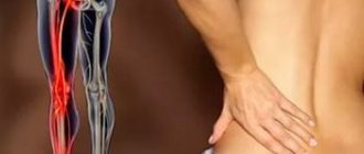
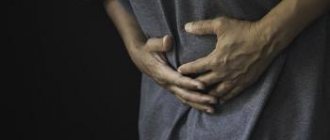
Symptoms of trauma.
A characteristic and primary symptom for all types of tendon injuries is the appearance of sudden severe pain. The following signs are added to it:
- redness and swelling in the area of injury
- increased sensitivity at the site of injury
- muscle cramps
- hemorrhage
- characteristic crunching sound at the time of injury
- limited movement of the injured limb
- complete ruptures are characterized by the appearance of dips when walking
Prevention of tendon inflammation
We will list some of the simplest ways to prevent inflammation of the tendons of the hand, and you will understand that since childhood you knew all these simple rules. But did you take them seriously?
- “We wrote, we wrote, our fingers were tired.” Remember that nursery rhyme they say during physics class in elementary school? The activities of many modern people are associated with long stays in the office, typing on the keyboard, while the wrist and fingers are in the same position for a long time, stagnation processes and salt deposition begin. To prevent this from happening, you should make active rotational and flexion movements with your fingers and wrist several times a day.
- Warm-up before the fight . Think about any sport. No trainer will allow you to start active exercises without a previous warm-up. You need to “warm up” your muscles, “stretch” your bones, and only then take on heavy loads. The same applies to everyday life. You cannot make sudden movements or overexert yourself when the body is not ready for it. Do exercises at least 2-3 times a week, and your body will always be ready for active movements!
- The healing power of massage. If “your bones ache,” “your body aches,” “your legs cramp,” etc., but you don’t have the opportunity to immediately start squatting and bending, then rub the sore area of your body. Stroking, pressing, pinching - all this will “disperse the blood” and stagnant salts, tone the muscles, ligaments and tendons.
For both therapeutic and preventive purposes, experts recommend using the shock wave therapy method.
At the medical center, experienced specialists will conduct an SWT course at a time convenient for you. The center treats inflammation of tendons and joints, arthritis, arthrosis, and relieves pain in the spine. Shock wave therapy specialists - doctors of the highest qualification category - have an impressive work experience (36 years!), among them: a full member of the association of shock wave therapy experts and a regular participant in symposia and congresses on shock wave therapy, a member of the Society of Radial Shock Wave Therapy and others. You can always go to the medical center that is closest to you - clinics are located in the Krasnopresnenskaya, Varshavskaya, Slavyansky Boulevard and Annino metro areas.
Diagnostics.
Diagnosis of these types of injuries includes:
- initial examination and collection of complaints by a traumatologist
- appointment of an x-ray examination (x-ray in lateral and direct projections)
- ultrasound examination (to detect partial or complete tears)
- CT and MRI
To sign up for a consultation and examination in Moscow, you can contact the following specialists from the Central Clinical Hospital of the Russian Academy of Sciences:
- Traumatologist
- Surgeon
- Rheumatologist
How to treat inflammation of the tendons of the hand
Depending on the diagnosis, the doctor prescribes treatment: medication, physiotherapy or surgery. You may be prescribed to wear a splint, a plaster splint, or any other device that has the effect of a bandage—that is, reducing the mobility of the device.
Drug therapy for inflammation of the tendons of the hand involves taking antibacterial, non-steroidal anti-inflammatory or restorative drugs, as well as the use of gels, ointments, and patches.
Physiotherapy has proven particularly successful in combating hand tendon inflammation. The following methods are used: microwave therapy, ultrasound, shock wave therapy (SWT), ultraviolet rays, physical therapy. SWT is particularly effective in combating this disease. Focused waves reach the tendon lesion, normalize its tone and reduce pain. After a course of shockwave therapy, patients return to their previous lifestyle and can bear the same loads as before.
Surgery is performed if a tendon rupture occurs. The surgeon makes a small incision (about 10 cm) that opens access to the tendon, processes the ends of the tendon and sews them together with a special strong thread. This type of treatment is carried out no later than 24 hours after the tendon is damaged. Otherwise, an irreversible process begins, leading to improper tissue fusion.
This is interesting In the 20s of the 20th century, Russian athlete and circus performer Alexander Zass invented a special course of exercises aimed at training tendons. He said this: “Muscles by themselves will not hold horses pulling in different directions, but tendons will, but they need to be trained, developed, and there is a way to strengthen them.” He himself was a man of remarkable strength, for which he received the nickname Iron Samson.
To avoid such unpleasant procedures, you should worry about your health in advance and take preventive measures to prevent tendon inflammation.
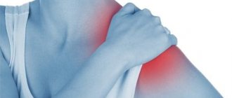
Shoulder tendonitis
Inflammatory-dystrophic diseases of the shoulder joint are based on neurodystrophic reflex disorders, which are closely related to cervical osteochondrosis. Tendinitis of the shoulder joint (ICD code 10 - M75) develops under the influence of direct or indirect trauma to the arm, shoulder against the background of osteochondrosis of the cervical and upper thoracic spine. The disease develops in the presence of the following factors:
- Occupational hazards;
- Microtrauma of the tendons of the muscles of the shoulder girdle, which are associated with monotonous and frequently repeated movements of the arm;
- Excessive loads on the upper limb;
- Hand inactivity for a long time.
Tendinitis of the supraspinatus muscle is based on microcirculatory disorders with the subsequent development of a focus of tendon necrosis. This causes reactive inflammation and swelling of the tendon segment near the location of the lesion. Due to the thickened segment of the affected tendon, it is difficult to move the arm to the side and upward. The patient experiences pain, most pronounced in the lateral part of the shoulder below the edge of the acromion process. The intensity of the pain increases at night. Sometimes pain does not interfere with daily activities, but it limits self-care.
Using palpation, the doctor determines pain in the anterior outer area of the shoulder under the edge of the acromion process of the scapula. The pain intensifies when the shoulder is moved to the side. For supraspinatus tendonitis, doctors identify a positive Dowborn's sign. It manifests itself as maximum pain under the edge of the acromion process on the outside of the left shoulder at the 15 o'clock level and the right one at the 9 o'clock level during active abduction of the arms to the sides and raising them upward. The pain disappears or weakens with further upward movement of the arm. When you lower your arm, severe pain appears again. It can cause a real arm block. Active movements of the affected upper limb become impossible.
Coracobrachialis tendinitis is characterized by anterior tenderness at the level of the coracoid process of the scapula. The arm is slightly abducted, extension is limited. When performing passive rotational movements, the patient does not feel pain. Pain appears during active movements of the affected upper limb.
A sign of tendovaginitis of the long head of the biceps muscle is Ergason's symptom: passive rotational movement of the forearm, bent at the elbow joint at an angle of 90°, outward, with resistance from the patient, causes pain in the front of the shoulder along the projection of the tendon of the long head of the biceps muscle. With tendonitis of the infraspinatus and teres minor muscles, pain occurs with external rotation of the shoulder. In order to determine the exact cause of shoulder pain, rheumatologists prescribe radiography of the shoulder joint, ultrasound and magnetic resonance imaging to the patient. With the help of clinical and biochemical studies, the inflammatory and autoimmune nature of tendonitis is excluded.
Why does my hand hurt with tendinitis?
The fact is that the muscles that work on the hand are mostly quite long and begin on the forearm just below the elbow. The contractile part of the muscle (belly) is located on the forearm, and the tendon (a thin cord connecting the muscle to the bone) passes past the wrist in special osteo-fibrous canals. There are six of them for the extensor muscles. The wrist that is most susceptible to tendinitis is the first one, which contains the abductor pollicis longus and extensor pollicis brevis muscles. With excessive load, chronic inflammation occurs and the channel becomes tight for the tendon to slide freely, which causes pain.
Pathogenesis
Tendons are formed in the form of dense, inelastic cords consisting of bundles of collagen fibers. Thanks to them, muscle tissue connects to bones. Tendons provide the transmission of movement from muscles to the skeleton and maintain stable joint function. Intense and monotonous movements interfere with the process of natural restoration of collagen fibers - the first signs of tendinitis appear.
The tendon structures swell and individual strands of collagen begin to break down. If the load remains high, foci of fatty degeneration, necrosis and deposition of calcium salts form in the tissues. Hardened calcifications lead to re-injury of the previously damaged area. The inflammatory process gradually spreads to the entire tendon.
Causes of the disease
Professional sports that require stress on the shoulder joint (tennis, volleyball, basketball, gymnastics, shot throwing and javelin) are the main cause of pathology in young people. Painters, plasterers and masons are also at risk. Contribute to the manifestation of pathology:
- shoulder injuries;
- immobilization for more than 3 weeks;
- bone and joint pathology (arthrosis and osteochondrosis);
- congenital pathology of the shoulder;
- cervical osteochondrosis.
Secondary tendonitis occurs with autoimmune, infectious, parasitic pathologies and endocrine diseases.
Anatomy of the Achilles tendon
Developed Achilles tendon
present only in humans and is an evolutionary adaptation to upright walking. It is believed that the human musculoskeletal system has not yet had time to fully adapt to upright walking, so its individual elements experience increased loads.
Although evolutionary selection gave the Achilles tendon
high endurance (its tensile strength reaches 400 kg) and has made it the thickest tendon in the human body, it is nevertheless considered one of the most vulnerable.
The Achilles tendon from above, in its widest and thinnest part, is attached to the fusion of the heads of the triceps muscle (formed by the gastrocnemius and soleus muscles), and from below, tapering at a level of 3-4 cm above the heel bone, it is attached to the calcaneal tubercle.
Due to its anatomical features, the Achilles tendon
is responsible for normal supination and pronation of the foot (the main biomechanical processes that occur during human movement). It also provides anatomically correct movement and stability in the knee, ankle and subtalar joints.
Mechanical properties of the Achilles tendon
fully correspond to these tasks: when running, it can withstand loads that are 8 times the weight of the person himself.
Symptoms
Symptoms of the disease appear gradually. In the early stages, the patient experiences pain at the time of maximum physical exertion. The rest of the time, the injured tendon does not cause any discomfort to the child or adult. Over time, the pain syndrome becomes more intense: discomfort occurs with minimal physical activity. If left untreated, the patient may be unable to perform everyday activities such as washing dishes, putting on clothes, fastening buttons, tying shoelaces.
The skin next to the source of inflammation turns red and swelling forms. Local temperatures may rise. Palpation of the tendon increases the pain. With sudden movements of the injured limb, the patient may hear a crunching or crackling sound.
Stages of tendinitis
Modern medicine distinguishes several stages of this disease. These include:
- Acute phase. The damaged area of the body causes severe pain. If a person feels discomfort in a calm state, then with physical activity it develops into unbearable acute pain;
- Subacute phase. The pain persists and is accompanied by swelling. Movements become difficult and limited;
- Chronic phase. Tissue degeneration occurs. Pain becomes a constant “guest”, as does stiffness.
Clinical picture
The pathology is characterized by gradual development. At the initial stage, discomfort arises, quickly disappearing after a few minutes of physical activity. A small load, for example, sports, helps to completely eliminate pain. When you feel the tissues affected by inflammation, a slight tingling sensation may occur. Lack of medical care is the reason for the chronicity of the disease. It is characterized by the following clinical manifestations:
- gradual increase in the severity of pain. It no longer disappears during training, and the intensity of the discomfort increases significantly;
- even after a long rest, the pain does not disappear; it also occurs in the morning;
- going up or down the stairs provokes the appearance of pain or its intensification.
When diagnosing Achilles tendonitis of moderate or severe severity, the doctor notes local hyperemia. In the damaged tissues, the temperature locally rises, the calf muscle is very tense, and there is no full flexion of the foot.
Treatment
Treatment of tendinitis (tenosynovitis, tenosynovitis) should be comprehensive and include conservative therapy (rest, cold, use of non-steroidal anti-inflammatory drugs), as well as physiotherapeutic methods.
It must be remembered that treatment of tendonitis should include limiting physical activity, the use of physical therapy, the action of which will be aimed at speedy healing of the damaged tendon, elimination of the inflammatory process, as well as strengthening and maintaining the tone of the whole body. In addition, if you have tendonitis, your doctor may recommend wearing special orthoses that will have a positive effect on the healing of the damaged tendon. If tendonitis is characterized by a severe course, then antibiotic therapy and even surgical treatment are possible. Surgical treatment is used only when the use of conservative treatment and physiotherapeutic procedures does not bring the expected results.
An important step in the treatment of tendinitis is to determine the cause of the development of this disease. At our Clinic, doctors use modern diagnostic methods that help make a diagnosis quickly and accurately. After identifying the causes of tendinitis, our specialists will be able to prescribe you the most effective treatment, which will be aimed at eliminating the symptoms, and will also allow you to forget about the pain in the shortest possible time. At the Bone Clinic, the doctor will select an individual treatment program, taking into account the stage and course of the disease and the presence of concomitant pathologies.
You will be guaranteed the best treatment for tendinitis in our Clinic. Here you can get advice from experienced traumatologists. In our Clinic, doctors are specialists, professionals in their field, working at the highest level, while achieving maximum heights and success.