Thoracalgia syndrome is a clinical symptom complex that is concomitant with most diseases of the spinal column. It is expressed in severe pain, stiffness, difficulty in mobility, and excessive tension in the muscular frame of the back. In this article we will understand what it is - thoracalgia syndrome and what treatment can be carried out without harm to the patient’s health, but to achieve complete restoration of damaged tissues.
Let's start with the fact that thoracalgia is a medical term that literally translated from Latin means chest pain. Thus, any unpleasant sensations in the sternum, ribs, or thoracic spine can be designated by this term. Most often it is present in various diseases of the musculoskeletal system and the autonomic nervous system. May accompany infectious inflammatory processes, traumatic lesions, etc.
To understand the pathogenesis, you should familiarize yourself with the anatomy of this part of the body. The spine in the thoracic region has the greatest extent. There are 12 vertebral bodies. Each of them has an additional pair of processes. These are the articular surfaces that form the costovertebral joints. These fasteners ensure mobility of the costal arches during inhalation and exhalation.
The vertebral bodies together with the arcuate processes form the oval foramen of the spinal canal. Inside is the spinal cord, from which paired radicular nerves arise. They exit through the lateral openings in the vertebral bodies. Further they branch and are responsible for the innervation of the chest, diaphragm, pancreas, kidneys, partly the liver, lungs, heart, intercostal respiratory muscles. These nerves are responsible for the performance of the muscular frame of the back and cervical-collar area.
Intervertebral cartilaginous discs provide protection from compression of the radicular nerves. They consist of a fibrous elastic membrane and an internal nucleus pulposus (jelly-like corpus pulposus). Discs do not have their own vascular network. They receive nutrition and fluid through diffuse exchange with paravertebral muscles and endplates.
In addition to protecting the radicular nerves, they are responsible for uniformly distributing the shock-absorbing load along the spine, maintaining the normal height of the intervertebral spaces, fixing the vertebral bodies, and preventing them from sliding relative to each other in the cavity of the spinal canal.
The radicular nerves branch out to form plexuses and large nerves. The large nerves then travel to the areas they are responsible for innervating, such as the upper extremities. As they move away from the spinal column, they break up into smaller structures. Finally, they are presented in the form of small nerve endings dispersed throughout the epidermis and muscles.
All root nerves carry three types of axons in their structure: motor (motor), sensory (sensitive) and mixed. The former are responsible for transmitting nerve impulses from the motor center of the brain to myocytes. In response, they contract or relax, and the muscle allows one or another movement. Sensory axons are responsible for transmitting the results of the interaction of body cells with the environment and each other. Therefore, with compression, mechanical damage, inflammation, and trophic disturbances, a feeling of pain occurs. It is the sensory types of axons and nerve endings that are responsible for this sensation.
Depending on the part of the nerve passage where pathological changes occur, the pain syndrome can change its location and intensity of manifestation. It may be accompanied by other symptoms.
But in any case, pain in the chest is always the result of a pathology of the sensory type of nerve fiber. But we’ll talk about what reasons may contribute to this later in the article. In the meantime, I would like to warn you that you need to consult a doctor. If such a syndrome appears, you will not be able to diagnose yourself. This is absolutely certain, since a number of special examinations are required. We recommend that you see a specialist as soon as possible.
In Moscow, you can make an appointment right now for a free appointment with a vertebrologist or neurologist at our manual therapy clinic. Experienced specialists work here. They will be able to give you an accurate diagnosis during the initial examination. They will give all the necessary recommendations for complex treatment.
What is vertebrogenic thoracalgia?
Thoracalgia – if translated literally: thorax from lat.
- chest, algos from Greek. - pain, so we get pain in the chest. This includes pain both in the back and along the entire perimeter of the chest: pain along the ribs, pain in the places of cartilaginous attachment of the ribs to the sternum, etc. The causes of thoracalgia can be divided into vertebrogenic (associated with pathology of the spine) and non-vertebrogenic (associated with pathology of the adjacent structures and organs).
Manifestations of thoracalgia cannot be ignored, due to the fact that pain in the thoracic region can be a manifestation of life-threatening conditions from the chest organs: aneurysm of the thoracic aorta, pulmonary embolism. For example, myocardial infarction can be falsely interpreted as vertebrogenic thoracalgia on the left.
In most cases, pain in the thoracic spine is associated with overstrain of the paravertebral muscles, i.e., the muscles lying along the spine, followed by degenerative-dystrophic changes in the spine (spondyloarthrosis, spondylosis and osteochondrosis), which include costovertebral and costotransverse joints. Due to the fact that the thoracic spine is less mobile and has a strong rib frame, herniated intervertebral discs rarely occur here.
Exercise therapy exercises
Physical therapy exercises are very important, but they are ineffective in the acute stage. After all, the thoracic region has limited mobility and muscle spasms can sometimes only be relieved with medications. However, as a means of preventing the recurrence of thoracalgia, exercise therapy exercises come first. In case of illness, sets of exercises are indicated to strengthen the muscle corset, swimming, and cardio exercises in order to reduce excess body weight. It is undesirable to engage in martial arts (risk of injury), weightlifting (overload), basketball (frequent vertical loads).
Causes of thoracalgia
Overstrain of paravertebral muscles
Another cause of pain in the thoracic spine is long-term overstrain of the paravertebral muscles, which is common among office workers who sit in one position in front of a computer for a long time. Prolonged muscle strain contributes to the development of myofascial pain syndrome, which is characterized by the presence of trigger points. A trigger point is a painful tightening of muscle fibers.
In most cases, trigger points can be found in the levator scapulae, rhomboid, and serratus anterior muscles. If there are trigger points in these muscles, palpation will cause increased pain, sometimes shooting pain. In addition to pain, the patient may experience a burning sensation, numbness, or tingling in the area. The pain may go away on its own after warm-up movements, for example, making circular movements with your shoulders, stretching your back muscles, bending backwards as much as possible, etc., but it can persist for a long time and be chronic.
Degenerative-dystrophic changes
Degenerative-dystrophic changes in the spine at the cervical level can quite often be accompanied by both unilateral and bilateral pain in the interscapular region. What is this connected with? With the development of arthrosis of the facet joints at the cervical level, a reflex spasm of the muscles occurs, which originate from the cervical vertebrae, and at the other end are attached to both the shoulder blades and the thoracic vertebrae themselves. These include the muscles of the superficial and deep layers: trapezius muscle, levator scapulae muscle, longissimus capitis and neck muscle, semispinalis capitis muscle, splenius capitis and neck muscle. Typically, the pain is chronic in nature with episodes of exacerbations and is localized at the level of the upper and middle thoracic region.
The cause of chronic vertebrogenic thoracalgia is also arthrosis of the facet (facet) joints and pathology of the costotransverse joints. As mentioned above, due to the peculiar structure of the thoracic region, degenerative changes in this area are much less common than in other parts of the spine. The development of arthrosis of the facet joints is caused by an increased load on the articular surfaces; in all likelihood, the muscular component may play a role - increased tone in the segmental muscles. The response to increased mechanical load is marginal bone growth of the facet joints to increase the area of support. The pain experienced by the patient is described as dull aching, without clear localization. Sometimes, when exposed to nerve roots, it can spread along the rib, both on one side, and be encircling in nature. As practice shows, relieving increased muscle tone in the superficial and deep segmental muscles using gentle manual techniques leads to a significant reduction in pain and increases the interval between exacerbations.
Mention should be made of such a pathology as posterior costal syndrome, in which intense acute pain is localized in the lower thoracic region and intensifies with rotation of the torso. This pain is often confused with renal colic. Any impact on this area causes pain. Often pain can manifest itself at the height of inspiration, so the patient tries to breathe shallowly. This is due to the fact that the XI-XII ribs are free, i.e. do not have a cartilaginous connection either with the sternum or with neighboring ribs, which leads to the possibility of displacement of the articular head of these ribs in the costovertebral joint, and can also be combined with local spasm in the quadratus lumborum muscle.
Osteoporosis
Vertebrogenic thoracalgia may be a consequence of osteoporosis. This reason is more typical for patients over 50-55 years old. Osteoporosis is a progressive loss of bone mass and changes in the internal structure of bone, resulting in increased bone fragility.
The thoracic spine is one of three points (radius, femoral neck) at which osteoporosis can be detected in its early stages using densitometry.
As bone mass decreases, the shape of the vertebrae changes. Due to the influence of the internal pressure of the intervertebral disc, the vertebral body takes the shape of a biconcave lens, and under the influence of gravity, its wedge-shaped deformation occurs and the height of the vertebral body decreases against the background of a compression fracture. A compression fracture in osteoporosis is typical for the middle and lower thoracic vertebrae; when it is localized at the upper thoracic level, oncological alertness is required.
Wedge-shaped deformation of the vertebrae leads to increased thoracic kyphosis, and in combination with a decrease in the height of the vertebral body, a decrease in human height becomes noticeable.
Acute back pain radiating to the abdominal area is often caused by a compression fracture of the vertebra, which can be caused by even a seemingly insignificant movement. This pain usually goes away within a few days to weeks, and the patient returns to his normal life.
Another cause of pain in osteoporosis is pinching of the nerve root in the intervertebral foramen with a decrease in the height of the vertebral body and its wedge-shaped deformity. The pain spreads along the intercostal spaces and can be one- or two-sided.
Scheuermann-Mau disease (juvenile kyphosis)
The first signs of the disease begin to appear before 10-15 years of age, but there is also a later onset, after 20 years. The disease is based on a wedge-shaped deformity of the thoracic vertebrae with multiple Schmorl's hernias. Schmorl's hernia is a depression of a section of the intervertebral disc into the vertebral body. Over time, dull aching pain appears in the interscapular area, intensifying at rest. Movements in the thoracic region bring relief. The disease, which steadily progresses, leads to the development of hyperkyphosis.
The diagnosis is made by the characteristic x-ray picture. The detection of only single Schmorl's hernias does not indicate the presence of this disease. In the population, Schmorl's hernias are detected quite often and, as a rule, are an accidental finding; in the vast majority of cases, they occur without pain and do not require any treatment.
The main treatment for Scheuermann-Mau disease: gentle manual therapy techniques, physical therapy aimed at strengthening the back muscles, wearing orthopedic corsets. Surgical treatment is required only in extreme cases.
Lumbodynia can turn into radiculitis (radiculopathy)
After some time, lumbodynia, if no measures are taken, may give way to the next, radicular stage of spinal osteochondrosis - radiculitis (radiculopathy). As a rule, radiculitis is caused by compression of the nerve root by an enlarged herniated intervertebral disc. In Latin, the spine is radicula (radicula).
Severe, persistent lumbodynia on the right or lumbodynia on the left occurs. Localization depends on which side the nerve root is compressed. Radiculopathy is more difficult to treat and takes longer. The help of a neurosurgeon is often required.
Diagnostics
Don't self-diagnose! This can harm your health and take away valuable treatment time!
In order to diagnose the disease and find out what the patient is sick with, it is necessary to correctly differentiate the symptoms, since they do not differ in specificity and may indicate various pathologies. Therefore, diagnostics requires a consistent, comprehensive approach.
At the initial appointment, the doctor must carefully examine the medical history. The patient’s medical notes may indicate the diseases that caused vertebrogenic thoracalgia, or the patient himself must tell about the diseases he has. Identifying the root cause, the key factor in the onset of pain, is essential in order to continue treatment correctly.
The thoracic spine must be palpated; during the examination, the patient must accurately describe his sensations. This will help determine the location of pain and identify additional symptoms if the patient forgot to indicate them. This is important for drawing up a complete picture of the disease.
If necessary, the patient is recommended instrumental research methods:
- Radiography;
- Computed tomography of the spinal column;
- Densitometry;
- Magnetic resonance imaging;
- Scintigraphy;
- Electrocardiogram;
- Electroneuromyography.
In most cases, no additional diagnostic methods are required!
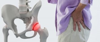
The mechanism of occurrence of lumbodynia
The mechanism of occurrence of both lumbago and lumbodynia is due to irritation of the sinuvertebral nerve. But there is some difference. Lumbago, as a rule, occurs as a result of infringement of part of the nucleus pulposus in the cracks of the fibrous ring of the damaged disc. And lumbodynia can also occur with less pronounced changes in the disc. It is also often found in patients with spondylosis of the lumbar spine, spondyloarthrosis, and spondylolisthesis. Chronic lumbodynia syndrome can also occur with anomalies in the development of the lumbar spine. These are “spina bifida”, “anomaly of tropism” and other congenital defects.
Treatment
In most cases, vertebrogenic thoracalgia requires conservative treatment. In everyday life, the patient can use orthopedic corsets to support and unload the spine.
In the treatment of back pain, gentle manual therapy techniques have proven themselves to be effective, helping the patient cope with the pain syndrome in the shortest possible time. According to the results of internal observation in our clinic, after the first session the patient feels an improvement in his condition, expressed in a decrease in pain, even if the pain tormented the patient for a long time. We have accumulated extensive experience in diagnosing and treating back pain.
In case of chronic vertebrogenic thoracalgia, in some cases, joint work of a vertebroneurologist, endocrinologist, psychotherapist and other specialists may be required.
Lumbodynia - the first signs of osteochondrosis
Lumbodynia syndrome (low back pain) is often the first clinical sign of osteochondrosis of the lumbar spine. Lumbar osteochondrosis of the initial stage in almost half of the cases manifests itself as subacute or chronic lumbodynia.
What causes lumbodynia
Lower back pain can occur after physical activity, lower back injury, hypothermia, and stress. But in some cases, the lower back does not begin to hurt immediately. Sometimes it takes days, weeks or even months before pain occurs.
At first, very mild lumbodynia may appear. Some patients note that their pain developed slowly and for no apparent reason. But this does not mean at all that there was no provoking factor. It’s just that this factor is not always obvious.
Then the lower back pain may intensify. Most often this happens gradually. But the pain usually does not reach the same degree of severity as lumbar pain - lumbago (acute lumbodynia). Patients walk independently and do certain work, although it is difficult for them to bend, but even more difficult for them to straighten up. At the same time, they often place their hand on the lower back, rubbing or massaging it a little. And only after that they can straighten up. Such people try to sit with a straight back.
