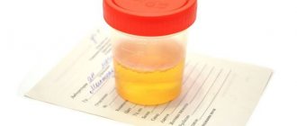
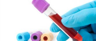
A biochemical blood test is a detailed study of a patient’s venous blood, carried out to assess the functioning of internal organs, identify deficiencies of vitamins, enzymes, macro- and microelements, as well as to diagnose metabolic pathologies. Blood biochemistry is more indicative than a general clinical analysis; the results of the analysis make it possible to detect many diseases at the initial stage. That is why this study is recommended not only as prescribed by a doctor, but also for preventive purposes at least once a year. The results of a biochemical blood test include many interrelated indicators and their interpretation should be handled by a doctor - self-medication is dangerous for your health!
How to prepare for the procedure?
- Blood sampling for biochemistry is always performed on an empty stomach, most often between 8 and 11 am. On the day of the procedure, it is allowed to drink still water, and the day before the procedure, heavy foods, carbonated drinks, strong coffee, tea and alcohol should be excluded from the diet.
- You should not smoke in the last hour before donating blood.
- Immediately before the procedure, try to avoid physical and emotional stress; for the last 10–20 minutes it is better to just sit near the manipulation room.
- If the date of the biochemical blood test falls during a course of drug treatment or a course of physical therapy, then it is worth consulting with a doctor - he may recommend rescheduling the test to another time or interrupting the course of treatment for several days.
Why does jaundice appear?
There are several mechanisms for the occurrence of jaundice.
| Cause of jaundice | What's happening? |
| Increased breakdown of hemoglobin | Due to the too active production of indirect bilirubin, the liver simply cannot cope with its processing and excretion |
| Liver diseases | The amount of bilirubin produced does not exceed the norm, but the affected liver cells are not able to fully function and cope with their work |
| Impaired flow of bile | Instead of entering the intestines, bile accumulates in the liver and provokes its destruction. Bilirubin then enters the blood again. |
Jaundice is an indication for a biochemical blood test to determine the level of bilirubin in the blood. These indications also include liver tumors and cirrhosis, hepatitis, and hemolytic anemia.
Decoding the results of a biochemical blood test
New generation laboratory analyzers are able to provide research results within two hours after blood sampling. As a rule, the patient receives the results within 2-3 days in the form of a printed or electronic table, which lists the studied indicators, their values and reference (average) normal ranges. Different laboratories offer different amounts of data, but this article will describe the most commonly tested blood parameters.
Squirrels
| Designation | Norma (women) | Norma (men) | Units | |
| Albumen | A.L.B. | < 14 years: 38–54 14–60 years: 35–50 > 60 years: 34–38 | g/l | |
| Glycated hemoglobin | HbA1c, A1c, glycosylated hemoglobin | < 5,7 | % | |
| Total protein | TP, TProt, Serum | < 1 year: 47–72 1–4 years: 61–75 5–7 years: 52–78 8–15 years: 58–76 > 15 years: 64–83 | g/l | |
| C-reactive protein | CRP, SRP | < 0,5 | g/l | |
| Iron binding capacity of serum | TIBC, IBC, OZhSS | 45,3–77,1 | µmol/l | |
| Myoglobin | Myoglobin | 12–76 | 19–92 | µg/l |
| Transferrin | Tf | 2,2–2,4 | 2–4 | g/l |
| Ferritin | Ferritin | 13–150 | 30–400 | µg/l |
Total protein characterizes the state of protein metabolism and reveals dysproteinemia (changes in the quantitative ratio of protein fractions in the blood serum). Decreased values may indicate poor nutrition, liver disease, consequences of burns, injuries and operations, while elevated values may indicate an infectious disease, non-infectious hepatitis, autoimmune diseases, dehydration, or may be caused by diarrhea and vomiting.
The protein albumin occupies up to 65% of the volume of blood plasma, is produced by the liver and performs the most important function of transporting many biologically active substances. The reasons for the decrease in albumin concentration coincide with those for total protein. The value increases quite rarely, for example, with dehydration, hemoconcentration or due to the use of anabolic steroids.
The iron-containing protein myoglobin is studied primarily for the purpose of early diagnosis of myocardial infarction. A high concentration of myoglobin may indicate myocardial infarction, heart failure, acute kidney damage, the consequences of thermal burns, and electric shock. Low myoglobin accompanies the course of rheumatoid arthritis and polio.
The value of glycated or glycosylated hemoglobin is very important for patients with diabetes mellitus and is also used for its diagnosis. Glycated hemoglobin gives an idea of the average level of glucose in the blood over a long period of time (1–2 months). If the concentration of this protein fraction does not exceed 5.7% of the total volume of hemoglobin in the blood, then we can speak of a compensated state. Values in the range of 5.7–6.4% indicate a risk of developing diabetes mellitus; values above 6.4% indicate severe decompensated diabetes.
C-reactive protein acts as an indicator of inflammation in the body. Exceeding the threshold of 0.5 g/l indicates acute inflammation or malignancy. This parameter is also important for assessing the effectiveness of antibacterial and anti-inflammatory therapy.
The studied values of transferrin , ferritin and iron-binding capacity of serum make it possible to diagnose the pathology of iron metabolism in the blood. Transferrin is the main carrier of iron; an increase in its concentration, as a rule, indicates the development of iron deficiency anemia, and a decrease indicates infections, liver cirrhosis, anemia of other etiologies, or protein starvation. In iron deficiency anemia, ferritin, on the contrary, decreases, and its increase indicates inflammatory processes, liver disease or oncopathology.
Lipids
| Designation | Norma (women) | Norma (men) | Units | |
| Triglycerides | TRIG | < 15 years: 0.40–1.48 15–30 years: 0.4–1.63 30–55 years: 0.44–2.63 > 55 years: 0.62–2.71 | < 15 years: 0.34–1.41 15–30 years: 0.45–2.81 30–55 years: 0.56–3.61 > 55 years: 0.65–3.29 | mmol/l |
| Total cholesterol | CHOL | 5,2 | mmol/l | |
| Cholesterol-HDL | HDL, HDL-C | 1,03–1,55 | mmol/l | |
| LDL cholesterol | LDL, LDL-C | 0–3,3 | mmol/l | |
Total cholesterol is used to identify primary and secondary disorders of lipid metabolism, assess the likelihood of developing atherosclerosis, and also to assess the effectiveness of treatment for atherogenic disorders of lipid metabolism. A decrease in value is caused by cachexia, starvation, malabsorption, severe acute diseases, liver failure, hyperthyroidism, and an increase is caused by primary and secondary dislipoproteinemia. The dangerous consequences of low cholesterol are psychophysiological disorders and reproductive dysfunction, while high cholesterol is diabetes and atherosclerosis. A biochemical blood test for triglycerides (products of carbohydrate metabolism in the liver) pursues the same objectives; the reasons for the increase and decrease in their concentration also coincide with total cholesterol.
and low-density lipoprotein cholesterol (HDL-C and LDL-C, respectively) are tested and interpreted in conjunction with total cholesterol and triglycerides for a more accurate diagnosis. HDL cholesterol increases in primary biliary cirrhosis, hepatitis, alcoholism, or its increase may be genetically determined. In patients with atherosclerosis, decompensated diabetes mellitus, chronic kidney disease, and cholestasis, the HDL-C value decreases. Low-density lipoproteins are involved in the processing and elimination of fats; a decrease in their concentration may indicate the development of chronic anemia, Raynaud's syndrome or myeloma, and an increase may indicate hypothyroidism, nephrotic syndrome, diabetes mellitus, porphyria, Cushing's syndrome, and the risk of developing atherosclerosis.
Carbohydrates
| Designation | Norma (women) | Norma (men) | Units | |
| Glucose | GLUC | 3,3–5,5 | mmol/l | |
| Fructosamine | FRA | 0–285 | µmol/l | |
Glucose is the main source of energy for all cells and tissues of the human body and, in particular, its only source for the brain. The glucose value in the biochemical analysis results reflects the blood sugar level. If this value is increased, then there is a possible risk of developing diabetes mellitus, damage to the central nervous system, and hormonal disorders. Glucose “falls” with the formation of tumors in the pancreas, with liver and adrenal insufficiency, hypothyroidism, malnutrition, or due to insulin intake.
fructosamine value reflects fluctuations in blood glucose levels in the 2–3 weeks preceding the test. If its concentration exceeds 280–285 µmol/l, then the doctor considers the likelihood of developing diabetes mellitus.
Inorganic substances and vitamins
| Designation | Norma (women) | Norma (men) | Units | |
| Vitamin B12 | 208–963,5 | pg/ml | ||
| Iron | Fe, IRON | < 2 years: 7–18 2–14 years: 9–22 > 14 years: 9–30 | < 2 years: 7–18 2–14 years: 9–22 > 14 years: 11–31 | µmol/l |
| Potassium | K | 3,5–5 | mmol/l | |
| Calcium | Ca | 2,25–2,5 | mmol/l | |
| Magnesium | Mg | 0,75–1,25 | mmol/l | |
| Sodium | Na | 136–145 | mmol/l | |
| Phosphorus | P | < 2 years: 1.45–2.16 2–12 years: 1.45–1.78 12–60 years: 0.87–1.45 > 60 years: 0.90–1.32 | < 2 years: 1.45–2.16 2–12 years: 1.45–1.78 12–60 years: 0.87–1.45 > 60 years: 0.74–1.2 | mmol/l |
| Chlorine | Cl | 98–107 | mmol/l | |
Vitamin B12 in the human body is involved, in particular, in the production of red blood cells. High levels of this vitamin may indicate liver disease, kidney disease, or leukemia. Diseases of parasitic etiology, inflammatory processes in the gastrointestinal tract and adherence to a vegetarian (vegan) diet, on the contrary, lead to a decrease in the level of vitamin B12 in the blood.
In one of the previous paragraphs it was mentioned that iron takes part in the process of oxygen transport. Its deficiency is usually explained by poor nutrition or metabolic disorders, and its excess is usually explained by functional intestinal disorders.
Potassium is responsible for regulating water balance and normalizing heart rate. Potassium deficiency occurs due to improper or insufficient nutrition, vomiting, renal failure, Cushing's syndrome, osmotic diuresis, chronic kidney disease, and also accompanies long-term use of steroid drugs. Potassium increases with acute dehydration, extensive injuries and burns, chronic adrenal insufficiency, diabetic coma, or due to taking potassium-sparing diuretics.
Calcium is involved in the formation of bone tissue and is extremely important for the normal functioning of muscles, nerves, heart muscle and blood vessels. Low blood calcium values indicate vitamin D deficiency, functional kidney disease, pancreatitis, impaired magnesium metabolism, or hypoparathyroidism. An increase in calcium levels accompanies hyperparathyroidism or is a symptom of cancer.
Magnesium performs the function of intracellular metabolism and transmission of impulses from nerve endings to muscles. Poor nutrition, impaired absorption, prolonged diarrhea, colitis, enterocolitis and dyspepsia reduce the concentration of magnesium in the blood. Its increase is caused by functional kidney disorders, hypothyroidism, lactic acidosis and neoplasms.
Along with magnesium, sodium , which also takes part in calcium metabolism, is responsible for transmitting impulses to muscles. The cause of sodium reduction may be hypothyroidism, Addison's disease, diabetes mellitus, kidney and gastrointestinal diseases, congestive heart failure, taking gentamicin, and less commonly, Parhon's syndrome or hypercalciuria. High sodium levels in biochemistry results indicate dehydration, oversaturation of the body with salts, diabetes insipidus or kidney disease with oliguria.
Normal functioning of the nervous and musculoskeletal systems is impossible without a sufficient amount of phosphorus in the body. The phosphorus content in the blood increases with hypoparathyroidism, excess vitamin D, rhabdomyolysis, bone diseases or poor diet, and less often with acromegaly. On the other hand, hypovitaminosis D, hyperparathyroidism, kidney transplantation, intravenous infusions of glucose, and respiratory alkalosis cause a decrease in the concentration of phosphorus in the blood.
Chlorine performs the functions of maintaining the acid-base balance of the blood and osmotic pressure. The most obvious reasons for a decrease in chlorine levels are profuse sweating, vomiting, diarrhea, incorrect treatment with diuretics; less commonly, a decrease is caused by nephrotic syndrome and hypokalemic metabolic syndrome. An excess of chlorine in the blood can be a consequence of dehydration, swelling, alkalosis and cardiac decompensation.
Example of blood biochemistry results
Low molecular weight nitrogenous substances
| Designation | Norma (women) | Norma (men) | Units | |
| Creatinine | CREA | 53–97 | 62–115 | µmol/l |
| Uric acid | U.A. | < 14 years: 120–320 > 14 years: 150–350 | < 14 years: 120–320 > 14 years: 210–420 | µmol/l |
| Urea | UREA | 2,2–6,7 | 3,8–7,3 | mmol/l |
Urea and creatinine are studied in combination; their values reflect the functional state of the patient’s kidneys, in particular, the degree of impairment of filtration and excretory functions. High values indicate kidney problems, but may be due to excessive exercise, a high-protein diet, prolonged fasting, or thyroid disease. Low urea values may be due to a low protein diet, pregnancy, and liver disease.
The level of uric acid , as an auxiliary parameter, reflects the body's ability to remove waste products from the metabolic processes of nucleic acids and purines. Of particular diagnostic interest for patients with gout. The main causes of increased uric acid are gout and alcoholism, and less commonly, kidney and liver pathologies. Low values of uric acid in the results of a biochemical blood test are much less common and, as a rule, indicate improper or insufficient nutrition.
Pigments
| Designation | Norma (women) | Norma (men) | Units | |
| Total bilirubin | BILT | 3,4–17,1 | µmol/l | |
| Direct bilirubin | BILD, D-BIL | 0–7,9 | µmol/l | |
| Bilirubin indirect | ID-BIL | BILT - BILD | µmol/l | |
The yellow pigment bilirubin begins to accumulate in the blood during diseases and hereditary pathologies of the liver and biliary tract, for example, with Gilbert's syndrome. Indirect bilirubin may also increase in some anemias and malaria.
Enzymes
| Designation | Norma (women) | Norma (men) | Units | |
| Alanine aminotransferase | ALT | < 31 | < 41 | units/l |
| Amylase | AMY | 28–100 | units/l | |
| Pancreatic amylase | AMY-P | 0–50 | units/l | |
| Aspartate aminotransferase | AST | < 32 | < 40 | units/l |
| Gamma glutamyl transferase | GGT | 6–42 | 10–71 | units/l |
| Creatine kinase | CK | 0–25 | units/l | |
| Lactate dehydrogenase | LDH | 250 | units/l | |
| Lipase | LIP | 0–190 | units/l | |
| Phosphatase alkaline | A.L.P. | 0–240 | 0–270 | units/l |
| Cholinesterase | CHE | 5860–11800 | 5800–14600 | units/l |
A liver enzyme with an unpronounceable name, alanine aminotransferase, is involved in amino acid metabolism. The indicator increases with myocardial infarction, acute hepatitis A and B, and other liver diseases.
The enzyme amylase is produced by the salivary glands and pancreas and is responsible for the digestion of carbohydrates. Exceeding the norm by 3–5 times may indicate acute appendicitis, peritonitis, gastric and duodenal ulcers, cholecystitis. In acute pancreatitis or exacerbation of its chronic form, the amylase value increases 10–30 times. An increased value of pancreatic amylase allows for timely detection of complications from operations on the abdominal organs and pancreatic diseases.
The concentration of the enzyme aspartate aminotransferase in a person’s blood increases greatly in case of damage to the liver or heart muscle; due to alcohol abuse, the levels can double the normal level.
High levels of the gamma-glutamyltransferase are observed in patients with acute hepatitis, extra- and intrahepatic cholestasis, alcoholism, pancreatic and prostate cancer, and primary liver tumors.
An increase in the concentration of creatine kinase in the blood may indicate myocardial infarction, muscle damage of various origins, and renal failure. Lactate dehydrogenase increases in diseases of the cardiovascular system, liver and kidneys, as well as in pregnant women. High lipase are observed in patients with acute pancreatitis, intestinal infarction, biliary colic, wounds, fractures, and breast cancer. The metabolism of phosphorus in the body is directly related to the content of alkaline phosphatase - its increase accompanies acute viral and alcoholic hepatitis, liver cirrhosis, mononucleosis, liver cancer or liver metastases. Cholinesterase decreases in liver cirrhosis, liver failure, hepatitis, myocardial infarction. The value of cholinesterase is also indicative for patients taking muscle relaxants.
Disturbed pigment metabolism
In the human body there are specifically colored proteins. These are peptides that contain a metal (for example, copper or iron). Such peptides include cytochrome, hemoglobin, myoglobin, etc. As a result of the breakdown of peptides, bilirubin and its fractions are formed.
Free bilirubin appears after the breakdown of red blood cells in the spleen. This bilirubin is very toxic, especially to the human brain. However, the body is not poisoned due to the rapid connection of free bilirubin with albumin. In liver diseases such as cirrhosis and hepatitis, the concentration of bilirubin in the body increases due to a decrease in the amount of albumin and the predominance of glucuronic acid.
Indirect bilirubin binds to this acid, which loses its toxicity. It then enters the bile and is converted into urobilinogen. This enzyme, in turn, penetrates through the small intestine into the blood and then into the kidneys. It is urobilinogen that colors urine yellow.
Taking blood from children from 1 day to 12 months
- Try to schedule blood sampling for infants during the break between feedings, closer to the second feeding.
- 30 minutes before the procedure, the child should drink 50 ml of the liquid that you usually give him.
- When taking blood, the child’s hands must be warm. If you come from outside or the room is not so warm, the child’s hands need to be warmed. This is a mandatory and very important condition, because the amount of blood that will be received by the health worker depends on its fulfillment.
- Immediately before blood is drawn, the child should be positioned so that he is as comfortable as possible. A couple of minutes should pass before the nurse begins drawing blood. This time will be enough for the baby to calm down and get used to the environment a little.
Alpha amylase
This enzyme is special. It breaks down complex carbohydrates into simpler compounds. Alpha amylase is concentrated in the salivary glands and pancreas. When examining blood, the doctor will definitely pay attention to the decrease and increase in the level of this enzyme.
Alpha-amylase exceeds the normal level in hepatitis, pancreatic cancer, pancreatitis, mumps, and renal failure.
A decrease in the level of the enzyme is observed with alcoholism, myocardial infarction, toxicosis during pregnancy, thyrotoxicosis, pancreatic necrosis, and taking steroid drugs.