In the picture: subluxation of the atlas (first cervical vertebra), view from the back. Atlas (atlas, atlas) is the first vertebra of the cervical spine, the function of which is to maintain the skull in a stable condition and regulate the position of the entire spine.
Displacement of the first vertebra is the name of a very common injury, subluxation of the atlas. The first cervical vertebra of the atlas (C1) is attached to the occipital region of the skull and connects to the second vertebra, the axis (C2), forming the atlantoaxial joint. It ensures head mobility, normal support of the skull, and high-quality blood supply to the brain and spinal cord.
In the picture: the first cervical vertebra (atlas) in the correct position, viewed from the back.
With the correct position of the C1 vertebra, when turning the head, it moves in concert with the skull relative to the C2 axis.
How is the first vertebra arranged?
Atlas is similar in structure to a ring, the lateral sections of which are denser compared to the back and front parts. These sections are connected to the occipital bone. The second vertebra has an odontoid process: it is this that ensures sliding along the inner surface of C1. More detailed information in this article.
General information
A dislocation is a displacement of the articular surfaces of two bones relative to each other, different from the anatomical norm, which is accompanied by a pronounced violation of the configuration of the joint and its function in the form of limited mobility.
Its variety is the condition of subluxation, in which there is a relatively slight displacement of the articular surfaces relative to each other while maintaining the point of contact and, in some cases, there is the possibility of spontaneous reduction. In most cases, subluxations are acquired, while congenital subluxations are relatively rare. Acquired subluxations are formed in the presence of risk factors such as:
- Traumatic effects (sharp jerks, falls, prolonged hanging, impacts) caused by injury or high physical exertion.
- Various diseases of the musculoskeletal system ( osteomyelitis , dysplasia /joint instability, poliomyelitis , osteochondrosis , etc.).
The most susceptible to subluxation are joints with a large range of motion, which have maximum mobility in various planes. Therefore, subluxation most often occurs in the joints of the upper/lower extremities (ankle joints, toes, subluxation of the patella), spine, jaw, dislocation of the upper extremities (shoulder and elbow joint).
Subluxations can be combined with rupture/sprain of ligaments and muscles, rupture of the joint capsule, damage to blood vessels and nerve fibers. Let's take a more complete look at the subluxation of the cervical vertebra (the so-called subluxation of the atlas), and also briefly touch on the most common subluxations of the shoulder, clavicle and joints of the upper (elbow) and lower limb - subluxation of the knee (patella), hip joint and ankle.
Subluxation of the cervical vertebra (subluxation of the atlas)
The craniovertebral region is the transition point of the spinal column into the skull and includes the upper cervical vertebrae (atlas, axis), as well as the basal part of the occipital bone. This vertebra performs an extremely important function, holding the cranium with its contents to itself.
The atlas is a slightly flattened ring-shaped bone formation, which is essentially a bone meniscus, a kind of washer between the lateral masses of the axis and the condyles of the occipital bone, providing the transition from flexion-extension to rotational movements. The wedge-shaped lateral masses of the atlas are connected by the anterior/posterior arches into a single ring-shaped formation and are connected by a powerful transverse ligament, which provides additional stability to the bone ring (Fig. below).
The atlas connects from above with the help of tiny joints to the occipital bones, and from below to the axis (second cervical vertebra). The weight of the head is transmitted through the condyles of the occipital bone to the lateral masses of C1 and is further transmitted to the upper articular facets of the axis, where it is distributed predominantly to the anterior structures (up to 80%) and to a much lesser extent to the posterior structures of C2.
The lateral masses of the atlas/body of the axis stabilize axial loads; shear loads - anterior arch of the atlas, tooth/lower articular processes of the axis; rotational, flexion-extension and dynamic shear load - ligamentous apparatus: the transverse ligament prevents anterior displacement of the atlas; The pterygoid ligaments stabilize axial rotation of the head, during which there are no bone restrictions on movement in the joints. The C1-C2 stabilization ligaments are ligaments of tension, which provide secondary stability in the joint. The primary/secondary stabilizers for the atlantoaxial and atlanto-occipital joints are different. When the function of different ligaments is impaired, the stability of the joint is affected differently.
One type of pathology of the craniovertebral segment is atlantoaxial instability, manifested by subluxation. There are three main types of atlantoaxial instability: distraction , flexion-extension and rotation , which can be isolated or complex in various combinations. Instability in the atlantoaxial joint is caused by damage to the structures of the craniocervical junction, mainly damage to the transverse ligament.
The most common occurrence in practice is rotational subluxation of the C1 cervical vertebra (Fig. below), which is a pathological asymmetrical displacement to the right/left of the articular surfaces of the ring-shaped C1 vertebra relative to the axis of the C2 vertebra (left-sided/right-sided subluxation).
Rotational subluxation of the atlas is a common injury of the cervical spine, accounting for about 30% of the total pathology of the cervical spine. Subluxations of the C1/C2 vertebrae are most often caused by isolated trauma. Rotational subluxations of the atlas are detected much less frequently in adults than in children.
A relatively rare occurrence is subluxation according to Kovacs (habitual subluxation), which manifests itself in the form of instability of one/several segments of the spine: when the head is tilted, the process of the underlying vertebra slips and layers on the posterior-inferior corner of the body of the overlying vertebra, and when the neck is extended, it returns to its original position. position.
In case of rotational atlantoaxial subluxations, spinal cord injuries are extremely rare, and compression/damage to the vertebral artery is practically never encountered.
The most common manifestation is dysesthesia (unpleasant sensations) caused by damage to the roots of the C2 vertebra. As a rule, after careful reduction of the C1-C2 vertebrae, restoration of all impaired spinal functions is observed.
Subluxation of the hip joint
Subluxation of the hip joint is a condition in which the surface of the articular head of the femur is displaced partially outward and upward in relation to the acetabulum, without leaving the limbus while maintaining contact between the elements of the joint. Due to the stretching of the capsule/teres ligament, as well as the upward displacement of the limbus, the support function is weakened, which causes the possibility of upward/partial displacement of the femoral head (Fig. below).
This pathology is based on joint dysplasia , caused by insufficiency of connective tissue, the development of which is based on mutations in different combinations of various genes and adverse environmental influences. Anatomical manifestations are underdevelopment of the joint: hypoplasia /flattening of the acetabulum, slow development of the bursal-ligamentous apparatus of the joint/head, which contributes to increased mobility of the joint.
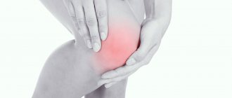
Subluxation of the knee joint
The most common pathology of this joint is external subluxation of the patella (synonymous with lateral patellar hyperpressure syndrome ), which is most often caused by high position of the patella, ligament weakness, hypoplasia of the lateral femoral condyle, valgus curvature of the knee , outward rotation of the femur/inward rotation of the tibia, and less commonly - congenital insufficiency/improper formation of the femoral-patellar joint. This is a common pathology of the patellofemoral joint, especially among children, adolescents and athletes (Fig. below).
It develops against the background of an imbalance between the lateral/medial stabilizers of the patella, congruent relationships between the articular surfaces of the patella and femoral condyles. This pathology is characterized by a redistribution of specific pressure in different zones of the articular surfaces, which contributes to overload of its lateral parts.
Since with long-term subluxation of the patella, the cartilage that covers the condyles/patella begins to suffer, there is a high risk of developing patellofemoral arthrosis . In addition, with long-term subluxation of the patella, synovitis (fluid accumulation in the joint) may develop, which will manifest itself as swelling of the knee joint.
Subluxation of the ankle joint
Subluxations of the talus are among the most common cases of intra-articular injuries of the ankle joint.
This pathology is most typical for a period of life with high activity (20-45 years). Depending on the nature of the translational movement of the talus (along, around) the longitudinal axis of the tibia/transmalleolar axis, 12 variants of subluxations are distinguished: anterior/posterior, upper/lower subluxations, internal/external, plantar/dorsal, supination/pronation, etc.
At the same time, each type of dislocation is characterized by its own mechanism of injury, typical anatomical changes in bones, ligaments, muscles, x-ray picture and specific treatment.
Subluxation of the ankle joint often occurs when the foot is twisted, loss of balance/fall, the foot touching a physical barrier, jumping, in the presence of ankle hypermobility, systemic connective tissue diseases, etc.
Subluxation of the shoulder joint
The shoulder joint is one of the joints that has the maximum range of motion in various planes.
Shoulder subluxation is a standing condition in which displacement of the humerus from the glenoid cavity of the scapula (forward/backward) occurs during movements associated with external rotation/abduction of the arm during active sports or awkward movements in everyday life. The shoulder joint is characterized by frequent development of articular instability, which is caused by degradation of the articular cartilage of the humeral head and the formation of a Hill-Sachs defect . As a rule, the patient eliminates dislocations with a certain movement independently.
Jaw subluxation
Caused by weakening of the temporomandibular joint ligaments. It is characterized by partial exit of the articular process of the jaw from the articular cavity, accompanied by pain/inability to close the mouth. The development of dislocations of the lower jaw is caused mainly by contraction of the muscles that open the mouth during various physiological acts during which the articular heads protrude forward and, stretching the capsules, partially emerge from the articular sockets (with excessive opening of the mouth, yawning, laughing, vomiting, less often with a blow to the chin, falling ). They are more common in females, which is due to the smaller size of the articular tubercle/depth of the articular fossa and the relatively weak ligamentous apparatus.
Depending on the cause, the following subluxations are distinguished:
- traumatic, caused by forced forced movement in a joint (excessive opening of the mouth, blow, falling on the chin, yawning, screaming, dental procedures);
- habitual, the development of which is based on stretching of the articular capsule/insufficient height of the articular tubercle, found mainly in older people, with rheumatism, deforming arthrosis of the lower jaw joint, gout, epilepsy.
Pathogenesis
Due to the impossibility of describing in a short article the mechanism of development of subluxations in various joints, as an example we will consider only the pathogenesis of subluxation of the atlas . The mechanism of development in relation to the occipital condyles/second vertebra depends on the cause that caused it. So, subluxation, caused by the action of a traumatic force, when the neck sharply bends/unbends during car accidents, develops as a result of damage to the ligamentous apparatus. Rotational/anterior subluxations of the atlas occur both under the influence of tensile forces and simultaneously during rapid labor/forceps/caesarean section, leading to tear/sprain of ligaments and subluxations of the vertebrae. The pathogenesis of uncoordinated sharp lateral movements of the head/neck in individuals with low muscle tone/congenital hypermobility is caused by the opening of the articular surfaces of the axis-atlas and pinching of the articular capsule, which develops as a result of spasm and pain in the group of adjacent cervical muscles.
With the development of inflammation in the lateral atlantoaxial joint due to the spread of infection from the nasopharynx to it, the pathogenesis of rotational subluxation of the atlas is caused by the accumulation of inflammatory fluid ( Grisel's disease ). In systemic diseases that cause connective tissue damage, the pathogenesis of subluxation of the atlas is caused by weakness of the neck muscles/extension of the ligamentous apparatus. With inflammation of the synovial membrane of the atlantoaxial joints, which develops against the background of rheumatoid arthritis , the mechanism of subluxation of the atlas develops due to weakening of the transverse ligament, which fixes the atlas to the odontoid process of the 2nd cervical vertebra.
Treatment of vertebral subluxations
The doctor realigns damaged elements of the spinal column. In case of acute pain, the patient is carefully transported to a medical facility, trying to move him as little as possible. A common and effective treatment technique is the Gleason loop. The patient is placed on a hard couch, and a small hard pillow is placed under the shoulder girdle. The head is secured with a loop that connects to the weight. Weight is selected individually, it all depends on the severity of the disease and its location.
When treating subluxations, a complex of medications is used. Muscle relaxants help relieve tension in the neck muscles and completely relax them. Pain syndrome is eliminated with injections and nerve root blockade.
Dr. Dlin’s clinic uses proprietary methods for treating vertebral displacements. During the recovery period, patients are shown manual therapy and osteopathy. In the first case, the effect is on joints, ligaments and bones, in the second, deep treatment of soft tissues and internal organs is provided. Kinesio taping is also used.
Classification
In this section we will consider only the classification of rotational subluxation. There are 4 types of rotational subluxation:
- Type A - stable rotational subluxation. It is a simple rotational displacement without damage to the transverse ligament, with the axis tooth being considered as the center of rotation.
- Type B - manifests itself as an anterior displacement of C1 to C2 by a distance of 3-5 mm with an incomplete transverse ligament with one lateral mass, which allows maintaining the function of axial rotation. There is potential for non-permanent subluxation with this type of injury.
- Type C - this type is characterized by anterior displacement of the atlas by a distance exceeding 5 mm. In this case, the lateral masses of the atlas are either subluxated or damaged, thereby fixing the resulting dislocation, significantly destabilizing the atlantoaxial joint.
- Type D is an extremely rare type of rotational subluxation characterized by posterior displacement of C1 to C2, often associated with a deficient/broken axis tooth.
The types of rotational subluxations at the atlantoaxial joint are shown in the figure below.
How to make an appointment with an atlas specialist
Leave a request for an appointment with a doctor using a special form.
A specialist will contact you as soon as possible. If you live in Moscow or St. Petersburg, you can agree on a visit time that is convenient for you. If you are in another city in Russia, doctors from the Atlas-Standard clinic will be able to travel to your city according to the schedule established on the website (updated every week).
Promotions and discounts are regularly offered to residents of certain cities. Check with the clinic specialists for information.
Causes
Only the causes of rotational subluxation of the atlas are given. The main cause of rotational subluxation of C1:
- Mechanical injury with damage to the ligamentous apparatus, resulting from sudden movements (turns), flexion/extension of the head, falls on the head.
- Birth injuries (rapid labor, caesarean section, forceps).
- Congenital anomalies (inferiority of connective tissue, accompanied by hypermobility of the joints).
- Rheumatoid arthritis.
- Imperfection of the muscular-ligamentous apparatus in children (weakness of the neck muscles in a child).
How to detect if the atlas is dislocated
Since all people could be exposed to birth trauma, every person needs a preventive examination to detect displacement. Some patients are diagnosed with signs of old diseases, the main cause of which is a displaced atlas. Therefore, the sooner the diagnosis is made and the atlas is reduced, the better for the person affected by the displacement of the vertebra.
When it is possible to eliminate the pathology, rapid sanogenesis (self-healing) of the body begins and the functioning of all its systems begins. But this does not mean that a victim of displacement can be cured of all the symptoms of existing diseases only by editing C1.
Symptoms
Symptoms of subluxations in joints of various locations, despite the specific symptoms inherent in each of them, also have common clinical manifestations in the form of acute/sharp pain in the area of the damaged joint; changes in the appearance/normal shape of the joint; restrictions on range of motion; redness of the skin around the joint and swelling.
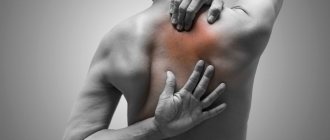
Symptoms of cervical vertebral subluxation
They manifest as pain in the upper parts of the neck, headache (in the back of the head, numbness in the hands). Rotational subluxation of C1 is characterized by a sharp increase in pain when trying to move: pain when turning the head to the left (with a right-sided subluxation) and, conversely, to the right - with a left-sided subluxation. In some cases, dizziness and loss of consciousness. Rotational subluxation of C1 due to uneven distribution of the load due to the spinal column being in an incorrect position affects the functioning of almost the entire musculoskeletal system, causing cervical osteochondrosis .
The back of the neck is almost constantly tense. With chronic subluxation, compression of the arteries may be observed to varying degrees, which leads to a deterioration in the blood supply to the cranial zone/brain and vertebrobasilar syndrome , vertebrobasilar insufficiency , vertebrobasilar arterial system syndrome , and vegetative-vascular dystonia .
In this case, the symptoms of subluxation of the atlas are manifested by neurosensory disturbances, dizziness , headaches , weakened hearing/vision and the appearance of “spots” before the eyes during sudden movements. There may also be an increase in blood pressure, numbness of the hands, dependence , tinnitus, fatigue, weakened memory, and insufficient adaptation to stress.
Clinically, with rotational subluxation of C1, there is marked tension in the neck muscles, a forced position of the head (rotation/tilting), limited mobility, and when pressing on the cervical vertebrae - pain, non-physiological protrusion of the vertebra. With the development of neurological symptoms, numbness, sensory disturbances, and, less commonly, urinary disturbances are noted.
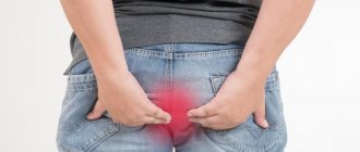
Symptoms of hip subluxation
Subluxation of the hip joint in adults is manifested by the appearance of rapid fatigue while walking, pain in the joint, a relatively high location of the greater trochanter, and impaired pelvic stability based on decreased function of the gluteus medius muscle (Trendelenburg's symptom), which contributes to a lateral tilt of the pelvis. The displacement of the pelvis is compensated by a large tilt of the torso (Duchenne's symptom), caused by strengthening of the oblique abdominal muscles. The lack of support for the femoral head, caused by an upward displacement of the femoral head, changes in the gluteal muscles and functional shortening of the leg disrupt the biomechanics of gait, which is manifested by a violation of the rhythm of walking, swaying of the pelvis and body when walking, and the phenomenon of lameness.
Jaw subluxation
Anterior bilateral subluxations are manifested by a wide open mouth, tense/flattened chewing muscles of the cheeks, and a posteriorly and downwardly displaced chin. Movements of the lower jaw are limited and are possible only in the direction of further opening of the mouth. The patient experiences pain, chewing is impossible, swallowing/speech is difficult, and saliva leaks from the mouth.
A characteristic sign is the retraction of tissue in front of the tragus of the ear. When palpating the articular head, its movements from the side of the external auditory canal are not determined. In cases of unilateral dislocation of the lower jaw, the mouth is slightly half-open, the chin is shifted to the “healthy” side and forward, slight movements of the lower jaw are possible due to the “healthy” joint. Chewing/swallowing and speech are significantly difficult, pain is minor.
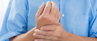
Symptoms of shoulder subluxation
Subluxation of the shoulder joint is manifested by stabbing/cutting pain in the area of the shoulder joint, the skin over which is hyperemic, swollen and becomes hot to the touch. The patient feels a burning sensation in this area. It is difficult or almost impossible to raise your arm or move it in any plane.
Unique diagnosis of atlas position
In almost all patients suffering from various diseases of the spinal column and spinal cord, seemingly not directly related to subluxation (headaches, migraines, cervical osteochondrosis, dizziness, back pain, lumbar pain, etc.), dislocation (rotation) is detected during diagnosis ) first cervical vertebra. Sometimes people have difficulty turning their heads since childhood: this is also a consequence of vertebral displacement. Correcting the situation (subluxation) simultaneously solves many health problems.
How subluxation is eliminated using the Swiss technique of adjusting the atlas
Setting C1 to a normal state is achieved through vibration effects on the muscles of the body that stabilize and support the vertebra, which have memory.
When vibrating at a certain frequency, which is produced by a special device developed in Switzerland, the muscles relax. The atlas falls into place, and the muscles are fixed in a new, correct position. First, a free initial examination is carried out and a diagnosis is made. The procedure is performed by a doctor specially trained in the nuances of the technique. You can read more about the method on the main page of our official website.
After undergoing the procedure, the patient is prescribed a special program to restore the functions of the musculoskeletal system, nervous and cardiovascular systems. Vitamin complexes are also prescribed and preventive recommendations are given depending on the initial condition and the presence of specific diseases.
In children
The formation of subluxations in children is based on excessive stretching of tendons/ligaments, which leads to disruption of the anatomical structure of the joint and its unclear fixation due to an unlimited/uncontrolled range of movements. That is, in fact, the child has a poorly fixed joint and therefore he can make movements with too large an amplitude, which are normally impossible, which forms a subluxation.
However, by adolescence, the musculoskeletal system acquires the properties of an adult and the mechanism of development of subluxations is similar to that of an adult. It is also important to take into account the high metabolic rate in children, which contributes to the development of inflammatory and degenerative processes in a joint with subluxation, causing the replacement of physiologically normal muscle-ligamentous tissue with connective tissue. These processes contribute to a decrease in the volume of the joint capsule and make it difficult to reduce the subluxation and restore full function of the joint. Therefore, timely diagnosis of subluxation and its reduction are extremely important in order to avoid surgical intervention to restore normal anatomical relationships in the damaged joint.
Let us dwell only on a few subluxations characteristic of children.
Subluxation of the cervical vertebra in a child
Subluxation of the atlas in children is often congenital, often arising during manipulation of the fetal head during the passage of the birth canal (extension, bending, sharp turns, the head against the background of immaturity of the ligamentous apparatus and birth weakness). In children under 1 year of age, there is a high risk of developing a subluxation of the atlas due to improper handling of the child - holding the mother in the arms in the absence of sufficient support for the head contributes to its tilting back and subsequently leads to subluxation. In older children, subluxations develop mainly during activity with sudden movements in the craniovertebral region (sudden forced turns, hits to the head with a ball, falling from a height, contact sports, sleeping in an uncomfortable position, causing muscle activation on one side and displacement of the vertebrae, etc. ).
Symptoms of subluxation in young children are tearfulness, anxiety, loss of appetite/bad breastfeeding, muscle hypertonicity in the neck, torticollis, and limited mobility in the upper spine. In older children, it manifests itself with headaches , poor sleep, dizziness, decreased concentration, memory, attention, attacks of depression / panic , etc.
Manual therapy plays a leading role in treatment in children, the methods of which can eliminate subluxation and normalize relationships in the joints. It should be taken into account that subluxation of the first cervical vertebra has a strong tendency to recur and can often recur, especially against the background of the development of instability of the ligamentous apparatus. Accordingly, it is important to pay attention to rehabilitation procedures, in particular, massage and exercise therapy, post-isometric relaxation, aimed at normalizing the muscular-ligamentous balance of the neck structures.
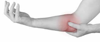
Elbow subluxation
The most common pathology of the elbow joint in children is subluxation of the radial head. It occurs mainly in preschool children aged 3-5 years, and then the frequency drops sharply. Develops with a sharp extension of an outstretched arm by the hand/lower end of the forearm along the longitudinal axis of the limb, mainly upward, less often forward (the arm was pulled when a child fell, during play, when getting dressed). It is characterized by a distal displacement of the head of the radial bone and a tear of the circular ligament of the joint, which falls into the interarticular space (Fig. below).
Subluxation of the hip joint
Symptoms of hip joint subluxation in a newborn with a slight displacement of the femoral head are extremely scarce. The leading symptom in this period should be considered an increase in passive mobility and excessive rotation in the hip joint. The main early clinical symptoms of subluxation in children of the first year of life are:
- Relatively shortened thigh.
- Limitations of passive hip abduction.
- Asymmetry of skin folds on the thigh and buttock folds.
- Marks-Ortolani slip symptom (click symptom) and Barlow modification (Barlow test).
- In children over one year of age, a limp or a characteristic “duck” gait is observed.
Treatment of subluxation of the hip joint in children is carried out using various types of devices/devices: splints, panties, pillows, stirrups designed to hold the leg in the abducted position, as well as special gymnastics in the form of abductor-circular movements in the hip joint, as well as massage of the gluteal muscles . Treatment of older children is carried out individually, taking into account the displacement of the head and the motor abilities of the child.
Consequences and complications
Common complications of atlas subluxation include:
- Transition from subluxation to dislocation.
- Headache.
- Increased blood pressure and intracranial pressure .
- Impaired blood supply to the brain ( vertebral artery syndrome , cerebral angiodystonia , disturbance in the vertebrobasilar region, deterioration of venous outflow, discirculatory disorders ).
- Damage to the roots of the C2 vertebra, which is accompanied by dysesthesia (distortion of sensitivity) in the occipital and parietal regions.
- Formation of disc protrusion and hernia .
- Formation of cervical kyphosis .
- Development of torticollis in children.
With subluxation of the atlas, the following complications are possible, but rare:
- Rotational atlantoaxial subluxations can cause damage to the spinal cord.
- Damage to the vertebral artery.
- Damage to the medulla oblongata due to vertical displacement of the axis tooth towards the foramen magnum.
Forecast
Most atlas dislocations/subluxations have a favorable outcome. An unfavorable prognosis occurs with instability of the articulation, which cannot be eliminated, or with vertical displacement of the axis tooth towards the occipital foramen. In this case, there is a risk of damage to the medulla oblongata and surgical intervention is necessary. Patients develop myelopathy . With timely jaw adjustment and proper rehabilitation, the prognosis is favorable. Sometimes there are repeated subluxations and joint stiffness .
The myth about safe caesarean section
In the photo: caesarean section is one of the common causes of subluxation of the atlas.
This is important for parents to know!
There is an opinion that a caesarean section relieves a woman in labor from painful labor and almost completely relieves the load on the cervical spine. In pursuit of bonuses and other privileges, some doctors advertise this method as the safest. This is very far from the truth! Caesarean section, like any other surgical intervention, should be done only if there are objective indications for it.
A Caesarean section is a method of delivering a baby through abdominal surgery, in which the newborn is removed through an incision in the uterus. The main problem in such childbirth is bleeding. To reduce it, the surgeon makes an incision as small as possible (so that only the arm goes through) and pulls the baby out by the neck with his fingers. To pull the fetus out through a small incision, a lot of force has to be applied, and the main part of this load in most cases falls on the child’s fragile neck. Due to the fact that the newborn is actually pulled by the head with great force, this method cannot be considered safe: it often leads to displacement of C1.
Also, during a caesarean section, there is a sharp change in intrauterine pressure to atmospheric pressure, which can lead to another type of birth injury - barotrauma.
Minor birth rotational subluxation (incomplete dislocation) of C1 is the most common type of birth injury of the cervical spine. The key word here is “minor.” This fact receives relatively little attention in medicine. Firstly, a slight displacement of C1 does not cause sharp and irreversible pathological reactions in the musculoskeletal system, and secondly, this phenomenon is so common that it is considered almost normal.
There is even a theory according to which the body itself compensates for the C1 shift with different bone thicknesses and leg lengths and seems to reduce the resulting imbalance to nothing. Whether this is good or bad, and what the consequences of such compensation are, one can guess even without a medical education.
Unfortunately, neither natural childbirth nor cesarean section are safe and do not avoid high stress on the newborn’s neck, and therefore do not exclude the possibility of birth injuries in infants.
List of sources
- Lutsik A. A., Ratkin I. K., Nikitin M. N. Craniovertebral injuries and diseases. - Novosibirsk, 1998. - 552 p.
- Selivanov V.P., Nikitin M.N. Diagnosis and treatment of dislocations of the cervical vertebrae. M., 1971.
- Kazakevich I.E. Unilateral subluxations and dislocations of the cervical vertebrae // “Orthopedics, traumatology and prosthetics”. 1986, no. 1, pp. 31-34.
- Tyazhelkov A.B., Zhila N.G., Belyaev Yu.A. About dislocations of cervical vertebrae in children // “Orthopedics, traumatology and prosthetics.” 1983, no. 7, pp. 58-60.
- Bondarenko N.S., Kazitsky V.M., Dovgan V.P. Dislocations and subluxations of the atlas in children and adolescents // Orthopedics, traumatology and orthopedics, 1988, No. 2, pp. 51-55.