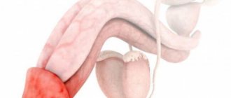
Hypospadias of the penis is a congenital anomaly of the development of the male penis, which is expressed in a violation of the structure of the urethra. In this case, the opening of the urethra in a man is not in its natural place, namely on the head of the penis, but on its back surface, on the scrotum or in the perineum.
Cost of urologist services in our clinic
| Initial consultation with a doctor with the highest category | 1000 rub. |
| Consultative appointment with a doctor based on test results and ultrasound results | 500 rub. |
| Ultrasound of the kidneys in standard mode and using Doppler techniques | 1200 rub. |
| Ultrasound of the bladder | 500 rub. |
| Ultrasound of the pelvis using Doppler techniques | 1200 rub. |
| Make an appointment by phone: 8-800-707-15-60 (toll-free) | |
| *The clinic is licensed to remove tumors |
This defect is formed during the intrauterine development of the fetus already in the first weeks. This disease occurs quite often. There is one case per 150-200 boys born.
Symptoms of the disease
Hypospadias is characterized by the main symptoms:
- the external opening of the urethra (meat) does not open at the apex of the glans penis, but is shifted proximally (towards the perineum). As a result, the meat is located in the region of the glans, coronary sulcus, shaft of the penis, scrotum or perineum.
- There is almost always curvature of the corpora cavernosa to a greater or lesser extent. The degree of curvature is determined during erection or during surgery, when an “artificial erection” test is performed.
- dysplasia of the foreskin - the foreskin is split, located on top of the penis, hanging over in the form of a “hood”.
Causes
All the causes leading to hypospadias have not yet been fully studied. It has been established that the leading role in the increase in the number of people suffering from this disease is played by the environmental situation, point mutations of genes, and consumption of food products containing disruptors: androgen destroyers and androgenic substitutes that change the hormonal status of the fetus in such a way that the process of sex formation is disrupted. Currently, disruptors include fungicides, phthalates, pesticides and herbicides, most of which are present in our daily lives. Often, the development of hypospadias can be caused by hormonal therapy prescribed to the mother when there is a threat of miscarriage, or hormonal contraception carried out before the expected pregnancy in less than 12 months. A fairly high risk of giving birth to a boy with hypospadias is possible with in vitro fertilization, since female sex hormones are used during pregnancy, which adversely affect the development of the child’s genital organs. Hereditary factors also play an important role. There are known cases of children being born with this diagnosis in several generations.
Urine drainage and wound dressing
Urine is drained using a transurethral drip stent or through a drainage tube placed above the pubis. Some surgeons do not perform drainage after repair of distal hypospadias. It is common to apply a circular bandage with light pressure, as well as prescribe antibiotics for prophylactic purposes. The duration of stenting and bandaging varied significantly between studies. Due to the low level of evidence, no recommendation can be made.
Concomitant pathology&
Often, hypospadias in boys is combined with other developmental anomalies: inguinal hernia, cryptorchidism, hydronephrosis, vesicoureteral reflux, myelomingocele, urogenital sinus, etc. Therefore, an ultrasound of the kidneys and bladder is recommended for all children to identify concomitant pathology of the urinary system before surgery. In the presence of hydronephrosis or vesicoureteral reflux, the first step is correction of the pathology of the upper or lower urinary tract, and after that, surgical treatment of hypospadias.
Use of anesthesia or general anesthesia
During surgery, the most modern method is considered to be combined anesthesia, when local conduction anesthesia is used. The drugs “Marcain” and “Naropin” make it possible to anesthetize the child from 4 to 9 hours after the operation.
If such a need arises, traditional analgesics can be used in the future. The use of local anesthesia can reduce the load on the nervous system, since the amount of general drugs administered is reduced.
Proximal hypospadias
The most common pathology of the penis in boys is undoubtedly hypospadias. The incidence of this anomaly, according to various authors, is increasing and amounts to 1:125. Proximal forms are less common with a frequency of 1:1200 newborn boys and are characterized by more pronounced curvature of the corpora cavernosa, often with dysplasia of the corpus spongiosum of the urethra. The external opening of the urethra is localized in the area of the penoscortical angle, scrotum or perineum. The foreskin is located on the dorsal surface. At an older age, such children urinate according to the female type.
Surgical treatment of proximal hypospadias is more complex than distal hypospadias. Since, in addition to moving the external opening of the urethra to the apex, it is necessary to straighten the corpora cavernosa and eliminate dysplastic tissues, which can lead to secondary curvature at an older age.
Depending on the surgeon's experience and preference, surgical correction is performed using either one-stage or two-stage techniques.
Patients with proximal forms, micropenia, or a non-palpable testicle often require a differentiated approach to treatment. Such children may require consultation with an endocrinologist, geneticist, and additional examination methods to determine the karyotype in order to exclude gender pathology.
Materials and methods
Over the past few years, a large number of patients with this pathology have been operated on. The age of the patients ranged from 5 months to 17 years. This group included patients with primary hypospadias that had not been previously operated on. Patients underwent Onlay-tube surgery and Bracka surgery using buccal mucosa. Before surgery, all patients were prescribed hormonal ointments on the glans penis and foreskin to increase the supply of plastic material. In both groups, PDS 7.0 was used for tissue suturing. The bladder was catheterized with an age-sized urethral catheter. In two-stage operations using the Bracka technique, after fixing the free flap, pressure was applied to it using a special “pellet” for the purpose of engraftment. The second stage was carried out after 5-6 months.
results
In the group of patients operated on using a one-stage technique, 10% of complications were observed. These complications included fistulas, urethral diverticula, and smegmal stones. A certain percentage of patients required a second stage, since they were able to form the urethra only up to the coronary sulcus or the size of the glans penis was insufficient for closure.
Patients operated on using the two-stage technique had about 4% complications. The vast majority of these were urethral fistulas. All patients were assessed for urodynamic characteristics, which showed a higher average urinary flow rate in patients after the Bracka procedure.
conclusions
Thus, the use of two-stage techniques for the correction of proximal forms of hypospadias allows us to obtain a lower percentage of complications and better urodynamic characteristics of urine flow.
Parents
The optimal age for hypospadias correction is 10 months.
Correction of hypospadias is most often performed in one operation.
You may need to use hormonal ointment before surgery. The ointment is applied 3 weeks before surgery in the morning and evening. The ointment should be applied to the penis, the size of a drop is the size of the nail plate of the parent’s thumb.
Before the operation, you need to collect the necessary tests.
In the postoperative period, 2 diapers are used, the catheter is removed into the second (external) diaper. This technique allows the child to be active throughout the entire period of hospitalization.
Antibacterial therapy is prescribed for the entire postoperative period.
After the operation, a special compression bandage is applied to the penis, and a urethral catheter is installed for 10-12 days. After this period, the bandage is removed and the catheter is removed.
To evaluate the result of the operation, it is necessary to videotape the process of urination.
Other forms of the disease
Forms of hypospadias:
- Features of the capitate shape
This form is characterized by the fact that the external opening of the urethra is located slightly proximal to the apex of the glans penis to the groove. The foreskin is usually dysplastic. With capitate hypospadias, the penis has a slight ventral curvature. Complaints include narrowing of the external opening in the genital canal, a thin stream of urine, and changes in the appearance of the penis.
- Crown shape
With coronal hypospadias, the external opening of the urethra is located in the area of the coronary sulcus. The foreskin is located on the dorsal surface in the form of a “hood”. A ventral curvature is noted. Complaints about a narrowed meatal opening. When urinating, the stream is directed at an angle to the urethra. The capitate and coronal forms are anterior anomalies. In the trunk form, the external opening of the urethra is located at different levels of the trunk. The penis has a more pronounced curvature, the stream is directed downwards. In order to urinate while standing, you have to pull your penis towards your stomach.
- Stem form
The stem is usually classified as a middle type of pathology.
- Scrotal
Scrotal hypospadias or penis-scrotal hypospadias refers to the posterior forms of damage to the genital organ. With such hypospadias, the external opening of the urethra is located in the area of the scrotum or at the border of the scrotum and the shaft of the penis. There is a pronounced ventral curvature of the corpora cavernosa or transposition of the penis. Urination is possible only in the female type while sitting. The external genitalia resemble the labia majora and an enlarged clitoris. In most cases, consultation with an endocrinologist is necessary.
- Perineal
Perineal hypospadias - with this anomaly, the external opening of the urethra opens at the perineum, the corpora cavernosa are significantly curved, the scrotum is split, and urination is female-type. The external genitalia have a mixed structure. Quite often a consultation with an endocrinologist or geneticist is required.
- Hypospadias without hypospadias or hypospadias of the chord type.
Chord type hypospadias or hypospadias without hypospadias is a special type of disease. With this pathology, the external opening of the urethra is located at the top of the head, but there is a curvature of the penis of varying degrees of severity. Curvature in such a disease can only be due to skin dysplasia on the ventral surface, a combination of skin dysplasia and the presence of connective tissue cords along the urethra, underdevelopment of the urethra itself.
Treatment is described in more detail on the page surgical treatment of hypospadias.
The optimal age for treatment is from 6 to 18 months of the child’s life. At this age, children tolerate the operation itself and the postoperative period much easier. Children operated on before the age of 3 usually do not even remember the fact of the operation.
Why is surgery necessary?
- Normally, boys should urinate standing up without straining. Surgery is required to restore this function.
- To form an anatomically correct penis.
- So that the penis has a correct, straight shape, without subsequently creating problems during sexual activity.
- To prevent psychological problems associated with the development of feelings of inferiority.
All these points determine the need for surgical intervention and correction of the anomaly, which is determined in any form.
Main goals of surgical treatment
Operations to correct this defect fall into the category of reconstructive plastic. If there is hypospadias, the main goals of surgical treatment are:
- elimination of curvature of the cavernous bodies;
- creation of the missing section of the urethra, without stenosis, fistulas and devoid of hair follicles. The formed urethra should grow along with the growth of the penis as the child grows older;
- the location of the external opening of the urethra at the top of the head and giving it a longitudinal direction to ensure a direct stream of urine without splashing and deviation;
- maximum elimination of all cosmetic defects to ensure complete psychosocial adaptation of the patient in society.
Surgical treatment of distal forms in a boy is carried out in one stage. The likelihood of achieving excellent functional and cosmetic results exceeds 95 percent, and hypospadias is highly likely to be correctable.
Treatment of proximal anomalies requires a more differentiated approach; preference is given to one-stage techniques.
For surgical treatment of our patients, we use modern surgical techniques, special suture material, magnifying equipment and microsurgical instruments. We do everything in our power to minimize the number of postoperative complications and obtain excellent functional and cosmetic results.
Forecast
The use of modern anesthesia, suture material, microsurgical instruments, magnifying equipment, and antibacterial therapy makes it possible to perform surgical treatment of hypospadias in just one stage with minimal risk of complications and obtain an excellent cosmetic result. Compliance with the optimal timing of surgical treatment allows you to eliminate the psycho-emotional component in the child. The desire to perform surgical treatment before 2 years of age is also due to the fact that the child will not remember the very fact of treatment and hospital stay.
Studies conducted on patients who underwent surgery for hypospadias showed that they were more satisfied with their sex lives than those who did not have surgery.
It is worth noting that treatment methods for hypospadias continue to evolve. New methods of tissue adhesion are being developed: tissue adhesives and the use of laser for adhesion, which leads to improved wound healing and a reduced risk of fistula formation.
Cellular technologies are also being actively developed and will soon make it possible to create an artificial urethra, especially in patients with severe forms of hypospadias. The identification of factors and understanding of the causes leading to hypospadias continues, which allows us to develop an approach to the prevention of this condition and carry out correction in utero.