Hello, friends!
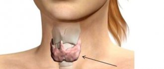
Cinquefoil is not just an ordinary ornamental plant. Its healing properties are also widespread. In particular, cinquefoil root is used to treat thyroid diseases.
The leaflet with five leaves of the shrub (with cinquefoil) is also called Dasiophorus shrub or Kuril tea. These are dense plants of small size with a small and elongated shape. The bast bush plant grows up to 1 meter in height and up to 1.5 meters in width.
Evergreen garden species with spotted edges develop elongated silver-green leaves. The upper leaves consist of 5 petals; on the underside the leaf blade is covered with silvery hairs. Occasionally, the leaves are triple, located on woody shoots.
From late summer to mid-summer, small flowers grow on the bushes, the color of which varies depending on the species:
- Snow White
- Yellowish
- Pink colors
- Peach, orange
- Red-red
- Red
Directions for use and doses
Preparation of the decoction. 10 g (1 tablespoon) of rhizomes are placed in an enamel bowl, poured with 200 ml of hot boiled water, covered with a lid and heated in a boiling water bath for 30 minutes, filtered without cooling, and squeezed. The volume of the resulting decoction is adjusted to 200 ml with boiled water.
Orally, 15 ml (1 tablespoon) 3 times a day. The course of treatment is 1–3 weeks.
Externally, for rinsing, use 1/2–1 glass. The course of treatment is 2–4 weeks.
It is recommended to shake the decoction before use.
Central pulse wave: pathophysiology and clinical significance
Evidence of the importance of antihypertensive therapy compared with placebo and the lack of differences between major classes of antihypertensive drugs in reducing the risk of outcomes was obtained from comparative randomized clinical trials measuring blood pressure (BP) in the brachial artery (peripheral BP). However, in some studies, the reduction in cardiovascular morbidity and mortality could not always be explained only in terms of the antihypertensive effect of the treatment regimens, and differences in the effects of antihypertensive drugs at the level of the macro- and microcirculatory bed, which cannot always be assessed with normal treatment, were discussed as additional reasons. blood pressure measurement [1-5]. The shape and amplitude of the blood pressure wave varies significantly depending on the measurement location. Systolic blood pressure (SBP) at the level of the brachial artery is higher than at the aorta, while differences in diastolic or mean BP are minimal [6]. Moreover, the reduction in SBP under the influence of antihypertensive drugs may differ in the central and peripheral segments of the vascular bed, and these differences are mainly due to the different pathophysiology of central and peripheral blood pressure [7].
The REASON and ASCOT-CAFE studies [7, 8] suggested that central BP, more than peripheral BP, is associated with regression of left ventricular hypertrophy (LVH) or cardiovascular outcomes. The results of these studies became the basis for studying the role of central blood pressure and the formation of ideas about the organoprotection of drugs not just “in addition to lowering blood pressure,” but “in addition to lowering peripheral blood pressure.”
The emergence of relatively simple technologies for non-invasive measurement of central blood pressure and the accumulation of data on its prognostic value raise the question of the possibility of optimizing the reduction of cardiovascular risk associated with elevated blood pressure by additional assessment of blood pressure directly at the level of target organs, especially in the aorta.
Pathophysiology of central blood pressure
The arterial bed is often thought of as a system of blood vessels characterized by stable cardiac output, mean hemodynamic blood pressure, and total peripheral resistance.
This model reflects the conditions of blood flow at the level of the microvasculature, which is characterized by minimal fluctuations in blood pressure - necessary conditions for the delivery of oxygen and nutrients to tissues. This model ignores the pulsatile nature of blood flow and the role of arteries in its modulation, assigning them an exclusively conducting role [9].
However, the biomechanics of large arteries is much more complex, and representatives of the domestic scientific school made a great contribution to its study [10, 11]. Professor M.V. Yanovsky owns the theory of the peripheral heart. The peripheral heart, according to Yanovsky, is an active systole-diastole of blood vessels, harmonized with the phases of cardiac activity, while rhythmic changes in arterial tone are of a peristaltic nature, spread to the periphery and help the heart in its propulsive work [10].
A huge contribution to the study of the biomechanics of arteries and hemodynamic processes in the human body, as well as to the development of alternative blood pressure measurements to the Korotkoff method, was made by the fundamental works of N.N. Savitsky [11].
The arterial bed has powerful adaptation mechanisms and has different elastic characteristics of the wall in different areas [12-18]. The aorta and large arteries are elastic vessels.
As we move towards the periphery, the proportion of elastic fibers in the vascular wall decreases, and muscle fibers increase.
The shock wave is generated by the left ventricle and propagates along the aorta and arterial tree. Due to its elastic properties, the aorta performs not only a conducting, but also a buffer function, ensuring continuity of blood flow. The ability to perform a buffering function decreases from the aorta to the peripheral arteries. Peripheral arteries perform mainly a conducting function.
The stiffness of the arterial wall increases from the aorta to the periphery. The stiffness gradient, branching of the arterial tree and the microvasculature serve as sources of the formation of numerous waves, summed up into a reflection wave. The reflected wave returns to the aorta in diastole. The main physiological function of the reflected wave is to maintain diastolic blood pressure in the ascending aorta at a level necessary to ensure coronary blood flow. Thus, the recorded pulse wave is the sum of the shock wave and the reflection wave, and the central blood pressure is the sum of the shock wave pressure (systolic blood pressure) and the reflected wave pressure.
To understand the factors influencing the level of central blood pressure, it is important to understand the differences between the central and peripheral pulse waves. Peripheral arteries are stiffer than central ones due to an increase in the muscle layer and a decrease in the elastic one. The stiffness of medium- and small-caliber arteries is influenced by vasomotor tone, which depends on the state of endothelial function, the activity of the renin-angiotensin and sympathetic nervous systems [12, 13, 15, 18]. At the level of peripheral arteries there are more branches, which serve as points of reflection of waves, they are located closer. An increase in stiffness, a large number and proximity of reflection points lead to a higher amplitude of the pulse wave and a higher level of blood pressure in the peripheral arteries compared to the central ones (Fig. 1). This phenomenon is called amplification. Systolic and pulse blood pressure undergo amplification, while mean and diastolic blood pressure remain relatively constant throughout the arterial bed. The physiological significance of pulse wave amplification is to prevent the extinction of the central wave and ensure adequate systolic blood pressure for the perfusion of peripheral organs and tissues.
A pulse wave is the sum of a shock wave and a reflection wave. Consequently, the amplitude of the central pulse wave and the value of central blood pressure can be influenced by changes in the amplitudes of these 2 components, as well as the time of appearance of the reflected wave. In turn, the reflected wave, which contributes to the level of central blood pressure, represents the sum of numerous waves reflected from various parts of the distal vascular bed.
The amplitude of the shock wave is directly proportional to stroke volume and inversely proportional to heart rate. The amplitude of the reflection wave depends on the proximity and number of reflection points. An inverse relationship between the reflected wave and height has been established, and its differences depending on gender have been described [18].
Constriction of arteries and arterioles leads to closer reflection points and an earlier appearance of the reflected wave in the aorta. However, the contribution of this mechanism to the increase in central systolic and pulse blood pressure is significantly less compared to the changes that result from a decrease in the elastic properties of the arteries.
An increase in arterial stiffness leads to an increase in the speed of propagation of the shock pulse wave and its earlier reflection. The return speed of the reflected wave also increases. As a result, the reflection wave appears in systole, and not in diastole, and the reflection wave superimposes on a new shock wave.
Quantitatively, this increase in blood pressure due to the early appearance of the reflection wave is characterized by an increase index (augmentation index, augmentation index), defined as the difference between the second and first systolic peaks, expressed as a percentage relative to pulse blood pressure in the aorta (Fig. 2).
The consequences of the early appearance of the reflection wave are an increase in central systolic blood pressure with increased left ventricular load and a decrease in diastolic blood pressure with a potential decrease in coronary blood flow (Fig. 3). Thus, central blood pressure is a kind of integrating indicator, which is determined by the state of the vascular bed throughout - from the heart, aorta to the microvasculature. Central blood pressure and growth indices are indirect indicators of arterial stiffness.
A clinically important aspect is the effect of age on central blood pressure. In young people, central arteries are much more elastic than peripheral ones. The pronounced gradient of stiffness and the resulting increase in pulse wave amplification in young people lead to significant differences between central and peripheral SBP, which can reach 20 mmHg. and more. With age, as well as with arterial hypertension (AH), the stiffness gradient between the central and peripheral arteries decreases due to a decrease in the elasticity of the central arteries .
Peripheral arteries are less susceptible to age-related changes due to the smaller proportion of elastic fibers in their wall [19, 20].
Hypertension, diabetes mellitus, dyslipidemia, and smoking lead to an acceleration of the loss of elastic properties of the central arteries. In people with hypertension or diabetes mellitus, the carotid arteries may become stiffer than the femoral or radial arteries, whose stiffness varies less with age or hypertension [20].
Young people may experience an increase in peripheral systolic and pulse blood pressure without an increase in central blood pressure. This phenomenon is called “false systolic hypertension.”
It is more often observed in tall, non-smoking young men with high levels of physical activity [21]. The estimated 20-year risk of coronary heart disease (CHD), taking into account brachial artery blood pressure and other risk factors, in such people is intermediate between the risks calculated for people with normal blood pressure or hypertension [22].
Thus, with a comparable level of peripheral blood pressure measured in the brachial artery using the Korotkoff method or an oscillometric device, the level of central blood pressure has a different value depending on age. In young people, the differences between central and peripheral pulse or systolic blood pressure are more pronounced than in older patients (Fig. 2). The reason for the increase in central blood pressure in the elderly is the early appearance of the reflected wave.
Methods for recording central blood pressure
Central blood pressure can be assessed invasively by aortic catheterization or non-invasively. It is clear that the clinical application of central BP measurement is related to non-invasive measurement methods.
Ideally, for non-invasive assessment of central blood pressure, it is necessary to analyze the pulse wave at the level of the aorta or the artery closest to it.
Direct recording of the pulse wave over the aortic region is difficult due to its deep location. Registration of the pulse wave over the carotid artery is considered a direct method for assessing central blood pressure, since the pulse wave in the carotid artery is closest in shape and amplitude to the pulse wave in the aorta.
The pulse wave in the carotid artery can be recorded with dynamic ultrasound by recording fluctuations in arterial diameter caused by changes in blood pressure, or by applanation tonometry using a special Millar sensor. Applanation tonometry ( applanatio
- flattening) is based on recording the pulse wave during flattening of the artery. This method is technically simpler and cheaper than ultrasound. However, the possibilities of applanation registration of a pulse wave on the carotid artery are limited in patients with obesity, atherosclerotic lesions of the carotid arteries, as well as with anatomical features of the location of the artery that do not allow high-quality recording of the pulse wave.
The most widely used methods for assessing central blood pressure are by converting peripheral waves recorded by various methods and on different arteries of the shoulder and forearm (Table 1). Arteries more distant from the aorta are not used due to significant changes in pulse waveforms compared to the aorta. The “gold standard” is the registration of the pulse wave on the radial artery using applanation tonometry with its subsequent transformation using a generalized transform function. Performing applanation tonometry of the radial artery is technically simple due to its convenient location and optimal conditions for applanation, since there is support for bone structures [18]. The transform function used has been validated against invasive measurements of central BP [23].
In addition to applanation tonometry, the pulse wave can be recorded oscillometrically on the upper arm, similar to traditional blood pressure measurements [24]. The central pulse wave in this case is also modeled by using a transform function. The advantages of this method are its simplicity and the possibility of integration into conventional oscillometric devices for measuring blood pressure and 24-hour blood pressure monitoring systems. The possibilities of 24-hour monitoring of central blood pressure and other parameters of arterial stiffness will apparently become a new stage in the development and clinical study of these parameters.
To assess the absolute values of central systolic and pulse blood pressure obtained by carotid applanation tonometry or the transformation of the peripheral pulse wave recorded by applanation tonometry or oscillometry, calibration is used relative to the level of blood pressure in the brachial artery measured by auscultation or using a validated electronic device [18]. The calibration of the pulse wave recorded over the carotid artery region is based on the assumption that there is no difference between mean and diastolic blood pressure in the carotid and brachial arteries. Calibration of the pulse wave recorded on the radial artery is based on the assumption of equal blood pressure levels in the radial and brachial arteries. These assumptions may cause errors in estimating central BP levels. In addition, the source of error when registering a pulse wave on the radial artery may be the transforming function itself.
It is possible to obtain the level of central blood pressure by analyzing the peripheral wave without using a conversion function using other mathematical approaches - from the second systolic peak (SBP2) of the peripheral pulse wave or by determining the moving average n
-th number of points.
Analysis of the central pulse wave, regardless of the method of its registration, includes an assessment of the central values of systolic and pulse blood pressure, as well as their growth indices. The growth index is a relative indicator and can be calculated without calibration.
Thus, there are sufficiently available validated non-invasive methods for measuring central BP.
Effects of medications on central blood pressure
Due to differences in the formation of central and peripheral pulse waves, the effects of antihypertensive drugs on aortic blood pressure may differ significantly from those on brachial blood pressure.
These differences are based on the different effects of drugs on the main factors determining the level of central blood pressure - arterial stiffness and reflected wave. There are no drugs in clinical practice that have a selective effect on arterial stiffness. Aortic stiffness may be partially reversible, and drug therapy (eg, statins) over a relatively short period of time can affect elastic structures and lead to a decrease in central BP [25–27].
The effects of antihypertensive drugs on central blood pressure may be unrelated to their effect on aortic elastic properties. The degree of change in vascular caliber, as well as the point of application of the effect in the vascular bed (elastic arteries, muscular arteries, arterioles, veins) varies significantly among antihypertensive drugs, and these differences may lead to differences in effects on central blood pressure due to a predominant effect on the reflected wave .
At the level of medium and small arteries, nitrates, calcium antagonists and ACE inhibitors lead to dilation of small and medium arteries (brachial and carotid) and reduce their stiffness by reducing hypertrophy of the muscular layer of the wall [28-31]. In other words, although drugs may have only a minor effect on the stiffness of the central arteries, they can significantly influence the intensity of the reflection wave from the peripheral parts of the vascular bed and, consequently, the augmentation of the central one and its magnitude.
An example is the results of the ASCOT-CAFE study, in which treatment regimens based on amlodipine/perindopril and atenolol/thiazide led to significant differences in central BP without significant differences in aortic stiffness, the velocity of the pulse wave from the carotid to the femoral region. arteries [8]. A less pronounced effect of atenolol on central blood pressure compared with angiotensin-converting enzyme (ACE) inhibitors, calcium antagonists and thiazide diuretics was also found in other studies [29-31].
Indirect evidence for the existence of an association between differences in treatment effects on central blood pressure and clinical outcomes was provided by the REASON study, in which the combination of perindopril and indapamide led to significant dynamics of the reflected wave in the carotid artery, leading to a decrease in central systolic and pulse blood pressure, and, as a consequence, , to regression of LVH. In the atenolol group, there was no decrease in pulse blood pressure in the carotid artery and no regression of LVH [7, 32].
Clinical and prognostic significance of central blood pressure
From a physiological point of view, central blood pressure better reflects the interaction of the left ventricle and the vascular bed than peripheral blood pressure. Central blood pressure correlates with the mass of the left ventricular myocardium and the state of its function; there is evidence of a stronger correlation with these indicators compared to blood pressure during 24-hour monitoring [33, 34]. An increase in central systolic blood pressure leads to an increase in myocardial oxygen demand, and the level of central diastolic blood pressure is responsible for ensuring adequate coronary blood flow during diastole [11]. Central pulse blood pressure (measured in the carotid artery) is an independent predictor of elastic vascular remodeling, and increases in carotid artery diameter and intimomedial layer thickness are strong markers of cardiovascular risk [35]. Finally, there is evidence that a decrease in central blood pressure (but not brachial artery blood pressure) is associated with regression of LVH [7].
Evidence has been obtained of a close relationship between central blood pressure and cardiovascular morbidity and mortality. Invasive studies have shown that aortic blood pressure (but not as measured by sphygmomanometry) is a predictor of CAD [36, 37]. There is also evidence that not peripheral, but rather central pulse blood pressure is an independent predictor of cardiovascular outcomes in men with coronary artery disease [38]. Central pulse BP, assessed by carotid applanation tonometry, was a stronger predictor of all-cause mortality than brachial artery PP in patients with end-stage chronic renal failure (ESRD) [39]. Data have been obtained on a similar circadian rhythm of central blood pressure and cardiovascular events with peaks in the early morning hours and evening [40].
The STRONG Heart Study supported the suggestion that central BP may be a stronger predictor of clinical outcomes than peripheral BP [41]. During a 4-year follow-up of 2,409 patients (mean age at inclusion: 63±8 years, 65% of patients were women), diabetes mellitus was diagnosed in 47%, hypertension in 52% of participants without a history of cardiovascular disease, every 10 mmHg. increases in central PP were accompanied by a significant increase in relative risk (1.11, 95% confidence interval (CI) 1.02-1.20; p
=0.012), while the increase in risk associated with the same increase in systolic blood pressure and pulse in the brachial artery was not significant (1.05, respectively, 95% CI 0.98-1.12;
p
= 0.176 and 1.06 , 95% CI 0.98-1.15;
p
= 0.130).
Central pulse blood pressure retained its predictive value after adjustment for age, gender and other traditional risk factors (1.11, 95% CI 1.02-1.20; p
=0.013).
The second independent predictor of cardiovascular outcomes was the aortic stiffness index (1.06, 95% CI 1.00-1.11; p
= 0.046).
However, some questions about the prognostic value of central BP are not yet clear. The results of the ANBP2 study did not show any superiority of carotid applanation tonometry compared with brachial pulse BP in predicting outcomes in women [42].
It has not been determined for which specific outcome, myocardial infarction or stroke, central BP carries stronger prognostic information. It is expected that further analysis of completed studies and new data will help to better understand the importance of central BP for specific clinical outcomes in different target groups. The question also needs further research: in what situations is central blood pressure more informative than well-studied markers of aortic stiffness, such as pulse wave velocity and growth index.
Interesting information regarding the question of whether central BP control is beneficial in addition to peripheral BP control is expected from the BP Guide study [43]. The design of this Australian study involving 284 patients with uncomplicated hypertension was to assess central BP in addition to clinical measurements, 24-hour monitoring and self-monitoring of BP in relation to the dynamics of left ventricular myocardial mass index (LVMI), the number of antihypertensive drugs taken and quality of life indicators. Patients were randomized into 2 groups, in one of which the decision to adjust therapy will be made not only on the basis of an assessment of clinical blood pressure, self-control data and 24-hour blood pressure monitoring, but also at the level of central blood pressure obtained using applanation tonometry of the radial artery using a transforming function.
The target values for clinical blood pressure are <140/90, for self-control - <135/85 mm Hg. according to measurements over 7 days, for average daily blood pressure - <130/80 mm Hg, daytime blood pressure - <135/85 mm Hg. Indicators taking into account the gender and age of patients were used as target values of central blood pressure [19]. The observation period was 12 months. The authors of the study expected that there would be no differences in the dynamics of LVMI, but perhaps the groups of patients would differ in the number of medications taken and quality of life.
Prospects for assessing central blood pressure
Obviously, for the clinical use of central blood pressure indicators, it is necessary to establish their standards. Such an attempt was made by analyzing a database of radial artery applanation tonometry results with subsequent pulse wave conversion in random samples of 3 European populations ( n
=534, among them 228 men, average age - 34.9 years). Patients with hypertension, diabetes mellitus and dyslipidemia were not included.
For men 40 years old, the following values of the 95th percentile of central and peripheral blood pressure were proposed: pulse blood pressure in the brachial artery - 60 mm Hg, central pulse blood pressure - 40 mm Hg, index of increase in pulse blood pressure in the brachial artery - 90%, in the aorta - 30%.
In women of all ages, the normative indexes for the increase in pulse peripheral blood pressure are 10% higher, and in the aorta - 7% higher compared to men [19]. The prognostic value of these cut-off points for central BP and reflected wave parameters continues to be studied.
In 2013, it is planned to publish central blood pressure standards obtained based on an analysis of data from almost 50 thousand people in various populations.
An argument in support of assessing central blood pressure may be the premature loss of elastic properties of the aorta in the presence of concomitant cardiovascular risk factors in people of relatively young age [44, 45].
The aortic wall accumulates the damaging effects of risk factors. A decrease in the elasticity of the aorta and large vessels leads to an increase in central blood pressure, i.e. An increase in central blood pressure indirectly reflects an increase in arterial stiffness, and assessing its level can help identify patients in whom the presence of potential risk factors translates into actual risk. The presence of other cardiovascular risk factors accelerates age-related changes in the structural and functional state of the arteries, and this may be more reflected in the level of central rather than peripheral blood pressure [46, 47]. Increases in central (but not peripheral) blood pressure are found in hyperparathyroidism, dyslipidemia, diabetes mellitus, and chronic inflammatory diseases (eg, rheumatoid arthritis) [48–51]. Finally, differences were found between the age-related dynamics of central blood pressure in men and women [52].
The idea of central blood pressure and the amplification phenomenon allows us to substantiate the results of the Framingham study on the different prognostic significance of systolic, diastolic and pulse blood pressure depending on age [53]. Before 50 years of age, systolic blood pressure in the brachial artery is not a strong predictor of risk, and in people over 50 years of age, this indicator significantly exceeds the level of diastolic blood pressure in its prognostic value. It can be assumed that the reason for the low prognostic value of systolic blood pressure in the brachial artery at a young age is the relatively low level of central systolic blood pressure. The reason for these differences is pulse wave amplification.
An extreme example of differences between central and peripheral BP is the phenomenon of false systolic hypertension in young men with very elastic arteries, which is characterized by high peripheral systolic BP but normal central BP [21, 22].
In other words, in young people, left ventricular afterload may be overestimated based on peripheral SBP measurements.
Unlike young people, in people over 60 years of age, the level of systolic and pulse blood pressure in the brachial artery has a higher predictive power.
At this age, due to the loss of elasticity of the central arteries, the amplification of pulse blood pressure is less pronounced, and the levels of central and peripheral blood pressure do not differ significantly. In other words, in older people, peripheral blood pressure indicators better reflect the level of central blood pressure, and the prognostic value of peripheral systolic blood pressure increases.
Thus, it can be assumed that in young and middle-aged people, knowledge of the level of central blood pressure is necessary for a correct assessment of the phenotype of peripheral blood pressure, which turns out to be similar in patients with false and true isolated systolic hypertension.
However, today, routine testing of central blood pressure, even with such an attractive goal as a more reliable diagnosis of hypertension in young and middle-aged people, seems premature.
Further prospective studies on the prognostic value and natural evolution of false isolated systolic hypertension are required. Further studies of the prognostic value of central BP in larger populations of different ages with different risks of cardiovascular events are needed to determine its value as a surrogate endpoint.
The results of the REASON, ASCOT-CAFE, STRONG Heart Study and others suggest that the assessment of central blood pressure opens up new perspectives and additional opportunities for stratifying the risk of developing cardiovascular complications in young and middle-aged people, as well as for assessing the effectiveness of antihypertensive drugs .
Unlike peripheral central blood pressure, it is modulated by the structural and functional characteristics of large and small arteries and is a kind of integrating indicator reflecting the remodeling of the vascular bed. The main contribution to the increase in central blood pressure is made by a decrease in the elasticity of the arterial wall. In 2007, the European Society of Hypertension and the European Society of Cardiology guidelines for the management of hypertension included measurement of pulse wave velocity between the carotid and femoral arteries as a method for assessing subclinical target organ damage, and during the discussion on the clinical and prognostic significance of central BP its routine definition was considered premature [54]. This attitude towards central blood pressure was preserved in the recommendations on hypertension in 2013 [55]. Further large-scale epidemiological studies are needed on the prognostic value of central BP, as well as comparisons of treatment strategies based on central BP measurements or traditional peripheral BP measurements on cardiovascular outcomes.
About the author
Yulia Viktorovna Kotovskaya
— Doctor of Medical Sciences, Professor of the Department of Propaedeutics of Internal Diseases, Faculty of Medicine, State Educational Institution of Higher Professional Education "Russian Peoples' Friendship University", Moscow; e-mail
Contraindications
White cinquefoil contraindications
has a little. The herb is safe for the body and is not a poisonous species, so you can take it without fear.
The herb is contraindicated in the presence of diseases:
- chronic hypotension – low blood pressure;
- chronic constipation;
- arrhythmia.
Also does not recommend taking products with cinquefoil
pregnant and lactating women, children under 12 years of age.
Elderly people are advised to consult a doctor before starting treatment. In some cases, the use of traditional medicine is not possible.
An absolute contraindication is individual intolerance. It occurs very rarely, most people tolerate the course of treatment well. But if nausea, vomiting, dizziness, tremors of the limbs, or increased sweating occur, the medication should be stopped.
Alcohol tincture is contraindicated for minors. To treat children, only decoctions and water infusions can be used.
Useful properties of Cinquefoil
Cinquefoil is not only an ornamental plant, but also a medicinal plant. Potentilla leaves and flowers contain more vitamin C than lemon. The complex of beneficial substances includes carotene, tannin, organic acids, flavonoids, and essential oils. An infusion of leaves and flowers (Kuril tea) helps get rid of many diseases. It has long been successfully used for gargling and mouthwash, treating wounds and burns, normalizing metabolism and intestinal motility, treating dysbiosis and intestinal infections. Kuril tea has a calming effect during stressful conditions.
Medicinal raw materials (leaves and flowers of cinquefoil) are harvested at the end of July - August, cutting off the one-year flowering tops and drying them in the shade. Brew at the rate of 1 tablespoon of crushed leaves and flowers per 250 ml of boiling water, then add honey and leave for half an hour.
Catalog Potentillas