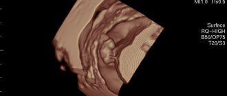
- At what time does fetal movement begin?
- Normal fetal movement
- Methods for assessing the “adequacy” of fetal movements
- Changes in fetal activity
- Determination of fetal condition
“Dear patients, we are pleased to welcome you to the website of the Center for Fetal Medicine - a medical center of expert level in the field of modern prenatal medicine.
We see our mission as making the expectation of a child and its birth a happy, calm and most comfortable period for every woman. By providing professional medical support, we help couples when planning a pregnancy, monitor its harmonious course, conduct prenatal diagnostics at an expert level, providing comprehensive care for the health of the expectant mother and baby.”
Bataeva Roza Saidovna
Head of the Center for Fetal Medicine in Moscow
From the very beginning of pregnancy, every expectant mother begins to carefully listen to the sensations inside her growing tummy. I really can’t wait to feel my baby’s movements. When does the fetus begin to move? At what age can a pregnant woman begin to listen carefully to herself, waiting for the first movements of her child? Should I worry if they are not felt or if the baby suddenly calms down? And can the movements carry any other information besides communication with the mother?
Sixth week
At the sixth week, the placenta begins to develop, but the blood circulation between it and the embryo itself does not function. The fetal brain is formed, as well as the facial muscles. The embryo's eyes are already more pronounced and eyelids are beginning to form. The upper limbs are developing, the legs remain unchanged. The division into chambers is completed in the heart; The first kidneys are formed, as well as parts of the gastrointestinal tract.
Around day 25, the development of the neural tube of the embryo ends. A pregnant woman may experience nausea, and sometimes vomiting, frequent urination, and constipation. During this period, it is already advisable to register with a gynecologist. The doctor will prescribe vitamins necessary for the development of the fetus and refer you for tests.
Starting point: how to find out when pregnancy started
The obstetrician calculates the date when a woman is expecting a baby during her first visit to the antenatal clinic.
- The doctor performs a manual examination to determine the size of the uterus. This will help him understand what stage of pregnancy the uterus corresponds to.
- Also, the local doctor must specify the date of the first day of the last menstruation. This point is taken into account, because The uterine mucosa begins to prepare for pregnancy from this period of time.
- You can find out the most reliable information about the duration of pregnancy using an ultrasound examination. An ultrasound examination can tell with precision down to the day when a small life was born. The examination, even at the earliest stages (starting from 4-5 weeks), assesses the size of the embryo, which allows the obstetrician-gynecologist to calculate the exact date of pregnancy.
In the first week after conception, the embryo actively moves along the fallopian tube. After six days of active “journey”, it enters the uterine cavity. Under the influence of progesterone (also called the pregnancy hormone), the unborn baby attaches to the lining of the uterus, this process is called implantation .
If the attachment of the embryo has taken place successfully, then the next menstruation will not occur - the pregnancy has begun.
Eighth - ninth weeks
The fetal body begins to straighten. Boys' testicles begin to develop. The hearing organs are formed. There is intensive growth of the head, upper and lower extremities. There is no longer a membrane between the fingers.
At the ninth week, the baby’s motor activity increases noticeably. The tail gradually disappears. The head is large in size relative to the entire body of the fetus. Movements become more intense due to muscle development, but so far only ultrasound can record them.
Seventh week
On days 29-35, the fetal genital organs are formed, the eyes and inner ear develop. During this period, a barely noticeable umbilical cord appears. The upper lip and nasal cavities are developed on the fetal face. Development of parts of the brain occurs.
Also during this period, the formation of the umbilical cord is completed and the uteroplacental circulation begins to function. The fingers are visible quite clearly, but they are not all separated from each other.
When exposed to external stimuli, the child spontaneously moves his arms. The eyes are well developed, already covered with eyelids. At the seventh week, the baby develops a tail, which will remain with him until the 10th week. A pregnant woman's uterus has doubled in size.
Stages of the IVF procedure
Before describing how fertilization will occur, it is worth noting the sequence of actions to prepare for it. The donor and recipient are prepared for upcoming procedures by synchronizing their menstrual cycles. To do this, hormonal drugs are used so that the processes of the reproductive system occurring in the bodies of two different women coincide.
This is followed by the introduction of another type of hormonal drugs to the donor in order to stimulate the maturation of several follicles at the same time, in contrast to the physiological menstrual cycle, when only one follicle matures completely.
After which a drug is administered that stimulates ovulation of all mature follicles. As a result, from three to four to eight or more eggs are obtained, which are subsequently removed from the donor under anesthesia by puncture. The entire procedure for preparing and obtaining eggs can take a considerable amount of time, up to several weeks.
And ultimately, fertility doctors begin the most important process - fertilization.
Embryo quality assessment
The central unit of the IVF clinic is the embryology laboratory. For each IVF attempt, which reflects the fate of each embryo from the moment oocytes are received until the moment of transfer or freezing.
Immediately after obtaining oocytes and sperm, their morphological assessment is carried out. Based on the results of the analysis of the ejaculate (spermogram), a decision is made on the method of fertilization - IVF or ICSI.
| № | Oocyte assessment | Fertilization | Embryo development stage | Type of ART (IVF/ICSI) | A comment | |||
| Day after puncture | 0 | 1 | 2 | 3 | 4 | 5 | Bottom line | |
| date | 07.03 | 08.03 | 09.03 | 10.03 | 11.03 | 12.03 | 13.03 | |
| Embryologist | Kalinina | Sergeev | Kalinia | Kalinia | Kalinia | Sergeev | ||
| 1 | Kalinina I.I. | MII | 2pN | 4 AB | 6 AB | Cryo (Bl Ab 3) | Cryo | ICSI |
| 2 | Kalinina I.I. | MII | 2pN | 4 AB | 6 BA | Cryo (Bl Bb 3) | Cryo | ICSI |
| 3 | Kalinina I.I. | MII | 2pN | 4 AB | 8 AB | Cryo (Bl Bb 2) | Cryo | ICSI |
| 4 | Kalinina I.I. | MII | 2pN | 4 AB | 8 AB | Cryo (Bl Ab 2) | Cryo | ICSI |
| 5 | Kalinina I.I. | 0-1 | 2pN | 4 AB | 7 AB | ET (Bl Aa 3) | ET | ECO |
| 6 | Kalinina I.I. | 0-1 | 1 pN | 4 AB | 8 AB | Cryo (Bl Bb 3) | Cryo | ECO |
| 7 | Kalinina I.I. | 0-1 | 1 pN | 4 AB | 8 AB | Cryo (Bl Bс 2) | Cryo | ECO |
| 8 | Kalinina I.I. | 0-1 | 2pN | 4 AB | 8 AB | ET (Bl Ab 4) | ET | ECO |
| 9 | Kalinina I.I. | 0-1 | 2pN | 6 AB | 8B | 8B | ECO | |
Description: Day 0. Immediately after puncture, the maturity of the resulting oocyte-cumulus complexes (OCC) is assessed. OCC with a transparent cumulus usually contains a mature oocyte and receives a score of “1-1.” During puncture, it is possible to obtain mature, immature, as well as degenerative and destroyed oocytes. An accurate assessment of the condition of the oocyte is possible only after it is cleaned before ICSI.
In mature oocytes ready for fertilization, the first polar body is detected. In the embryological protocol, the mature oocyte is designated MII.
If the process of oocyte maturation in the follicle is disrupted or the trigger (CG) is incorrectly introduced, then there is a high probability of obtaining immature cells, designated MI and GV (Fig. 1). Complete degeneration of the oocyte (Deg) is also possible.
Fig. 1. Oocytes obtained by puncture of the ovaries after removal of the cumulus (denudation). A - MII, B - MI, C - GV. Pb – polar body, ZP – zona pellucida
18-20 hours after the addition of sperm or ICSI (1st day), during normal fertilization, two pronuclei are formed. These are the precursors of the nuclei of future blastomere cells, into which the fertilized egg begins to divide. With proper fertilization, both pronuclei are clearly distinguishable. In this case, they are assigned a score of 2pN. If no pronuclei are visible, which is usually due to lack of fertilization, 0pN is recorded in the protocol. If fertilization is incorrect, several pronuclei may appear, which is reflected in the record, for example, 3pN, 6pN, etc. (Fig. 2). “Incorrectly” fertilized oocytes are not suitable for further work and are disposed of.
Fig 2. The first day of development in vitro – the formation of the zygote and the formation of pronuclei. A – 2pN (normal fertilization) – two pronuclei B – 3pN (abnormal fertilization) – more than two pronuclei.
Further development of the embryo, fragmentation, occurs within five to six days. The quality of embryos is assessed 40-42 hours (2 days), 72-74 hours (3 days) and 120 hours (5 days) after fertilization. The fragmentation of the embryo must be symmetrical (blastomeres of the same size are obtained) and uniform (all blastomeres undergo division). For clarity, embryologists use a numerical-letter quality assessment system, where the number indicates the number of blastomeres, and the letter their quality. To designate an embryo of good quality, the letter index “A” is used, average “B”, low “C”. Intermediate options are also possible, for example AB, BA, BC, SV, when it is difficult to give an unambiguous assessment (Fig. 3).
2nd day (4A) 3rd day (8A) 4th day (comp)
Fig. 3. Fragmentation of the embryo during the first three days in vitro and compact morula on the 4th day of development. Embryos of excellent (A) quality are presented. B – blastomere ZP – zona pellucida.
For the second day of cultivation, embryos with four blastomeres - 4A, 4AB, 4B - are considered promising. For the third - eight-cell (8A, 8AB, 8B). Poor quality embryos (BC, SV, C) are usually not tolerated; they are left until the fifth day and when a normal blastocyst is formed, they are frozen or transferred to the uterus. In the case of uneven fragmentation (the presence of blastomeres of different sizes), the potential of the embryo for implantation is reduced, and this is denoted by the prefix “un”, for example 3Bun. The presence of cytoplasmic fragments is designated “fr”, for example 8ВСfr (Fig. 4). The presence of vacuoles is also assessed. When visualizing them, an o. Normally, each blastomere carries one nucleus; if more than one nucleus is visualized in at least one blastomere, this is called multinucleation (mN) and indicates a significant probability of chromosomal pathology of this embryo.
Fig. 4. Cleavage of the embryo during the first three days in vitro. Embryos of good (BA, B) and satisfactory (BC) quality are presented. In the second row are embryos with uneven division (3Bun and 4BAun).
By the end of the third and fourth days of cultivation, the embryo begins to compact (the boundaries of its cells become indistinguishable) and prepares to form a blastocyst. Its cells can be partially (p.comp) or completely compacted (comp) (Fig. 5). Usually, embryos are not assessed on the fourth day due to the low information content of this stage of development. However, the presence of compaction on the second day may indicate abnormal development and must be reflected in the embryological protocol.
Fig 5. Compactization stage 4th day of cultivation A – p.comp B – comp
On the fifth day, approximately 120 hours after fertilization, the embryo forms a blastocyst (Bl) (Fig. 6). Assessing the quality of a blastocyst involves its size, which is reflected in numbers from 1 to 5; the state of the inner cell mass (ICM) (from “A” to “C”) and the surrounding cells - the trophoblast (from “a” to “c”). The best for transfer will be blastocysts of size from 3 to 5, having a multicellular ECM and trophoblast - Bl4Aa, Bl4Ab. Blastocysts of average quality are designated as – Bl2Bb, Bl3Bb, and of poor quality – BlCc.
Figure 6. Blastocysts on the fifth day of development in vitro. e.bl – early blastocyst, Bl3Ab – expanded blastocyst, Bl5Ab – blastocyst before hatching, ICM – inner cell mass.
Further development of the embryo occurs in the uterus after implantation. For successful implantation, the blastocyst must emerge from the surrounding zona pellucida. This process is called hatching, or hatching. In the case when the embryologist notes changes in the zona pellucida and after cryopreservation of the embryos, the process of independent hatching of the blastocyst can be difficult and can be facilitated by using assisted hatching. At our center we use the most effective laser assisted hatching.
The highest fertilization rate was obtained when embryos were transferred at the blastocyst stage. So, in our center it is 53%. While during transfer at the stage of 6-8 blastomeres - 47%.
Freezing and PGD are also best performed at the blastocyst stage. However, not all embryos reach the blastocyst. Therefore, the following approach is generally accepted: if there are less than 5 embryos on the 5th day of cultivation, at least one is transferred, the rest are left until the 5th day. Of those that reach the blastocyst, one more can be transferred (double transfer), the rest are frozen. If there are 5 or more embryos on day 3, they are not transferred and left to be cultured until day 5. On day 5, one, or less often two, blastocysts are transferred, the rest are frozen.
In conclusion, it should be noted that the described morphological criteria for assessing the quality of embryos are basic, necessary, but not always sufficient for selecting an embryo for transfer. Thus, often after the transfer of embryos of the highest quality, implantation does not occur and, on the contrary, there are cases of pregnancy occurring and successfully developing when embryos of poor quality are transferred. Therefore, there are additional methods for predicting embryo implantation (time-laps, PGS, metabolite analysis, etc.), which complement morphological criteria and collectively ensure the selection of the best embryo.
Blastocyst in the process of hatching (exit from the zona pellucida for implantation)
A – light microscopy HMC contrast B – Scanning electron microscopy – ZP – zona pellucida TE – trophectoderm ECM – inner cell mass
Bottom line
The development of a child from conception to birth is a rather complex and labor-intensive process. The first trimester is the most important period of gestation, since at this time all the baby’s systems are laid and formed. At the end of the first trimester, a study is carried out that helps to track the correspondence of embryo development to the gestation period and prevent pathologies. For the expectant mother, screening is an opportunity to see the baby for the first time.
The first abdominal image is one of the most touching moments of pregnancy, what do you think?


