Determination of blood group in combination with the Rh factor - this study is one of the first carried out after confirmation of pregnancy. Scientists in the field of immunology have long studied the influence of compatibility or, conversely, conflict between blood groups and Rh factors of parents on the intrauterine development of the fetus.
In case of compatibility, there is nothing to worry about, but if there is a conflict between the blood groups of the mother and child, then the pregnant woman will need constant medical supervision in order to prevent adverse effects on the newborn. Let's explore in detail what constitutes a blood group conflict during pregnancy.
What is the Rh factor?
Rh factor (Rh factor) is a blood protein that is found on the surface of blood cells - red blood cells.
If this protein is present, then this means that the person has a positive Rh factor, but if it is not there, then it is negative. Rh factor is determined by antigen. There are five main antigens, but it is the D antigen that indicates Rh. 85% of the world's population have positive Rh factors. How to determine your Rh factor? It is enough to donate blood from a vein just once. This indicator does not change throughout life. The Rhesus status of the embryo is formed already in the first trimester of pregnancy. Determining this indicator is very important for the expectant mother, since in the case of an Rh-negative mother and an Rh-positive child, various pregnancy complications are possible. In this case, it will be especially important to follow the doctor’s instructions, avoid infectious and colds, as well as stress. Also on various websites there are so-called calculators that determine the Rh factor of the unborn child. It must be remembered that blood is donated on an empty stomach. A rapid Rh test can be taken at any independent laboratory where blood is taken (for example, Invitro). The price depends on the price list of the clinic itself. You can find out about the cost of the analysis immediately before delivery. You can also donate blood and find out your Rh factor for free if you become a donor. To do this, you need to fill out a form to register yourself as a blood donor at the appropriate institution.
The Rh factor also plays a big role in blood transfusions. A transfusion involves two people: the recipient (the one who receives the blood) and the donor (the one who donates the blood). If the blood is incompatible, the recipient may experience complications after the transfusion.
The most common myth among couples is that blood type (like the Rh factor) is inherited from a man. In fact, the inheritance of the Rh factor by a child is a rather complex and unpredictable process, and it cannot change during life. But it is worth remembering that in rare cases (about 1% of Europeans) a special type of Rh factor is determined - weakly positive. In this case, Rh is determined either positive or negative. This is where questions arise on forums: “why did my Rh minus change to plus?”, and legends also appear that this indicator may change. The sensitivity of the testing method plays an important role here.
An equally popular search on the Internet is “horoscope by blood type.” For example, in Japan, great attention is paid to deciphering blood type. Believe it or not - it's up to you.
In the world there is such a thing as a medical tattoo, photos of which can be easily found on the Internet. What do these tattoos mean and what are they for? Its designation is quite pragmatic - in case of a serious injury, when an urgent blood transfusion or surgery is required, and the victim is not able to give the doctor information about his blood type and Rh. Moreover, such tattoos (a simple application of the blood type and Rh factor) should be in places accessible to the doctor - shoulders, chest, arms.
Sources
- Ceniceros LC., Capitanio JP., Kinnally EL. Prenatal Relocation Stress Enhances Resilience Under Challenge in Infant Rhesus Macaques. // Front Behav Neurosci - 2021 - Vol15 - NNULL - p.641795; PMID:33854420
- Raguz MJ., Prce Z., Bjelanovic V., Bjelanovic I., Dzida S., Mabic M. 20 Years of Follow-up Alloimmunization and Hemolytic Disease in Newborn: Has Anything Changed in the Field Over the Years? // Klin Padiatr - 2021 - Vol232 - N6 - p.314-320; PMID:33063311
- Shirtcliff EA., Lubach GR., Mooney R., Beck RT., Fanning LK., Coe CL. Transgenerational propensities for infant birth weight reflect fetal growth history of the mother in rhesus monkeys. // Trends Dev Biol - 2021 - Vol12 - NNULL - p.55-65; PMID:32616989
- Deere JD., Chang WLW., Villalobos A., Schmidt KA., Deshpande A., Castillo LD., Fike J., Walter MR., Barry PA., Hartigan-O'Connor DJ. Neutralization of rhesus cytomegalovirus IL-10 reduces horizontal transmission and alters long-term immunity. // Proc Natl Acad Sci USA - 2021 - Vol116 - N26 - p.13036-13041; PMID:31189602
- Lee DS., Ruiz-Lambides AV., Higham JP. Higher offspring mortality with short interbirth intervals in free-ranging rhesus macaques. // Proc Natl Acad Sci USA - 2021 - Vol116 - N13 - p.6057-6062; PMID:30877247
- Aljuhaysh RM., El-Fetoh NMA., Alanazi MI., Albaqawi AS., Alanazi WM., Alanazi NS., Alanazi RM., Alanazi AM., Alnemer EM., Alenezi RA., Alabdullatif TK., Alanazi RA., Alanazi SS., Alsultan KS., Alanazi IM., Alsunayni DS. Maternal-fetal Rhesus (Rh) factor incompatibility in Arar, northern Saudi Arabia. // Electron Physician - 2021 - Vol9 - N12 - p.5908-5913; PMID:29560141
- Bishop CV., Stouffer RL., Takahashi DL., Mishler EC., Wilcox MC., Slayden OD., True CA. Chronic hyperandrogenemia and western-style diet beginning at puberty reduces fertility and increases metabolic dysfunction during pregnancy in young adult, female macaques. // Hum Reprod - 2021 - Vol33 - N4 - p.694-705; PMID:29401269
- Krendl C., Shaposhnikov D., Rishko V., Ori C., Ziegenhain C., Sass S., Simon L., Müller NS., Straub T., Brooks KE., Chavez SL., Enard W., Theis FJ ., Drukker M. GATA2/3-TFAP2A/C transcription factor network couples human pluripotent stem cell differentiation to trophectoderm with repression of pluripotency. // Proc Natl Acad Sci USA - 2021 - Vol114 - N45 - p.E9579-E9588; PMID:29078328
- Dettmer AM, Woodward RA, Suomi SJ. Reproductive consequences of a matrilineal overthrow in rhesus monkeys. // Am J Primatol - 2015 - Vol77 - N3 - p.346-52; PMID:25382028
- Dizik GM., Pavliuk RP. . // Klin Khir - 2011 - Vol - N9 - p.69-72; PMID:22168031
Rh factor and pregnancy
Compatibility of Rh factors during pregnancy is one of the tests that is carried out in the antenatal clinic. When a woman registers with a gynecologist, she will need to donate blood to determine her group and Rh factor. It can significantly affect the course of the next nine months. If the baby inherits the positive Rh of the father, and the mother has a negative Rh, then the protein in the child’s blood is unfamiliar to the mother’s body. The mother's body "considers" the baby's blood a foreign substance and begins to produce antibodies, attacking the baby's blood cells. If there is a Rhesus conflict during pregnancy, the fetus may experience anemia, jaundice, reticulocytosis, erythroblastosis, hydrops fetalis and edematous syndrome of the newborn (in the latter two cases there is a high probability of death of the child).
Possible consequences of an immune conflict
Maternal antibodies, penetrating the fetal bloodstream, destroy its red blood cells. This is how hemolytic disease of the fetus and newborns occurs, which leads to the development of anemia, intoxication, and disruption of the functioning of various organs.
The edematous variant of hemolytic disease can be diagnosed during pregnancy by performing an ultrasound of the fetus. Ultrasound examination reveals:
- swelling of the placenta;
- polyhydramnios;
- accumulation of exudate in the abdominal and pleural cavity;
- hypertrophy of the liver, spleen, myocardium, thickening of the intestinal walls;
- “Buddha pose” - the fetal abdomen is enlarged in size, and the legs are placed in different directions
With the icteric variant of hemolytic disease, clinical symptoms appear after the birth of the child:
- a blood test reveals a high level of indirect bilirubin;
- the baby's skin becomes jaundiced;
- a newborn may experience an enlargement of the spleen, liver, heart, and lymph nodes.
Blood type and Rh factor: compatibility
The cause of incompatibility may be not only Rh blood type, but also blood type.
What are the different blood types? They are distinguished by the presence of specific proteins.
Four groups:
- the first (occurs most often) - O - there are no specific proteins in it;
- the second - A - contains protein A;
- the third - B - contains protein B;
- the fourth (the rarest of all) - AB - contains both type A and type B proteins.
First
(Rh negative) can provoke a conflict in the mother:
- for protein of the second group (A);
- for protein of the third group (B);
- for Rh protein (positive).
Second
(Rh negative) can provoke a conflict in the mother:
- for protein of the third group (B);
- for protein of the fourth group (B);
- for Rh protein (positive).
Third
(Rh factor is negative) the mother can provoke a conflict:
- for protein of the second group (A);
- for protein of the fourth group (A);
- for Rh protein (positive).
Fourth
does not conflict with any other group. The only case when an immune reaction is possible is if the mother has group IV and is Rh negative, and the father is positive.
Table 1. Statistics
| Blood groups parents | Possible blood type of the child (probability, %) | |||
| I+I | I (100%) | — | — | — |
| I+II | I (50%) | II (50%) | — | — |
| I+III | I (50%) | — | III (50%) | — |
| I+IV | — | II (50%) | III (50%) | — |
| II+II | I (25%) | II (75%) | — | — |
| II + III | I (25%) | II (25%) | III (25%) | IV (25%) |
| II + IV | — | II (50%) | III (25%) | IV (25%) |
| III+III | I (25%) | — | III (75%) | — |
| III + IV | — | II (25%) | III (50%) | IV (25%) |
| IV + IV | — | II (25%) | III (25%) | IV (50%) |
Rh positive during pregnancy
If you are Rh positive, then your husband’s Rh negative will not affect the course of your pregnancy. In the case when a child inherits a negative Rh factor, there is no protein in his blood that is “unfamiliar” to the mother’s immune system, and a conflict will not arise.
- Rh-positive mother + Rh-positive father = Rh-positive fetus
The child has inherited the positive Rh factor of the parents, and the pregnancy will pass without complications. - Rh-positive mother + Rh-positive father = Rh-negative fetus
Even if the parents' Rh factor is positive, the baby can be negative. In this case, we can still talk about the compatibility of Rh factors during pregnancy: the mother’s body is “familiar” with all the proteins in the child’s blood. - Rh-positive mother + Rh-negative father = Rh-positive fetus
Both the mother and the fetus are positive, and there is no conflict during pregnancy. - Rh-positive mother + Rh-negative father = Rh-negative fetus
Although mother and fetus have different Rh blood factors (mother and child have positive and negative, respectively), there is no conflict.
As already mentioned, Rh blood is a protein. And since the mother’s body already has this protein, the fetal blood does not contain components unfamiliar to the mother’s immune system.
Let's sum it up
When conceiving a child, the compatibility of the parents according to the Rh factor does not matter, but this is one of the important criteria that affects the successful course of pregnancy. Therefore, it is so important to thoroughly prepare the female body for bearing a child.
Pregnoton, which contains folic acid, iodine, vitamins E, C, B6, and zinc, will help with this. It eliminates the lack of micronutrients important for conception and early pregnancy, and also normalizes the functioning of the reproductive system, reduces prolactin levels in hyperprolactinemia, increases the thickness of the endometrium in women with thin endometrium and increases the likelihood of pregnancy (more information about the product can be found here).
THIS IS NOT AN ADVERTISING. THE MATERIAL WAS PREPARED WITH THE PARTICIPATION OF EXPERTS.
Rh factor negative during pregnancy
Rh negative during pregnancy is not always a death sentence for the baby. The main thing is that it is the same for both the baby and the mother.
- Rh-negative mother + Rh-negative father = Rh-negative fetus
The baby inherited the Rh factor of his parents. And since both the mother and the fetus have no protein (Rhesus) in their blood and their blood is similar, then a conflict does not arise. - Rh-negative mother + Rh-positive father = Rh-negative fetus
This is one of the cases when the Rh factor is very important: the compatibility of the blood of the mother and the fetus affects the next nine months of intrauterine life. Although a woman is Rh negative during pregnancy, it is good that the fetus is also Rh negative. There is no Rh in either the mother's blood or the fetus's blood.
What are the consequences of blood flow group mismatch?
An immunological conflict occurs when the mother’s blood does not match the child’s blood substance. A conflict in the blood substance group during pregnancy can lead to hemolytic pathology of the baby. Hemolytic disease of the newborn, abbreviated as HDN, threatens the development of hemolysis of red blood cells, resulting in a huge risk of fetal death. In any case, erythroblastosis disrupts the full functioning mechanism of the baby’s circulatory system. With such a pathology, there can be no question of any proper development of the embryo.
When does Rh-conflict pregnancy occur?
Rh negative mother + Rh positive father = Rh positive fetus
Please note: no matter what group the mother has, negative Rh during pregnancy becomes a cause of conflict. In this case, the embryo inherits it from the father and brings the “new protein” into the body of the Rh-negative mother. Her blood “does not recognize” this substance: there is no such protein in the body. Accordingly, the body begins to defend itself and produce antibodies. They penetrate the placenta into the baby's blood and attack his red blood cells. The fetus tries to defend itself: the spleen and liver begin to work hard, and they increase significantly in size. If a child has few red blood cells left, he develops anemia, or anemia.
Treatment of the consequences of Rh sensitization
The most effective way to treat severe Rh incompatibility is intrauterine blood transfusion, which is performed if the fetus has severe anemia. During the procedure, red blood cells are injected into the fetal umbilical cord vein under ultrasound guidance.
The procedure allows you to:
- improve the condition of the fetus by maintaining the volume of red blood cells above critical;
- smooth out the immune reaction of the female body.
However, even intravenous blood transfusion is not a panacea for hemolytic disease of the fetus. In the future, this procedure must either be repeated, or the question of early delivery must be raised.
After the birth of the child, a neonatologist treats him. If necessary, the baby is placed under a special photo lamp. Ultraviolet rays penetrate the skin and promote the formation of a water-soluble form of bilirubin, which is quickly eliminated from the body.
If a baby has hemolytic disease, breastfeeding is prohibited for several weeks. If there is no hemolytic disease, then the mother is allowed to breastfeed after an immunoglobulin injection. The drug is administered once, which helps prevent the development of Rh conflict in the next pregnancy.
Rh negative during pregnancy
There is a vaccine - anti-Rhesus immunoglobulin, which prevents Rh-conflict during pregnancy. It binds the antibodies that the mother’s body produces and brings them out. Vaccination can be carried out during pregnancy. If you are Rh negative and your husband is positive, this is not a reason to give up motherhood. Over the course of 40 weeks, you will have to donate blood from a vein several times:
- up to 32 weeks - once a month;
- from the 32nd to the 35th week - 2 times a month;
- from the 35th to the 40th week - once a week.
If Rh antibodies appear in your blood, your doctor can detect the onset of a Rh conflict in time. In case of conflict pregnancy, immediately after birth, the newborn is given a blood transfusion: the group and Rh factor must be the same as that of the mother. This is especially important in the first 36 hours of the baby’s life - the mother’s antibodies that enter the child’s body are neutralized when they “meet” familiar blood.
Immunological incompatibility of maternal and fetal blood
the Rh factor may occur.
, less often according to
the AB0 system
and even less often according to some other (
Kell, Duffy, Lutheran, Lewis system, PP system
) blood factors. As a result of this incompatibility, hemolytic disease of the fetus and newborn occurs.
Rh factor and hemolytic disease
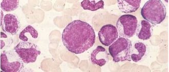
Rh factor (Rh) is a protein found primarily in human red blood cells and in much smaller quantities in white blood cells and platelets. There are several types of Rh factor. So antigen-D is present in the blood of 85% of people. Antigen C (Rh') is found in the blood of 70% of people, and antigen E (Rh") is present in 30% of people. Blood that lacks the listed antigens, which are types of the Rh factor, is Rh negative.
Most common hemolytic disease of the fetus
and the newborn arises due to an immunological conflict due to the incompatibility of the blood of mother and fetus for factor D. In this case, the maternal blood must be Rh-negative. And the fetal blood contains the Rh factor. Hemolytic disease of the fetus and newborn can also occur when the blood of the mother and fetus is incompatible according to the blood group (according to the ABO system). The grouping of human blood is determined by the presence of certain antigens in it. Thus, the presence of antigen “A” in the blood determines blood group II (A). The presence of antigen “B” - III (B) blood group. The combined presence of antigens “A” and “B” determines blood group IV (AB). In the absence of both antigens, a person’s blood group is determined to be I (0). Immunological incompatibility most often manifests itself if the mother has blood group I (0), and the fetus has blood group II (A) or III (B). Fetal blood cells containing the Rh factor or group antigens and having the corresponding antigenic activity penetrate into the maternal bloodstream. As a result, the woman is immunized, which is accompanied by the production in her body of either anti-Rh antibodies or antibodies against group antigens.
Hemolytic disease of the fetus and newborn develops as a result of immunization of the maternal body
, which occurs either already during the first pregnancy with a fetus with Rh-positive blood or with the presence of corresponding group antigens in its blood, or after a transfusion of Rh-positive blood. Most often, elements of the fetal blood containing the corresponding antigens enter the maternal bloodstream during childbirth, especially during surgical interventions, when a cesarean section or manual separation of the placenta and discharge of the placenta are performed. The entry of fetal blood elements into the maternal bloodstream and subsequent immunization of the woman is also possible during pregnancy if the integrity of the placental villi is disrupted, which happens with toxicosis of pregnant women, threatened miscarriage, placenta previa, placental abruption, intrauterine fetal death.
Taking into account that the Rh factor is formed from the very first days of pregnancy, and red blood cells are formed in the first weeks of pregnancy, their entry into the maternal body and immunization are possible during abortion or ectopic pregnancy. After the first pregnancy, about 10% of women are immunized.
Immune antibodies
, which are produced in the woman’s body, penetrate back into the bloodstream of the fetus and affect its red blood cells. In this case, red blood cells are destroyed, which entails the formation of indirect toxic bilirubin, anemia and oxygen starvation (hypoxia). The fetus develops hemolytic disease. The structure and function of the fetal liver is disrupted, protein production in the fetal body is reduced, and blood circulation in the fetal body is disrupted with symptoms of heart failure. In the fetus, excess fluid accumulates in the body, which manifests itself in the form of edema and ascites. Brain tissue is often affected. The development of hemolytic disease of the fetus is possible as early as 22-23 weeks of pregnancy.
Diagnosis of hemolytic disease
Diagnosis of hemolytic disease must be comprehensive, using a number of diagnostic techniques, and is based on identifying signs indicating maternal immunization, determining antibodies and their titer, assessing the condition of the fetus and amniotic fluid indicators. The results of just one test cannot serve as an objective and reliable indicator of a possible complication and require supplementation with the results of other research methods.
If in a conversation with a woman with Rh-negative blood
or with I (0) blood group, it turns out that she was given donor blood without taking into account the Rh or group affiliation, and also if she had spontaneous abortions in the late stages or there was fetal death in previous pregnancies, or the birth of a child with icteric anemic or edematous form, then these factors are prognostically unfavorable from the point of view of a high risk of fetal damage during this pregnancy.
One of the simple ways to determine the nature of the course of pregnancy, the rate of growth and development of the fetus and the increase in the volume of amniotic fluid is to measure the height of the uterine fundus (UF) above the pubic symphysis and measure the length of the abdominal circumference. It is advisable to carry out these measurements over time every two weeks from the second trimester of pregnancy. An excessive increase in these indicators in comparison with the normative ones for a given stage of pregnancy, together with the results of other studies, allows us to suspect the presence of an existing pathology.
Determination of antibodies in the blood of a pregnant woman
has a relative diagnostic value and should be used to identify this pathology only in combination with other diagnostic tests. An important sign is the magnitude of the antibody titer and its change as pregnancy progresses. The antibody titer corresponds to the highest serum dilution at which it is still able to act (agglutinate) Rh-positive red blood cells (antibody titer can be 1:2, 1:4, 1:8, 1:16, etc.). Accordingly, the higher the titer, the more antibodies and the less favorable the prognosis.
During pregnancy, the antibody titer may increase, remain unchanged, decrease, and there may also be an alternating rise and fall in the antibody titer. In the latter case, the likelihood of fetal disease is greatest.
Assessment of the condition of the fetus and feto-placental complex
To assess the condition of the fetus and feto-placental complex, instrumental diagnostic methods such as cardiotocography (CTG) and ultrasound can be used. According to CTG data, in case of fetal suffering, there are clear signs of fetal heart rhythm disturbances, slowing of heartbeats, monotonous rhythm, etc. An ultrasound examination evaluates the size of the fetus, the condition of its internal organs, the structure and thickness of the placenta, and the volume of amniotic fluid. The functional activity of the fetus is also determined and, using Doppler sonography, the intensity of blood flow in the mother-placenta-fetus system is studied.
Ultrasonography
in women with expected hemolytic disease of the fetus should be carried out at 20-22 weeks, 24-26 weeks, 30-32 weeks, 34-36 weeks. and shortly before delivery. Until 20 weeks of pregnancy, signs of hemolytic disease are not detected echographically. Repeated studies are necessary to exclude dynamic hemolytic disease of the fetus. For each pregnant woman, the timing of repeated scans is determined individually; if necessary, the interval between studies is reduced to 1-2 weeks.
Whereas in hemolytic disease of the fetus, swelling of the placenta occurs
, then its thickening by 0.5 cm or more may indicate a possible disease of the fetus. However, other complications should be excluded, in which thickening of the placenta can also occur (for example, intrauterine infection or diabetes mellitus).
For hemolytic disease of the fetus, along with thickening of the placenta, an increase in the size of its abdomen is possible in comparison with the chest and head of the fetus. This increase is due to the excessive size of the fetal liver, as well as excessive accumulation of fluid in its abdominal cavity (ascites). Swelling of the fetal tissues manifests itself in the form of a double contour of its head. With severe fetal hydrops, there is also an accumulation of fluid in other cavities (hydrothorax, hydropericardium), and signs of swelling of the intestinal walls.
Ultrasound scanning also evaluates the behavioral reactions of the fetus (motor activity, respiratory movements, fetal tone). If its condition is disturbed, corresponding pathological reactions from these indicators are noted. According to Doppler ultrasound, there is a decrease in the intensity of blood flow in the feto-placental complex.
Determination of the optical density of bilirubin in amniotic fluid is of great diagnostic importance.
, fetal blood type, antibody titer. On the eve of delivery, it is advisable to examine amniotic fluid to assess the degree of maturity of the fetal lungs.
To study amniotic fluid, starting from 34-36 weeks of pregnancy, amniocentesis is performed, which is a puncture of the uterine cavity to take a sample of amniotic fluid. The procedure is carried out under ultrasound control with anesthesia and in compliance with all rules of asepsis and antisepsis.
Amniocentesis
can be performed either through the anterior abdominal wall or through the cervical canal. The puncture site is chosen depending on the location of the placenta and the position of the fetus. Possible complications in this case may be: increased uterine tone, placental abruption, bleeding, rupture of amniotic fluid and the onset of labor, development of inflammation.
Based on the optical density of bilirubin in amniotic fluid, if it is more than 0.1, the possible severity of fetal hemolytic disease is assessed. If this indicator is more than 0.2, one can expect the birth of a child with a moderate to severe form of hemolytic disease.
Detection of fetal blood type
amniotic fluid test is one of the tests that determines the prognosis for the fetus. More often, one can expect the development of hemolytic disease due to incompatibility of the Rh factor, with the same blood group of the mother and fetus.
It is important to determine the blood group if the development of an AB0-conflict pregnancy
. A significant diagnostic test is the determination of antibody titer in amniotic fluid.
To study fetal blood, puncture of the umbilical cord vessels is used ( cordocentesis
) under ultrasound control. A study of the fetal blood obtained in this way makes it possible to determine its blood type and Rh status, assess the level of hemoglobin, serum protein and a number of other indicators that reflect the presence and severity of the complication.
As part of pregnancy management, all patients and their husbands should be determined to have their Rh and blood type determined when they first see a doctor, regardless of whether childbirth is imminent or termination of pregnancy is planned. In pregnant women with the first blood group, if their husbands have a different blood group, it is necessary to conduct a test to determine group antibodies. Pregnant women with Rh-negative blood must be examined for the presence of antibodies and their titer once a month until 20 weeks, and then once every 2 weeks. If a pregnant woman with Rh-positive blood has an indication of the birth of a child with a hemolytic disease, the blood should be examined for the presence of group immune antibodies, as well as for “anti-c” antibodies if the husband’s blood contains the “c” antigen. The presence of Rh sensitization in pregnant women is not an indication for termination of pregnancy.
Prevention and treatment of hemolytic disease of the fetus
All pregnant women with Rh-negative blood, even in the absence of antibodies in their blood, as well as in the presence of AB0 sensitization, should undergo 3 courses of nonspecific desensitizing therapy
lasting 10-12 days, in periods of 10-22, 22-24, 32-34 weeks.
As part of nonspecific desensitizing therapy, the following is prescribed: intravenous administration of 20 ml of 40% glucose solution with 2 ml of 5% ascorbic acid solution, 100 mg of cocarboxylase; orally rutin 0.02 g 3 times a day, theonicol 0.15 g 3 times a day or methionine 0.25 g and calcium gluconate 0.5 - 3 times, iron supplements, vitamin E 1 capsule. Antihistamines are used at night: diphenhydramine 0.05 or suprastin 0.025.
In case of complicated pregnancy: threat of miscarriage, early toxicosis of pregnant women, gestosis, etc., patients should be hospitalized in the department of pathology of pregnant women, where, along with treatment of the underlying disease, a course of desensitizing therapy is carried out.
Pregnant women who have a high titer of Rh antibodies
, and they have a history of spontaneous miscarriages or delivery of a fetus with an edematous or severe form of hemolytic disease, it is recommended to use
plasmapheresis
. This procedure removes antibodies from the blood plasma. Plasmapheresis is carried out once a week under the control of antibody titer, starting from 23-24 weeks. pregnancy, before delivery.
Hemosorption is also used to treat hemolytic disease of the fetus.
using activated carbon to remove antibodies from the blood and reduce the degree of Rh sensitization. Hemosorption is used in pregnant women with an extremely complicated obstetric history (repeated miscarriages, stillbirth). Hemosorption begins at 20-24 weeks in the hospital, with an interval of 2 weeks.
Treatment of hemolytic disease of the fetus during pregnancy is also possible by blood transfusion to the fetus under ultrasound guidance
.
Pregnant women with Rh sensitization
hospitalized in the maternity hospital at 34-36 weeks. pregnancy. With AB0 - sensitization, hospitalization is carried out at 36-37 weeks. If there are appropriate indications indicating a pronounced nature of the complication, hospitalization is possible at an earlier date.
In the presence of hemolytic disease of the fetus, early delivery is advisable due to the fact that towards the end of pregnancy the flow of antibodies to the fetus increases. In case of severe hemolytic disease of the fetus, pregnancy is terminated at any time during pregnancy. However, in most cases, pregnancy can be prolonged until an acceptable delivery date. As a rule, delivery is carried out through the natural birth canal. Caesarean section is performed in the presence of additional obstetric complications.
During childbirth, careful monitoring of the condition of the fetus is carried out, hypoxia is prevented
. Immediately after birth, the baby is quickly separated from the mother. Blood is taken from the umbilical cord to determine the content of bilirubin, hemoglobin, the child’s blood type, and his Rh status. A special test is performed to identify the newborn's red blood cells associated with antibodies.
Treatment of hemolytic disease of the newborn
In hemolytic disease of the newborn, there are three forms: anemic, icteric and edematous.
.
When treating the anemic form of hemolytic disease of newborns with hemoglobin levels less than 100 g/l, transfusions of Rh-positive erythromass corresponding to the blood type of the newborn are performed. To treat the icteric-anemic form of hemolytic disease of newborns, exchange transfusions, phototherapy, and infusion therapy
. In severe cases of hemolytic disease of the newborn, exchange transfusions are performed repeatedly in combination with therapy aimed at reducing intoxication caused by indirect bilirubin.
The action of phototherapy or light therapy is aimed at destroying indirect bilirubin in the superficial layers of the newborn's skin under the influence of a fluorescent or blue lamp (wavelength 460A). When performing infusion therapy, solutions of hemodez, glucose, albumin, and plasma are injected intravenously in various combinations.
Treatment of the edematous form of hemolytic disease of the newborn consists of immediate replacement transfusion of single-group Rh-negative red blood cell mass. At the same time, dehydration therapy is carried out aimed at eliminating edema.
To prevent hemolytic disease of the fetus and newborn, it is necessary that with any blood transfusion it is of the same group and similar in Rhesus status.
First pregnancy and Rhesus conflict
It is important to maintain the first pregnancy in women with Rh-negative blood
.
It is necessary to prevent and treat complicated pregnancy, as well as carry out desensitizing therapy. For primigravid women with Rh-negative blood who are not yet sensitized to the Rh factor, anti-Rh immunoglobulin is administered
. After childbirth, the administration of immunoglobulin is repeated in the first 48-72 hours.
Make an appointment with specialists by calling a single call center:
+7(495)636-29-46 (metro stations “Schukinskaya” and “Street 1905 Goda”). You can also make an appointment with a doctor on our website, we will call you back!
When can immunoglobulin prophylaxis be carried out?
To prevent conflict in subsequent pregnancies, women with a negative Rh factor should undergo prophylaxis. This is done after:
- childbirth (within three days);
- abortion;
- analysis of amniotic fluid;
- spontaneous miscarriage;
- ectopic pregnancy;
- placental abruption;
- transfusion.
Remember: if you and your baby’s group and Rh are different, this is not an indication that there will definitely be problems. Group and Rh are just the presence or absence of specific proteins in the blood. The reaction of the body and the development of pathologies in our time can be successfully controlled with the help of medications. Your attentiveness to your body, as well as an experienced doctor, will help you bear a healthy baby.
The likelihood of an immune reaction in a pregnant woman with Rh-
If a woman is pregnant for the first time, then the risk of her developing antibodies tends to zero: the likelihood of contact between the blood of the fetus and the mother is too small. As a rule, contact occurs during the birth of the baby, after which the immune reaction starts. With each subsequent pregnancy, the immune reaction increases and the risk of developing Rh sensitization increases.
In addition, the likelihood of sensitization increases:
- induced and spontaneous abortions;
- ectopic pregnancy;
- premature placental abruption;
- amniocentesis (examination of amniotic fluid);
- chorionic villus biopsy (examination of the tissue of the outer membrane of the embryo);
- cordocentesis (examination of fetal cord blood);
- some obstetric manipulations. For example, external obstetric rotation (turning over the fetus, which is in a position unfavorable for childbirth);
- increased permeability of the blood-placental barrier;
- abdominal organ injuries.
How do your chances of conceiving depend on your blood type?
Quite a lot is already known about the influence of blood groups, for example, on the likelihood of developing Alzheimer's disease, cancer, blood clots, etc. However, virtually nothing was known about the effect on fertility. And finally, thanks to the efforts of Turkish doctors, research has appeared in this area.
A study published last week found that men with type O are four times less likely to develop impotence compared to guys with other blood types. Experts from Ordu University in Turkey noted that blood type is as important a risk factor as smoking, excess weight, and high blood pressure. The reason is not clear, but scientists have said that in people with type A blood, the penis has a large number of veins, the lining of which can become damaged, leading to erectile dysfunction.
Blood type also affects female fertility. Girls with the second group are more likely to bear a healthy child for a long time than with the first. Studies have shown that women in the first group quickly deplete their egg reserves early in life. But at the same time, women with type 0 have a lower risk of developing preeclampsia - high blood pressure during pregnancy, which can be dangerous for mother and baby.
Naturally, representatives of the rest of humanity (which, by the way, are a little more than half, because people of the 1st group account for a little more than 40%) should not panic either - a higher probability does not mean a 100% chance. Likewise, representatives of the “happy” group should not relax ahead of time - reduced risk does not mean zero.
Rh factor, blood group, blood protein, antibody production, anemia, immune reaction, Rh compatibility, anemia, hydrops, fetal edema, immunoglobulin
Forms of hemolytic pathology:
| Forms | Description |
| Anemic | When a baby has a low level of hemoglobin and an increased level of unconjugated bilirubin. The condition does not entail any serious consequences and is easily normalized. |
| Jaundice | Occurs due to severe bilirubin intoxication. |
| Edema | The most dangerous form of HDN, leading to the death of the fetus in the womb. If the child manages to survive, then a number of pathologies are detected, including damage to the brain and central nervous system. |
The state of red blood cell hemolysis is characterized by the destruction of the red blood cell membrane and the release of hemoglobin into the plasma fluid. Released hemoglobin in large quantities has a poisonous effect on the body.
Therefore, simultaneously with poisoning and overload of the fetal body with bilirubin and other substances, such a pathology also entails anemia.
The liver does not have time to neutralize free bilirubin on a large scale. And this substance in a neurotoxic form wanders throughout the body of the embryo, disrupting oxidative processes in organs and tissues. This leads to irreversible, destructive consequences, including the death of the child. For this reason, the danger of blood substance group conflict during pregnancy should not be underestimated.