Classification
The upper limit of normal platelet count varies from 350,000 to 400,000 per microliter depending on the reference intervals of the specific laboratory performing the analysis. According to the degree of increase, the following types of thrombocytosis are distinguished:
- Soft
: from 350-400 to 700 thousand. - Moderate
: from 700 to 900 thousand. - Heavy
: from 900 to 1000 thousand. - Extreme
: over 1,000,000.
The cause of extreme and severe thrombocytosis is oncohematological pathologies. According to the origin of thrombocytosis there are:
- Primary
(tumor, clonal). They account for approximately 10-15% of all cases of thrombocytosis. The cause is tumor diseases of the blood system. - Secondary
(reactive). The most common variety (about 85%). The cause is infectious, systemic inflammatory processes, and anemia. - False
(pseudothrombocytosis). The reason is an error in the hematology analyzer, which mistakes tumor cell fragments during treatment with chemotherapy drugs, small red blood cells, or red blood cells that have undergone hemolysis as platelets. Pseudothrombocytosis is also observed with cryoglobulinemia. - Hereditary
(family). This is a rare genetic disease, the cause of which lies in a mutation of the genes encoding the synthesis of thrombopoietin and its receptors (THPO, MPL).
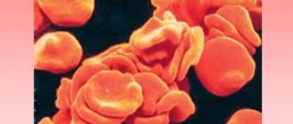
Blood test for platelets
A complete blood test can determine the platelet count. The main indications for its implementation are the following:
- Increased bleeding of gums.
- Heavy menstruation.
- The appearance of bruises from minor impacts.
- Frequent nosebleeds.
- Difficulty stopping bleeding from minor injuries.
The number of platelets in the blood is measured in thousands per 1 microliter of blood. Counting is carried out in specialized laboratories in various ways that guarantee high accuracy.
The normal platelet count in the blood depends on gender and age and is:
- For men, 200–400 thousand.
- In women, 180–320 thousand, during menstruation the amount can decrease to 75–220 thousand, and during pregnancy to 100–310 thousand.
- In children, indicators depend on age, and the corresponding values are given in special tables.
To conduct a general blood test, blood is taken from a finger. No special preliminary preparation is required before this. To ensure accurate results, it is better to donate blood in the morning on an empty stomach. At the same time, 12 hours before the procedure it is not recommended to consume fatty spicy foods, carbonated drinks, and alcohol.
Additionally, to determine blood clotting indicators, Sukharev and Lee-White tests are performed. They are informative and allow you to obtain the necessary additional data about the pathological condition. This will allow you to carry out correct treatment measures and avoid dangerous consequences.
Causes of thrombocytosis
Physiological conditions
An elevated platelet level does not always indicate pathology. There is physiological (short-term, transient) thrombocytosis, caused by various circumstances, for example, stress, intense physical activity. The reason is the mobilization of blood platelets, or rather their transition from the marginal position to the central blood flow in the vessels of the spleen and lungs.
In addition, minor physiological thrombocytosis is observed in children from the neonatal period to 11 years. There is also a so-called hemoconcentration thrombocytosis, which is caused by dehydration. This phenomenon is due to a decrease in the volume of the liquid part of the blood (plasma) and a relative increase in formed elements (platelets, erythrocytes, leukocytes). In this situation, it is necessary to focus on the hematocrit - with dehydration it is increased.
Infections
This is the most common cause of thrombocytosis (about 40%). An increase in the level of blood platelets develops when:
- Bacterial infections
. In adults, thrombocytosis occurs mainly with local (pneumonia, pyelonephritis, meningitis, endocarditis) and systemic (sepsis, tuberculosis) bacterial infections. - Viral and fungal infections
. Thrombocytosis occurs less frequently during infection with viruses (hepatitis B, C) and pathogenic fungi (aspergillosis). - Helminthiasis.
In children, parasitic infestations (toxocariasis, ascariasis) are recognized as a common cause of an increase in the number of blood platelets.
There are two pathogenetic mechanisms for the development of thrombocytosis in response to an infectious disease. First, during the fight against pathogens, leukocytes produce a large number of inflammatory mediators, including interleukin-6, which stimulates bone marrow megacaricytopoiesis (the formation of platelet precursors). Secondly, platelets themselves are part of the anti-infective immunity - they are able to produce bactericidal substances, capture, neutralize, and even phagocytose certain types of bacteria, viruses, and foreign particles.
Platelets facilitate the migration of leukocytes to the site of infectious inflammation by interacting with endothelial cells of the vascular wall. Thrombocytosis during infections occurs abruptly, correlates with the severity of the disease, and quickly resolves after the pathogen is eliminated from the body and the inflammatory process subsides. Thrombocytosis is usually mild or moderate; in septic conditions it can reach a severe degree; in children it is somewhat more pronounced than in adults.
Autoimmune diseases
Another common cause of thrombocytosis is considered to be chronic rheumatological pathologies that occur with autoimmune inflammation. The mechanism for increasing the content of blood platelets is the overproduction of substances such as interleukin-6, colony-stimulating factors, which activate bone marrow platelet formation. The degree of thrombocytosis corresponds to the activity of inflammation (minimal during remission, maximum during relapse).
In diseases such as rheumatoid arthritis, systemic lupus erythematosus (SLE), inflammatory bowel diseases (Crohn's disease, ulcerative colitis), a mild or moderate degree is observed. In systemic vasculitis with necrotizing destruction of vessel walls, severe thrombocytosis occurs, since in vasculitis, in addition to other inflammatory mediators, tumor necrosis factor is synthesized in large quantities, which also has a stimulating effect on platelet formation. High platelet counts are especially common in children.
Anemia
Iron deficiency anemia is often the cause of thrombocytosis, especially in children. The exact mechanisms of this phenomenon have not yet been clarified, but an inverse relationship has been clearly established between reduced levels of iron metabolism (ferritin, iron-binding capacity of serum) and an increased level of blood platelets. Iron is thought to have an inhibitory effect on the maturation of megakaryocytes (precursor cells).
In addition, some pluripotent stem cells in the bone marrow are unable to transform into red blood cells under conditions of iron deficiency. As a result, a kind of “bypass” occurs and a larger percentage of stem cells begin to mature along the megakaryocyte pathway. Thrombocytosis in iron deficiency anemia is mild, sometimes moderate. Platelets quickly return to normal after correction of iron deficiency and an increase in hemoglobin.
However, as anemia worsens, platelet levels drop to a state of thrombocytopenia. The cause of iron deficiency can be a lack of iron in food, increased iron consumption (growth period in children, pregnancy, lactation) or chronic blood loss (long menstruation, bleeding from the gastrointestinal tract due to gastric ulcer).
Malignant blood diseases
The cause of approximately 15% of all thrombocytosis is hemoblastosis - chronic myeloid leukemia, Ph-negative myeloproliferative pathologies (essential thrombocythemia, polycythemia vera, and primary myelofibrosis). The increase in the number of blood platelets in these diseases is due to clonal (tumor) transformation of the megakaryocyte lineage of the bone marrow due to various mutations, which leads to hyperproduction of platelets.
These diseases are more common in adults and older people, in children - only in exceptional cases. Initially, thrombocytosis is moderate, as it progresses it increases, reaching a severe or extreme degree, which is why microcirculation disorders, arterial and venous thrombosis of various localizations often occur. The concentration of blood platelets normalizes very slowly, only after courses of specific myelosuppressive treatment.
Splenectomy
The spleen, being an organ that deposits blood, retains a large number of formed elements, including platelets. The spleen is also directly involved in thrombocytopoiesis, secreting the hormones thrombocytopenin and splenin, which suppress bone marrow maturation of megakaryocytes. Therefore, thrombocytosis after splenectomy is caused by two mechanisms: the release of platelets, normally located in the splenic depot, into the circulating blood, and the phenomenon of “disinhibition of the bone marrow,” i.e. increased platelet production.
An increase in the number of blood platelets does not occur immediately, but approximately a week after splenectomy, reaches a maximum by the 13-14th day (up to 700-800 thousand), often becoming the cause of venous thrombosis of the portal vein, and then slowly returns to normal over several weeks or months .
Injuries and surgeries
Massive tissue damage (wound during abdominal surgery, fracture, extensive burns) causes activation of the blood coagulation system, namely the vascular-platelet unit, which is the first stage of hemostasis. It involves vasospasm, as well as adhesion and aggregation of platelets at the site of damage to the vascular wall. The consumption of platelets stimulates their active release from the depot and a compensatory increase in their bone marrow production. The extent of damage correlates with the degree of thrombocytosis. This type of thrombocytosis usually does not require treatment.
Oncological diseases
The cause of thrombocytosis in solid (non-hematopoietic) tumors is the ability of cancer cells to produce interleukin-6, which stimulates thrombocytopoiesis. This feature was found in small cell lung cancer, colon adenocarcinoma, and malignant mesothelioma. In addition, tumor disintegration often causes bleeding, leading to iron deficiency anemia. The degree of thrombocytosis is usually moderate, in children it can be severe, and regresses after long-term treatment with chemotherapy.
Rare causes
- Functional asplenia
: sickle cell anemia, chronic alcoholism, celiac disease. - Use of medications
: vincristine, adrenaline. - Rebound phenomenon
: development of thrombocytosis 1-2 weeks after treatment of thrombocytopenia or discontinuation of medications that cause thrombocytopenia (methotrexate, vitamin B12, prednisolone).
Summarizing
Our study made it possible to describe a new mechanism of cell redistribution within the thrombus: during contraction, “slippery” procoagulant platelets are mechanically squeezed onto the surface of the thrombus, forming a heterogeneous structure of its outer part.
But the point has not yet been made in determining the role of procoagulant platelets in hemostasis. The formation of a weakly adhesive, that is, unsuitable for the adhesion of new cells, layer of dying cells and fibrin on the surface of the thrombus can help stop its growth by reducing the efficiency of fixation of non-activated platelets brought by the blood stream. However, this hypothesis requires further research.
It is pleasant to note that an important contribution to this work was made by young co-authors - students of the Department of Biophysics of the Faculty of Physics of Moscow State University - Roman Kerimov and Alexandra Yakusheva, as well as a student of the Faculty of Fundamental Medicine of Moscow State University Taisya Shepelyuk (Fig. 4). The results of the work were published in one of the leading journals of the American Cardiovascular Association and reported at several international conferences, including the Gordon Conference on Hemostasis.
Figure 4. Roman Kerimov, Alexandra Yakusheva, Taisya Shepelyuk and Dmitry Nechipurenko
This version is a modification of a note that was published in the physics department newspaper “Soviet Physicist”.
The author expresses gratitude to Anastasia Masaltseva and Yuri Nechiporenko for their assistance in editing the article, as well as to all his colleagues - co-authors of the original work.
Diagnostics
Thrombocytosis is detected in a clinical blood test. Although very high platelet counts are more common in hematologic diseases, platelet levels alone cannot determine the cause of thrombocytosis. Therefore, if it is detected, you should visit a therapist. The doctor carefully asks about the patient’s complaints, how long ago the symptoms occurred, and conducts a general examination of the patient. Then, based on the data obtained, an additional examination is prescribed, including:
- Blood tests
. In a general blood test, the content of other formed elements (erythrocytes, leukocytes) is determined, and the leukocyte formula is calculated. The concentration of inflammatory markers (ESR, CRP) is measured. The indicators of serum iron, TBC, ferritin are assessed. The presence of autoantibodies (RF, ACCP, antibodies to the cytoplasm of neutrophils) is checked. In case of endocarditis and sepsis, an analysis for procalcitonin and presepsin is performed. - Pathogen identification
. To identify the pathogen, microscopy, bacterial culture of urine and sputum are performed. If tuberculosis is suspected, an intradermal test with tuberculin is prescribed. Using an enzyme immunoassay, antibodies to viruses, parasites, and fungi are detected, and using the polymerase chain reaction method, their DNA and RNA are detected. To diagnose meningitis, a cerebrospinal fluid analysis is informative. - Genetic research
. In patients with myeloproliferative pathologies, mutations of Janus kinase (JAK2V617F), thrombopoietin receptors (MPL), and erythropoietin are determined using fluorescent hybridization (FISH) and PCR. Sometimes chromosomal abnormalities are detected - trisomies, deletions. In chronic myeloid leukemia, cytogenetic analysis reveals the Philadelphia chromosome (Ph). - X-ray
. On an X-ray of the lungs, in case of pneumonia, foci of darkening and infiltrates are noted, in case of tuberculosis - enlargement of the mediastinal lymph nodes, expansion of the roots of the lungs, rounded shadows (cavities) of the upper lobes of the lungs. In patients with arthritis, x-rays of the joints show a narrowing of the joint space, areas of erosion, and marginal osteoporosis. - Ultrasound
. Ultrasound of the abdominal organs in case of pyelonephritis determines compaction and expansion of the pyelocaliceal system, and in case of blood diseases - splenomegaly. In bacterial endocarditis, cardiac echocardiography reveals vegetations of the valves and sometimes effusion into the pericardial cavity. - Endoscopy
. In patients with inflammatory bowel pathologies, fibrocolonoscopy is performed, which reveals hyperemia of the mucous membrane, lack of vascular pattern, erosion, and ulcerative defects. Crohn's disease is characterized by the "cobblestone pavement" symptom - alternating deep ulcers with unchanged mucous membrane. - Histological studies
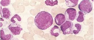
. In bone marrow aspirate for malignant hematological pathologies, hyperplasia of the megakaryocyte lineage of hematopoiesis is noted (in polycythemia vera - all three lineages), a large number of blast cells (in myeloid leukemia), proliferation of reticulin and collagen fibers (fibrosis). In case of vasculitis, a biopsy of a vessel reveals pronounced perivascular infiltration with lymphocytes and plasma cells.
Platelet count according to Fonio
Treatment of thrombocytopenia
Thrombocytopenia is treated by a hematologist. The treatment regimen is developed individually for each patient: the patient’s age, the presence of any diseases, the degree of deficiency, and the amount of bleeding are taken into account. It is necessary to determine the cause of thrombocytopenia so that treatment is as effective as possible. Standard drug therapy consists of:
- Corticosteroids – have anti-inflammatory and analgesic effects;
- Immunoglobulins – correct the functioning of the immune system;
- Immunosuppressants – stop the destructive effect of the immune system on the body;
- Agonists to thrombopoietin receptors – promote more active production of this hormone.
In case of thrombocytopenia caused by bleeding, it is necessary to identify the source of this bleeding and stop it. Only after this the platelet correction is carried out. The patient will have to take medicinal iron and drugs that promote the production of red blood cells.
If drug treatment does not produce any results, splenectomy is indicated - surgical excision of the spleen. For hereditary diseases that prevent the body from producing enough platelets, a bone marrow transplant is necessary. If the platelet level is critically low, a blood transfusion of platelet mass from a donor is performed. If a large volume of blood is lost, a transfusion of fresh frozen plasma and red blood cells is necessary.
Correction
Conservative therapy
In most cases, to correct thrombocytosis, it is enough to eradicate the cause, i.e. treatment of the underlying disease. Short-term thrombocytosis that develops due to stress or drug administration does not require intervention. In case of persistent long-term thrombocytosis, consultation with a hematologist is necessary to identify the cause and prescribe appropriate treatment. Therapy for thrombocytosis has several areas, including:
- Fighting infection
. To eliminate the infectious agent, antibacterial (amoxicillin), antifungal (fluconazole), and antiparasitic agents (mebendazole) are used. Treatment of viral hepatitis requires long-term use of peligated interferon in combination with antiviral drugs. - Treatment of iron deficiency anemia
. Correction of iron deficiency is carried out with tablet preparations (iron sulfate). For children, there are forms of syrup and drops for oral administration. The addition of ascorbic acid promotes better absorption. - Therapy of autoimmune diseases
. Treatment of autoimmune diseases is carried out using medications that suppress inflammation - glucocorticosteroids (prednisolone), immunosuppressants (cyclophosphamide). - Targeted therapy
. For myeloproliferative diseases, specific targeted treatment is prescribed to slow down the progressive growth of the malignant tumor. These drugs include Janus kinase inhibitors (ruxolitinib), tyrosine kinase inhibitors (imatinib, dasatinib). - Symptomatic treatment
. To relieve high thrombocytosis, medications are used that suppress the activity of the megakaryocyte lineage, and, consequently, the production of platelets - anagrelide, interferon-alpha, hydroxyurea. For polycythemia, regular bloodletting is successfully used as a treatment method to remove excess formed elements. - Blood thinning
. In case of high thrombocytosis, antiplatelet agents (acetylsalicylic acid) are prescribed to prevent thrombosis. In case of contraindications (peptic ulcer of the stomach, duodenum), platelet receptor blockers (clopidogrel, ticagrelor) are used. In people at high risk of thrombosis (elderly, patients with diabetes mellitus or atrial fibrillation), anticoagulants (warfarin, dabigatran) are used.
Specialized treatment
The only method that allows achieving complete recovery from a malignant hematological disease is allogeneic bone marrow transplantation. This requires HLA typing to select a compatible donor. However, due to the high risk of developing life-threatening complications, this method is used only if conservative treatment is ineffective.
Prognosis and prevention
Thrombocytopenia is a dangerous disease that poses a huge threat to the life and health of the patient. Anemia often develops, and vision loss can occur due to hemorrhages in the retina. Advanced forms can provoke hemorrhages in the internal organs and in the brain. In most cases this leads to death.
Thrombocytopenia has no specific prevention. Experts give the following recommendations:
- Take a general blood test every year and undergo a general medical examination;
- Eat properly and nutritiously: eat as much meat and fresh vegetables as possible;
- Play sports and lead an active lifestyle;
- Avoid self-medication: taking aspirin and steroid drugs should be under the strict supervision of a doctor;
- Try to avoid any procedures where there is a risk of cutting (shaving, manicure);
- Treat any infectious processes in a timely manner;
- Take precautions when working with chemicals;
- Give up bad habits: drinking alcohol slows down platelet production.
Thrombocytopenia is a disease that requires constant medical supervision. If you suspect you have it or know about the pathology and want to get rid of it, contact the Medscan medical center. Experienced doctors will perform the necessary diagnostics and develop an individual treatment plan.
Forecast
The outcome depends on both the underlying pathology and the degree of thrombocytosis. For example, acute viral infection and iron deficiency anemia are characterized by a benign course. Patients with essential thrombocythemia, with proper selection of pathogenetic and symptomatic treatment, can live more than 80 years. People with chronic myeloid leukemia, on the other hand, live about 5-10 years from diagnosis.
Since reactive thrombocytosis almost always occurs in children, their prognosis is favorable. Thrombosis is not typical for mild to moderate thrombocytosis. In extreme or severe cases, there is a very high probability of fatal thrombosis leading to myocardial infarction, pulmonary infarction, and ischemic stroke.
What does this mean during pregnancy?
Among the physiological causes of low platelets in women, pregnancy is named, and for the early stages this is indeed a normal situation, if we are talking about acceptable low values. In the early stages, pregnant women experience an increase in the volume of circulating blood due to plasma, while the mass of red blood cells and platelets is produced at a less rapid pace. As a result, the effect of hemodilution is observed - blood thinning, accompanied by a reduced content of red blood cells and platelets.
Typically, this transient phenomenon does not have any significant impact on hemostasis. If platelets are reduced to a level of 50 units per microliter or lower, emergency measures may be required to stabilize the indicators. Especially if there are few platelets a couple of weeks before giving birth, this means that the body is not preparing for the upcoming blood loss and can suffer greatly during childbirth.
- Thrombocytopenia - symptoms and treatment
This is when the value of a general blood test becomes obvious - thanks to it, it is possible to promptly detect a low platelet count, suggest the reasons for the deviation and take action.
The range of conservative methods for increasing low platelets during pregnancy is limited, but the expectant mother will not be left without help, since the arsenal of medicine includes drugs with minimal effects on the fetus.
MPV reduced
If the average platelet volume is low, this may indicate the following pathological conditions:
- enlarged spleen (splenomegaly);
- cirrhosis of the liver;
- anemia;
- some hereditary diseases lead to a decrease in MPV (for example, this indicator is low in Wiskott-Aldrich syndrome).
- oncological diseases: sarcoma, leukemia, lymphoma, lymphogranulomatosis, carcinoma;
- inflammatory diseases;
- autoimmune diseases (rheumatoid arthritis, hemorrhagic vasculitis and others);
- myocardial infarction;
- uremia, renal amyloidosis, glomerulonephritis;
- hypothyroidism, hyperthyroidism;
- •hypoproteinemia;
- taking certain medications.
Low MPV may be found during pregnancy. If the platelet level is also low, then there is a risk of miscarriage and premature birth.