General information
Edwards syndrome (also called trisomy 18 syndrome) is a chromosomal disease characterized by multiple malformations and trisomy 18 chromosomes. This disease was first described by John Edwards in 1960. This disease is rarely diagnosed - as statistics show, in the world it occurs with a frequency of 1:5000. It has been noted that children with this pathology are more often born to mothers of mature age. Thus, the probability of giving birth to a baby with this pathology for a woman over 45 years old is 0.7%. The risk increases if the expectant mother has diabetes .
Moreover, girls with this pathology are born three times more often than boys. Edwards disease is a very severe pathology, in which survival after a year is 5–10%. How this disease is diagnosed and what causes its development will be discussed in this article.
Normal indicator for pregnant women in the first trimester
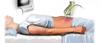
To identify genetic disorders, pregnant women must systematically undergo ultrasound diagnostics. The study allows you to determine the presence of anomalies in the early stages.
During the examination, phonometric indicators are assessed. In this case, the main criterion is to determine the thickness of the collar space (TN). Its norms are 0.7-0.8 mm.
The structure of the facial structures and skull is also studied, parameters and the presence of the nasal bone are determined (can be detected by the 10th week of pregnancy). Closer to the 12th week, its size ranges from 2 to 3 mm.
Additionally, the fetal heart rate is measured and the blood flow in the venous duct is assessed, since disturbances in it may indicate the development of a pathology such as Down syndrome.
The determining values are the parameters of the coccygeal-parietal and biparietal region:
| Index | Gestational age | Norm |
| Coccyx-parietal size | 11 week | 34-42 mm |
| 12 week | 42-51 mm | |
| Week 13 | 51-63 mm | |
| Nasal bone | 11 week | Considered but not measured |
| 12 week | 2-3.1 mm | |
| Week 13 | 2-3.1 mm | |
| Heart rate (HR)/min. | 11 week | 153-165 |
| 12 week | 150-162 | |
| Week 13 | 147-159 | |
| BDP (biparietal size) | 11 week | 13-17 mm |
| 12 week | 18-21 mm | |
| Week 13 | 20-24 mm | |
| LZR (fronto-occipital size) | 11 week | 19-21 mm |
| 12 week | 22-24 mm | |
| Week 13 | 26-29 mm | |
| hCG (ng/ml) | 11 week | 17,4-130,4 |
| 12 week | 13,4-128,5 | |
| Week 13 | 14,2-114,7 | |
| PAPP-A (mU/l) | 11 week | 0,46-3,73 |
| 12 week | 0,79-4,76 | |
| Week 13 | 1,03-6,01 |
Pathogenesis
Trisomy 18 is a genetic pathology. Each human cell contains 46 chromosomes - these are normal values. During fertilization, 23 chromosomes of female and male germ cells combine and give a total of exactly this number of chromosomes. Sometimes, for unknown reasons, genetic mutations occur, and as a result, an extra 47th chromosome appears, additional to the 18th pair. An extra chromosome appears in gametes as a result of chromosome nondisjunction during meiotic division. In most cases - 95% - it is the extra chromosome that causes the development of Edwards disease. But sometimes - in about 2% of cases - the sum of chromosomes remains normal, but chromosome 18 is abnormally lengthened, which leads to the development of congenital pathology. In another 3% of cases, mosaic trisomy is observed, in which an additional chromosome is not present in all cells of the body, but only in some part of it. All three forms of the syndrome follow the same type. But still, a more severe course is observed in the first form of the disease.
Cytological diagnosis
Fluorescent hybridization is used to detect submicroscopic chromosomal deletions and duplications that are too small to be identified by conventional cytogenetics. It is also used to identify “soft” translocations and marker chromosomes. RI8N analysis is performed on a preparation of metaphase chromosomes isolated from cultured lymphocytes, amniocytes, chorionic villi, interphase nuclei from blood, tissues, chorionic villi, amniotic fluid for prenatal detection of fetal developmental anomalies and the need for screening for aneuploidy (trisomy 21, etc. ). RI8N analysis is used for the purpose of preimplantation genetic diagnosis to identify balanced translocations or deletions. The RI8H method provides information on the analysis of a specific chromosome or several chromosomes, but is not performed to determine the karyotype.
Classification
Complete trisomy - if a whole additional chromosome appeared at the stage of one cell, then the set of chromosomes is disrupted in each cell of the fetus. With this form, the child has numerous developmental defects that are incompatible with life. This is the most severe version of the disease.
Partial trisomy is diagnosed if an extra chromosome does not appear at the earliest stages of fetal development. In this case, a violation of the genetic makeup is not observed in all cells, and some of them will remain healthy.
Can a chromosomal mutation be cured?
Chromosomal abnormalities cannot be clinically cured. The vast majority of children die in the first months of life. With proper care and medical assistance, the chances of extending the child's life increase. However, constant monitoring, complex operations and care for such patients are necessary.
The most dangerous condition for a child's life is Down syndrome. With this disease, the chances of living longer than 30-40 years are several times higher than with other chromosomal abnormalities.
At the moment, the issue of defects in the chromosomal series is being studied by specialists, but effective methods for treating pathologies have not yet been developed.
Trisomy in the 18, 21 or 13 chromosome series is a life-threatening disorder for the child, therefore, before conception and during pregnancy, the expectant mother should take into account all the risks of developing pathology and undergo systematic research to identify signs of abnormalities in the early stages.
Causes
Trisomy 18 develops as a consequence of the presence of an additional 18th chromosome in the zygote’s karyotype. The occurrence of this pathology is accidental and cannot be prevented. But some risk factors for the development of this pathology are determined:
- Maternal age – the older the woman, the higher the expected risk of trisomy. The likelihood of developing Edwards disease increases after 35 years of age - every year the frequency of manifestation of such mutations in a child is higher.
- Genetic predisposition - the likelihood increases if there have already been cases of this disease in the family.
- Taking certain medications that affect embryonic cell division.
- Past infectious diseases.
- Smoking and alcohol consumption.
The main causes of chromosomal mutation
At the moment, the exact causes of the development of disorders are being studied. However, experts identify several predisposing factors that increase the chances of having children with chromosomal abnormalities.
Trisomy 18/21/13
Possible reasons:
- age over 30 years (for women);
- age over 40 years (for men);
- hereditary factor (there are relatives in the family with genetic disorders in the chromosomal series);
- problems during a previous pregnancy - frozen pregnancy, miscarriages, abortions;
- infectious diseases that arose during the period of conception of a child or directly in the first stages of pregnancy.
An altered number of chromosomes (aneuplody) is observed in children born to women over 35 years of age. At the same time, the risk of developing such disorders is 1%.
Symptoms
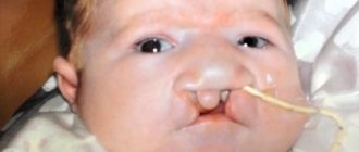
Photos of newborns with Edwards syndrome
Edwards syndrome manifests itself in the fetus during intrauterine development. During this period, signs of such pathology can be detected during ultrasound examination. During such a study, some of them may be discovered. 9 months of pregnancy is a period during which a woman should undergo ultrasound examinations several times. The following symptoms may indicate Edwards syndrome:
- Significant intrauterine growth retardation - it can last several weeks.
- Polyhydramnios.
- Malformations of the brain.
- One umbilical artery in the fetus.
- Very weak fetal movements.
- Long duration of pregnancy - up to 42 weeks. In this case, the child is born with a very low weight.
Signs of Edwards syndrome in newborns are noticeable immediately after the baby is born. The following characteristic symptoms are noted:
- Low body weight.
- Low forehead, wide nape.
- Dolichocephalic skull.
- The ears are underdeveloped and located very low.
- Short upper lip, small mouth, high palate.
- "Hare" lip, "cleft" mouth.
- Pathological changes in the musculoskeletal system - narrow pelvis, dislocated hips, too wide chest, clubfoot .
- Fused toes, they can also be webbed.
- Uneven placement of fingers.
- Distorted face shape.
- Very small head.
A number of symptoms associated with damage to internal organs, as well as disorders of neuropsychic development, are highlighted:
- Scoliosis , muscle atrophy .
- Strabismus.
- Central nervous system disorders: smoothing of brain convolutions, underdeveloped cerebellum and corpus callosum, mental retardation.
- Pathologies of the genitourinary system: segmented or horseshoe-shaped kidney, duplication of the ureters, underdeveloped ovaries, cryptorchidism in boys, clitoral hypertrophy in girls.
- Pathologies of the gastrointestinal tract: atresia of the anus or esophagus, malpositioned intestines, gaseosophageal reflux disease, impaired sucking and swallowing reflex.
- Inguinal and umbilical hernias.
- Congenital heart defects, large vessels.
As a rule, the mental development of children with this disease corresponds to a severe degree of mental retardation . However, with the mosaic form of the syndrome, patients may have certain achievements in mental development.
What varieties are there?
The pathology is classified depending on the affected pair of chromosomes. Each type of disorder has its own name, manifestations, characteristics and other distinctive features.
Trisomy 13, Patau syndrome
This disorder is rare and occurs with equal frequency in both sexes. Moreover, the risks of developing the syndrome increase with increasing age of the parents. The presence of the disease can be determined from the 9th week of pregnancy using ultrasound or blood tests.
Causes
Trisomy 21, 18, 13 can occur as a result of various provoking factors, which are almost impossible to determine exactly.
At the moment, the exact origin of the anomaly is unknown. Most experts are inclined to the version of a random genetic disorder in chromosomal pairs. Pathology develops as a result of the formation of an additional chromosome in the 13th pair. The influence of somatic disorders, infectious diseases, bad habits and other factors has not been established.
Signs
Symptoms of the pathology are diverse and affect many different systems and internal organs.
Signs:
- small body weight – up to 2.5 kg;
- asphyxia (suffocation);
- abnormal brain development;
- defects in the structure of the face and skull;
- disorders of the central nervous system - hydrocephalus, underdevelopment of the cerebellum, cataracts, deafness, damage to the optic nerve;
- congenital heart defects;
- focal absence of hair or skin;
- altered structure of the foot - absence or presence of toes;
- abnormalities in the urinary system;
- malformations of the digestive tract;
- underdevelopment of the musculoskeletal system;
- abnormal structure of the reproductive and reproductive systems.
Children with Patou's syndrome are developmentally delayed (mental and physical delays).
What newborns look like
Pathology can be determined by the appearance of the baby. Children with the disease are characterized by a small head circumference, a low and sloping forehead, and narrow eye openings. The bridge of the nose is flat and falls slightly into the inside of the skull.
At the same time, the development of other facial defects (cleft palate, cleft lip) is also noted. The ears are also subject to deformation and are located lower than in healthy children.
Forecast for children
The prognosis for the disease is unfavorable. More than 95% of all newborns die in the first months after birth. The remaining percentage of children have severe disorders of psychomotor development, including complex forms of idiocy.
Trisomy 18, Edwards syndrome
Trisomy 18 differs from disorders in chromosome pairs 21 and 13. The pathology ranks 2nd among chromosomal disorders and is characterized by abnormalities in the structure of various internal organs and systems.
According to statistics, the majority of girls are affected by the disease.
Pathology is divided into 3 main forms - complete, partial and mosaic.
Causes
Pathology occurs when an additional pair of chromosomes is formed. This factor may be influenced by various reasons. At the same time, a relationship has been established between the frequency of development of the disorder and the age of the mother - the older the woman, the greater the risks.
Possible factors:
- mother's age over 40 years;
- bad habits (smoking and alcohol);
- taking medications (especially in the 1st trimester of pregnancy);
- infectious diseases of the reproductive system (research not conducted);
- radiation.
In some cases, the disease develops as a result of a genetic predisposition.
These factors are only an assumption, since at the moment the exact causes of the pathology are unknown. It is believed that when a child is born with Edwards syndrome, the risk of having a subsequent child with similar anomalies is 2-3%.
Signs
With this pathology, pregnancy proceeds with disturbances. During pregnancy, oligohydramnios, low fetal activity, and small placenta are observed.
After birth, the child himself has various anomalies in the structure of systems and internal organs.
Signs:
- low body weight (up to 2170 g);
- asphyxia;
- congenital heart defects;
- pathologies of the gastrointestinal tract (hernias, fistulas, atresias);
- abnormalities in the genitourinary system;
- malformations of the central nervous system - hydrocephalus, hypoplasia, cysts;
- problems with swallowing, sucking, breathing;
- mental retardation, imbecility, idiocy.
In most cases, additional disorders develop due to complications of the pathology.
What newborns look like
External signs of the disease are characterized by abnormalities in the structure of the head and limbs. Skin signs also predominate.
External signs:
- dolichocephaly – long and narrow skull;
- microcephaly – discrepancy between the size of the head and the body (skull size is too small);
- low location of the ears - cartilage reliefs are little pronounced or completely absent, narrowing of the ear canal, absence of a lobe and nodule;
- non-fusion of the soft or hard palate, lips;
- changes in the foot and toes - abnormal length of the toes, clubfoot, fusion of the toes;
- underdevelopment of the penis and hypertrophic changes in the clitoris;
- flexor position of the hands.
Also, a child with this syndrome exhibits inadequate emotional reactions, sometimes even to the point of their complete absence.
Forecast for children
The prognosis for pathology is almost always unfavorable. About 55% of children with anomalies do not survive beyond 3 months of age. About 10% of newborns survive to 1 year.
Children who live to older ages have health problems and need constant care. To prolong a child’s life, it is necessary to perform complex operations on the heart, kidneys or other organs. Children with Edwards syndrome have virtually no chance of a full and long life.
Trisomy 21, Down syndrome
Trisomy 21, 18 and 13 are diagnosed in most cases in children born to parents over 40 years of age. Among the most common genetic disorders, the 1st place is occupied by the pathology of the 21st chromosome series.
Down syndrome is a genetic disorder that affects 1 in 700 babies.
Pathology occurs as a result of nondisjunction of chromosomes in the male reproductive cell. However, according to statistics, the additional chromosome is transmitted from the woman in 88% of cases, from the father - in only 8%. In other cases, the anomaly occurs as a result of improper cell division after fertilization.
Causes
At the moment, there are only 2 main reasons why the disease develops. The first of these is the age of the parents. The larger it is, the higher the risk of the syndrome.
For parents aged 35-40 years, the risk of having a child with a genetic abnormality is 1 in 1000. After 40 years, these risks increase significantly and amount to 1 in 60.
The second reason for the development of the disorder is heredity. If there are or have been relatives in the family with similar pathologies, the risk of developing this disease increases significantly. The disease can also occur in children born from marriage with blood relatives.
It is believed that factors such as poor environmental conditions, bad habits, and radiation do not affect the birth of children with Down syndrome. The exact mechanisms and causes are currently being established.
Signs
Children with a genetic disorder have characteristic external signs. The presence of pathology can be determined even at the stages of bearing a child during an ultrasound.
Symptoms:
- disproportionate size of the head compared to the rest of the body (skull size is too small);
- low body weight at full gestation;
- delayed physical, mental and mental development;
- cardiac disorders - heart defects (heart failure), shortness of breath, hyperhidrosis, presence of murmurs;
- disorders of the digestive tract - intestinal obstruction, dyspeptic disorders, obstructive disorders in the duodenum;
- concomitant diseases - eye diseases, hearing problems, skin manifestations;
- tendency to obesity, the occurrence of Alzheimer's disease, and the development of infertility.
In the first days and years of life, children with pathology develop various respiratory and inflammatory diseases, which is why they die in the absence of timely help.
What newborns look like
Trisomy 21, 18, 13 has similar external and internal signs, so additional research is required to make an accurate diagnosis. Externally, children with Down syndrome have significant differences from healthy children. Thus, they have various defects in the structure of the body, limbs, and head.
External signs:
- in profile the child's face is flat;
- microcephaly (not observed in all cases);
- flat nose;
- formation of skin folds around the back of the neck;
- narrow section of the palpebral fissures (Mongoloid type);
- Brushfield spots (white spots on the iris);
- slightly open mouth;
- protruding tongue;
- monkey fold on the palms;
- shortened fingers;
- the presence of 2 phalanges on the fifth finger.
A shortened neck, small ears and wide hands are also noted.
Children experience hypermobility (joint mobility). This is due to underdevelopment of connective tissue.
In rare cases, some newborns show signs of chest and spinal deformities.
Forecast for children
The disease has the most favorable prognosis among all chromosomal disorders. This is due to the fact that the disorder does not have such critical defects and anomalies in the structure of internal organs and systems.
Qualified medical care and care in the first years of a child’s life can reduce the risk of mortality, since during this period of time the newborn is susceptible to the development of respiratory diseases, to which a child with Down syndrome is predisposed.
With properly organized child care, life expectancy is 40-45 years.
Tests and diagnostics
It is possible to determine trisomy syndrome in an unborn child already at the stage of intrauterine development. If such a pathology is confirmed in the fetus, this is considered an indication for termination of pregnancy. After all, a child with such a disease will not be able to live a full life, and his health will deteriorate every day. This is why it is very important to diagnose the disease as early as possible.
Currently, non-invasive and invasive methods for studying biomaterials are used.
Non-invasive techniques include prenatal screening of a woman's blood. A pregnant woman donates blood during the period from 11 to 13 weeks of gestation, after which a biochemical blood test is performed. The risk of developing trisomy is determined by the level of human chorionic gonadotropin and plasma protein A.
Invasive techniques make it possible to identify Edwards syndrome at the earliest stages of development. These methods include the following:
- Amniocentesis - a sample of amniotic fluid is examined. This technique allows you to determine pathology from the 14th week of pregnancy.
- Chorionic villus biopsy - a previously taken piece of the placenta is examined, since its structure coincides with the structure of the fetal tissue. Conducted from 8 weeks.
- Cordocentesis - the umbilical cord blood of the fetus is examined. It is carried out from 20 weeks.
- Ultrasound - Edwards syndrome is determined during this study only in late stages of pregnancy. Characteristic signs of this pathology according to ultrasound are: the presence of defects of the cardiovascular and genitourinary systems; pathologies of soft tissues of the head, skull; developmental disorders of musculoskeletal structures.
In addition, indirect signs of the disease determined during ultrasound examination are:
- one artery in the umbilical cord;
- developmental delay;
- hernia in the peritoneum;
- bradycardia;
- absence of nasal bones.
Deviation rates
Trisomy 21, 18, 13 is determined using various diagnostic methods, so testing them during pregnancy is considered mandatory.
The presence of abnormalities can be determined using ultrasound. However, diagnosis in the first weeks of pregnancy is ineffective. In this regard, it is recommended to undergo other research methods.
The most informative and frequently prescribed examination is a blood test and fetal biopsy.
Blood is drawn from the mother. When assessing indicators, accepted standards are taken into account. The condition can be assessed using indicators such as pregnancy-associated plasma protein A (PAPP-A) and human chorionic gonadotropin (hCG).
Low levels of PAPP-A and hCG clearly indicate one of the chromosomal abnormalities. An increased level of indicators indicates problems with bearing a child.
Prevention
Speaking about the prevention of the disease, it should be noted that the risk of its development in the unborn child always exists. But it still increases in parents after 40 years of age.
Therefore, it is very important for a pregnant woman to follow all the instructions of the gynecologist observing her and undergo an antenatal screening procedure for the timely detection of pathologies.
If one of the spouses has an unfavorable family history, then at the pregnancy planning stage you should definitely visit a genetic specialist and follow all his advice.
Material and methods
As part of modern obstetric monitoring in Russia, mass early prenatal screening (EPS) of pregnant women is carried out for frequent CA, trisomies on chromosomes 21, 18, 13 and early manifesting malformations in the fetus. The RPS algorithm is based on the international standard developed by the independent medical organization “Fetal Medicine Foundation” and recognized in most countries of the world [1]. In the period of 11-14 weeks of pregnancy, at the expert level of PD, ultrasound examination (US) and biochemical screening (BCS) are simultaneously carried out to identify pathological prenatal markers, as well as calculation of the individual risk of detecting CA based on a single software that allows maintaining a database and conducting internal individual audit of all prenatal measurements with assessment of the quality of RPS in the region as a whole.
The source of data for this study was information on surveys conducted within the framework of the RPS in the Moscow region over 5 years, from 2011 to 2015. A total of 270,087 pregnant women were examined, and 266,779 cases of singleton pregnancies were analyzed due to the existing differences in approaches to calculating risks and interpreting results in multiple pregnancies.
In the RPS program, databases were created where ultrasound results were entered (including the presence and description of markers of CA and VPR), indicators of a biochemical study of maternal serum markers of CA (PAPP-A and free β-subunit of hCG) [11], calculated risks of CA taking into account the cut-off threshold value of 1:100, the results of medical genetic counseling (MGC), invasive prenatal diagnosis (IPD): types of manipulations and laboratory data on the analysis of fetal material, as well as pregnancy outcomes in women who underwent RPS.
IPD (chorionic villus aspiration, amniocentesis) to make a final diagnosis was performed in 3053 cases: in 61% of cases - chorionic villus aspiration, in 39% - amniocentesis. Based on the results of a laboratory study of fetal material, performed through molecular cytogenetic and cytogenetic studies, 916 different chromosomal abnormalities were identified with an overall efficiency of prenatal karyotyping of 30%. The structure of prenatally diagnosed CAs during the study period is presented in Fig. 1. The number of common aneuploidies included in the study (trisomies on chromosomes 21, 18 and 13) was 663.
Rice. 1. Structure of prenatally detected CA in the Moscow region (2011—2015).
Diet
Diet when planning pregnancy
- Efficacy: no data
- Timing: 3-5 months before planned pregnancy
- Cost of products: 1800-2000 rubles. in Week
Children with this pathology require special nutrition. In severe cases, in the absence of swallowing and sucking reflexes, feeding is carried out through a tube.
Expectant mothers before and after conception need to eat healthy, without consuming harmful foods or alcohol.
Down syndrome: causes and risk factors
Down syndrome occurs due to an error in the production of reproductive cells - eggs or sperm.
Eggs and sperm arise from the division of precursor cells with a normal double set of chromosomes. This diploid or double set of chromosomes includes 22 pairs of autosomes and 1 pair of sex chromosomes (XX in women and XY in men). So, there are 46 chromosomes in total.
During the process of division, genetic information is usually evenly distributed among the developing sex cells, each of which then has a haploid or single set of chromosomes - 22 autosomes and one sex chromosome = 23 chromosomes. During fertilization, the fusion of an egg and a sperm produces a zygote cell with a normal double set of chromosomes. A child develops from it as a result of numerous cell divisions.
Errors can occur when the 46 chromosomes are divided into the sex cells that are created—sometimes two copies of a chromosome accidentally end up in the same new sex cell. Then the gamete has only 24 chromosomes instead of 23.
If it later fuses with another "normal" germ cell during fertilization, the result is a trisomic cell - it contains the three chromosomes in question - for a total of 47 chromosomes. In Down syndrome, an extra chromosome, that is, 3 instead of 2, is present in 21 pairs.
Forecast
Edwards syndrome, like other genetic diseases (trisomy 16, trisomy X), is an incurable disease.
Like any other genetic disease, Edwards syndrome has no cure. However, symptomatic treatment is possible to make life easier for this child, including surgical treatment of defects accompanying the syndrome.
Children with the full form of the syndrome rarely live beyond the age of one year. According to statistics, about 60% of children with this disease die before the age of 3 months. Only 5-10% of such patients survive to one year of age, and only about 1% of children survive to 10 years of age. On average, boys live 2-3 months, girls - 10 months. Patients remain oligophrenic forever.
But if these children are given good care and treatment, in some cases they can live longer and have a higher quality of life. They can learn to recognize loved ones, eat on their own, and smile.
Children with the mosaic form of the disease have a higher chance of living a more fulfilling life.
What is prenatal screening
Prenatal screening, diagnosis and treatment is a relatively new problem in obstetrics. The origin of prenatal screening was perhaps the era of ultrasound in obstetrics, which began about two decades ago. With the discovery of new genes and their phenotypes, prenatal genetic diagnosis is becoming increasingly possible. It is necessary to distinguish between the concepts of screening and diagnosis.
Prenatal screening identifies individuals at high risk for complications from a population of individuals at low risk for complications. The specificity and sensitivity of screening tests are very important given the possibility of false-positive and false-negative screening results.
Prenatal diagnosis is, of course, more specific than screening (for example, amniocentesis or chorionic villus sampling), but also has a greater risk of complications. The first step in determining the risk to the fetus is to screen the mother for certain conditions or diseases.
The question often arises about the likelihood of an increase in the incidence of congenital defects in the offspring of couples who received treatment for infertility. Severe oligospermia and azoospermia are associated with balanced chromosome translocations (3-5%), Klinefelter syndrome (47, XXY), abnormalities and microdeletions of the Y chromosome.
X chromosome abnormalities (XXY, XXX, X mosaicism in Turner syndrome) are associated with reduced fertility (subfertility), as well as an increased risk of chromosomal abnormalities in offspring. In 2/3 of patients with congenital absence of the vas deferens, there is at least one mutation of the gene that is responsible for the development of cystic fibrosis. So, these patients should be screened for cystic fibrosis. In these patients, intracytoplasmic sperm injection is usually indicated, although the presence of a cystic fibrosis gene mutation may influence reproductive intentions.
Down syndrome: risk factors
In principle, with every pregnancy there is a chance that the baby will be born with Down syndrome or another genetic disorder. However, as the mother ages, the likelihood of this happening increases. In a 20-year-old mother it is lower, but then increases with the age of the pregnant woman, especially noticeably in mothers over 35 years of age.
The likelihood that a child will be born with Down syndrome
| Mother's age | The likelihood of having a child with Down syndrome |
| 20 years | 1: 1500 (0,06%) |
| 25 years | 1: 1350 (0,075%) |
| from 30 years old | 1: 900 (0,11%) |
| from age 32 | 1: 700 (0,14%) |
| from 34 years old | 1: 500 (0,2%) |
| from 35 years old | 1: 360 (0,27%) |
| from 36 years old: | 1: 300 (0,33%) |
| from 38 years old | 1: 200 (0,5%) |
| from 40 years old | 1: 100 (1%) |
| since 42 | 1:65 (1,5%) |
| at 44 years old | 1:37 (2,7%) |
| at 46 years old | 1:21 (4,8%) |
The decisive factor is the age of the mother at the birth of the child.
Scientists suggest that egg division is more susceptible to disruption as we age. This can make it easier to make mistakes when dividing chromosomes. Whether paternal age plays a role remains controversial.
Researchers also discuss other factors that contribute to the development of Down syndrome. These include:
- endogenous or internal factors - certain gene variants;
- exogenous or external factors: harmful radiation, alcohol abuse, excessive smoking, use of oral contraceptives or viral infection during fertilization.
However, the importance of such factors is controversial.
Down syndrome symptoms
Excess genetic material affects physical and mental development.
Typical symptoms:
- short head – brachycephaly;
- flat back of the head;
- short neck;
- round and flat face;
- slanted eyes with a thin fold in the inner corner of the eye - epicanthus;
- increased interpupillary distance;
- bright white spots on the iris - “brush spots” - they disappear with age and with the accumulation of colored pigments in the iris;
- flat wide bridge of the nose;
- mostly open mouth and increased salivation;
- flattened tongue, it is often too large and protrudes from the mouth - macroglossia;
- narrow, high sky;
- underdeveloped jaws and teeth;
- small, deep-set, round ears;
- excess skin on the back of the head, short neck;
- short, wide hands with short fingers;
- four-finger groove - a transverse groove on the palm, starting under the index finger and continuing below the little finger;
- large space between the first and second toes;
- poorly developed muscles – low muscle tension and delayed reflexes;
- growth is slow and below average - short stature, severe weakness of connective tissue makes the joints excessively mobile.
Possible consequences:
- Heart defects. They occur in about half of all people with Down syndrome. A common heart defect is the AV canal or atrioventricular canal. This is a defect in the septum between the atria and ventricles. This causes shortness of breath, growth disturbances, and recurrent pneumonia. In many cases, the heart septum between the chambers of the heart is also not completely closed - a ventricular septal defect.
- Malformations of the digestive tract, such as narrowing of the small intestine or malformations of the rectum.
- Orthopedic problems. These include, for example, incorrect posture in the neck and shoulders, as well as hips - hip dysplasia, unstable kneecaps and foot defects, such as flat feet.
- Hearing and vision impairments.
- Increased susceptibility to infections because the immune system is underdeveloped. For example, many children with Down syndrome are prone to ear infections, bronchitis, and pneumonia.
- Sleep-disordered breathing – obstructive sleep apnea. It is accompanied by snoring: the upper airways relax and narrow during sleep, which leads to short pauses in breathing. The oxygen saturation in the blood drops every time. The brain reacts to this with an alarming impulse - the person wakes up. However, those affected quickly fall asleep again and usually cannot remember brief periods of wakefulness the next day. However, they are often tired during the day due to lack of restful and continuous sleep.
- Increased risk of leukemia. It is 20 times greater than in children without this chromosomal abnormality. There are several genes on chromosome 21 that play an important role in the development of leukemia. So-called acute myeloid leukemia is more common in Down syndrome than acute lymphoblastic leukemia.
- Increased risk of epilepsy.
- Increased risk of autoimmune diseases:
- type 1 diabetes mellitus;
- celiac disease;
- chronic rheumatic disease in childhood - juvenile rheumatoid arthritis or juvenile idiopathic arthritis;
- thyroid diseases, such as Hashimoto's thyroiditis.
- Limited intellectual capabilities. Down syndrome is the leading cause of congenital mental retardation. Children with trisomy 21 often learn to speak later than other children, in part because they are less able to hear. This is why their language is sometimes difficult to understand. In many cases, people take longer to understand the problem. They often have difficulty remembering what they have already learned if they have to learn something new.
- Motor development is delayed - children begin to crawl or walk late.
- Increased risk of behavioral problems or mental illnesses such as ADHD, autism, anxiety disorders and emotional problems including depression.
- Reduced fertility. Boys or men with trisomy 21 are usually infertile. Affected girls and women are fertile to a limited extent. The chance of them passing the chromosomal abnormality to an unborn child during pregnancy is about 50%.