16 June 2020
48468
2
4 out of 5
Hemangiomas are vascular tumors that can occur in the skin and any internal organs that contain a network of blood vessels. Thus, such formations cannot form only in the articular surfaces and optically transparent environments of the eyes. The spine is one of the places where it can form. In such cases, a hemangioma of the spine, or more correctly, a vertebra, is diagnosed.
A tumor of this kind in 100% of cases is a benign neoplasm and never becomes malignant, i.e., does not turn into malignant. Vertebral hemangioma is localized in its thickness, which is due to the peculiarities of the blood supply to the structural elements of the spine, and is, so to speak, an advantageous location for a tumor of this type. Growing inside the spongy tissue of the vertebra, the vascular formation is reliably protected from external influences by the bone, which reduces the risk of injury to a minimum. At the same time, hemangiomas formed in the parenchyma of internal organs are easily damaged and can become a source of severe bleeding.
In recent years, the frequency of diagnosing vertebral hemangiomas has increased significantly, but not due to an increase in the number of cases of their formation, but due to the introduction of modern diagnostic methods: CT and MRI.
Definition
Any hemangioma, including a vertebral one, is a vascular tumor. The name of this tumor can be deciphered as heme (blood) + angio (vessel) + “-oma”, that is, a tumor originating from blood vessels. The only place where hemangioma cannot occur is a place where there are no vessels: the optically transparent media of the eye and articular surfaces, where there is cartilage tissue. What cells are the source of the tumor? This is the endothelium, the inner lining of blood vessels.
Patients with a hemangioma should not worry that the tumor will become cancerous. Hemangioma is one of the few neoplasms that never malignize, that is, do not turn into malignant ones.
Hemangioma of this localization is always located deep in the spongy bone substance of the vertebral body, saturated with blood vessels. This is an advantageous localization, since the vascular tangle is protected on all sides by bone. Hemangiomas, which are located in the parenchyma of internal organs, for example, inside the liver, are not protected from external influences, and in case of injury they can be a source of massive bleeding.
From the late eighties of the 20th century to the present day, the number of detected hemangiomas of internal organs, including vertebrae, has increased significantly due to the massive introduction of new imaging diagnostics - computed tomography and magnetic resonance imaging. Almost always it is an accidental finding when research is carried out for a completely different reason.
Types of acquired red moles on the body
- Simple (capillary). Proliferation of newly formed capillaries, small venous and arterial vessels. Looks like a red spot.
- Cavernous. A spongy cavity with blood - a red or bluish nodule. Often forms under the skin.
- Branched (racellose). A plexus of tortuous dilated capillary trunks. They pulsate, noise and trembling are detected. It is rare and occurs on the extremities or face. If injured, life-threatening bleeding may occur.
How to identify hemangioma? Press on top of it and it should fade or disappear.
Causes of hemangiomas and statistics
Modern medicine cannot name a single exact cause of the appearance of hemangiomas, including those with vertebral localization. Most often, this is a congenital appearance of tumors associated with a genetic predisposition. In this case, the hemangioma always begins at the stage of intrauterine development, or immediately after birth.
Occasionally, in newborn children, more than three different localizations of hemangiomas on the skin can be counted. In this case, there is a high probability of tumors appearing in internal organs, including the vertebrae. This multiple condition is called hemangiomatosis.
It is assumed that acquired hemangiomas can arise, but exactly how is unclear. Possibly with prolonged exposure to ultraviolet rays from the sun, but no convincing evidence has yet been presented. Therefore, if you accidentally found a hemangioma during a CT or MRI, it is impossible to say when it formed. It could appear in the last months of intrauterine development, or a year ago.
In both men and women, these formations occur with the same frequency, in every 10th person. If we assume that each of us has 24 vertebrae, not counting the sacrum and coccyx, then the chance that any vertebra will be affected by a hemangioma during our lifetime will be 1/240. Since the causes are unknown, the methods of prevention are unknown, as well as possible, as yet unstudied and undiscovered risk factors.
Preventive measures
Having learned what spinal hemangioma is, you can now prevent the development of complications. Preventive measures will prevent the disease from starting and not worsening the condition. It is necessary to see a doctor annually and monitor tumor growth. Benign neoplasms react very poorly to sunlight. With this disease, it is recommended to avoid solariums, long stays on the beach, avoid engaging in traumatic sports, and stop using immunostimulants.
What is the danger of hemangiomas?
Hemangioma can be growing, or it can be stable. A stable small hemangioma does not pose any danger. Before X-ray and magnetic resonance imaging were introduced into clinical practice, hemangiomas were also identified during X-ray examination as a light spot against the background of the vertebral body, as a loss of bone tissue. There were no specific signs of hemangiomas, and therefore an intravital diagnosis was rarely made, only in the case of a growing tumor leading to fractures of the vertebral bodies. The diagnosis was made during surgery, after a biopsy of intraoperative material and histological examination.
Since hemangioma is not capable of malignancy, it carries only the danger of mechanical destruction of vertebral tissue, and the occurrence of three complications:
- compression fracture of the vertebral body (most common);
- compression of the central canal with the development of compression myelopathy and the development of corresponding neurological symptoms, depending on the level (less often);
- compression of the canal with injury from bone fragments of the dura mater and (or) penetrating injury to the spinal cord (very rarely).
Fortunately, hemangioma of the vertebral body never produces profuse bleeding, since, unlike parenchymal tissue of the liver, kidneys or spleen, bone requires a small volume of blood supply. A complication such as bleeding is not recorded with vertebral hemangioma. But if a fracture has already occurred, but no operation followed, then the hemangioma can bleed in a closed volume, damaged by vertebral fragments.
Treatment information
If a tumor process is detected, but there are no symptoms or progress, no special treatment is required. The patient needs to be examined by specialized specialists 2 times a year and undergo an MRI at least once a year. The pathology is treated by:
- vertebrologists;
- neurologists;
- oncologists.
You can make an appointment with leading specialists and undergo examination using the latest equipment at JSC “Medicine” (clinic of Academician Roitberg). A complete diagnosis can improve the patient’s prognosis for recovery.
The treatment method for spinal hemangioma is determined depending on the form of the process, the location of the tumor, the condition and age of the patient. In modern clinical practice, the following methods of treating spinal hemangioma are used: .
- Vertebral resection. Removal of deformed soft tissues is ensured, followed by resection of the affected areas of the bone. Now the method is considered obsolete due to the high likelihood of complications during the operation. There is a risk of massive bleeding from vessels and cavities. Resection is performed when there is severe compression of the spinal cord and nerve roots.
- Radiation therapy. The practice consists of irradiating tumor vessels. The soft component degenerates, and the affected bone tissue, penetrated by capillaries, does not change its structure. The use of this method is justified if the risk of malignancy is established.
- Alcoholization. A new technique developed at the end of the 20th century. Its principle is based on the introduction of alcohol into the area of the tumor. The component causes vascular thrombosis, provokes destruction of the endothelium, which leads to a decrease in tumor size. Despite its high effectiveness, the practice is rarely used. This is associated with a high risk of complications in the form of myelopathy, paravertebral abscess, and pathological fractures.
- Emobilization. Emboli are injected into the vessels located next to the tumor, or directly into the formation. The main goal is to stop the feeding of the hemangioma and restrain its growth.
- Percutaneous puncture vertebroplasty. Bone cement is injected into the tumor. Vessels and cavities are compressed and destroyed. The cavity is filled with a durable material that ensures the integrity of the vertebra. This minimally invasive practice is recognized as the safest. Patients can return to their normal lives within a few days after exposure. The pain characteristic of the pathology disappears immediately.
The prognosis for recovery for the patient is favorable. With timely diagnosis and a correctly defined treatment regimen, complete recovery is possible, eliminating the risk of relapse of the disease.
Dangerous dimensions
There are critical tumor sizes at which the risk of the above complications significantly increases. But a single, abstract size is not enough. Therefore, if you or your loved ones have a hemangioma measuring 1 cm, then this is not enough to determine the prognosis. If a large, massive man has this hemangioma in the fourth or fifth lumbar vertebra, then there is nothing to worry about. And if a young girl has a tumor of the same size in the third cervical vertebra, then this is extremely dangerous, and in this case it is necessary to be treated without waiting for complications.
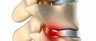
Large hemangioma with compression of the spinal canal.
The situation is aggravated by the fact that with a hypothetical fracture of the 4th lumbar vertebra, no spinal cord at this level no longer exists, and, in extreme cases, cauda equina syndrome is possible, with the development of shooting pains in the legs, numbness of the perineum, the development of erectile dysfunction, and urinary disorders. In the case of a fracture of the third cervical vertebra, and progressive myelopathy or spinal cord injury, we are talking about quadriplegia, that is, complete paralysis of the arms and legs, severe disability and the same urination disorders. This is what “1 cm” means for prognosis without an exact indication of the level of hemangioma.
The danger of hemangioma also increases in proportion to its growth rate. Therefore, if a tumor of non-critical size is detected, only one thing is required: a re-examination after a year, if there are no complaints, then after another year, then, possibly after 3 years, then after 5 years. And if the tumor is truly stable, then there is no need to worry.
Another important criterion for danger is the location in the vertebral body. If the hemangioma is small, and it is located approximately in the center, and is surrounded on all sides by undestroyed bone beams that make up a single mass, then there is a high chance that the vertebra will remain strong for a long time, even if it grows evenly outward. But if the hemangioma is located close to the vertebral border, if only a thin bone edge separates it from the external space, then the risk of a compression fracture is much higher.
Cervical hemangioma with compression of the spinal canal
Finally, you should always keep in mind the condition of the bone tissue. If we are talking about a vertebral hemangioma in a young man, then there is no need to worry about bone density. But if a hemangioma is found in an elderly woman after menopause, then she will already have a high risk of osteoporosis, but in any case, the fragility of her spongy bone will be much higher. Therefore, even a small hemangioma in a patient with a high risk of osteoporosis will be more prognostically dangerous than a slightly larger tumor in a healthy person.
It follows from this that the main factor that frightens patients (namely, the size of the hemangioma taken by itself) does not play a decisive role in determining the degree of risk. But if you still focus on this indicator, then if the tumor begins to occupy a volume equal to 55-60% of the vertebral body, and at the same time there is a tendency for it to grow, then it is necessary to think about surgical treatment.
The doctor must explain to the patient that the slow growth of a hemangioma does not mean malignant growth, and even more so, has nothing to do with metastasis. If this is not done, the patient can be frightened and depressed, and, as is known, chronic depression and anxiety are one of the risk factors for chronic back pain. A hemangioma hidden inside a vertebra never hurts until bone destruction occurs. But if the patient is under stress, he may constantly complain of false back pain, which will significantly worsen the quality of life.
Pathogenetic features
As a rule, hemangioma forms in the vertebral body. However, there is also a small percentage of cases where the tumor appeared in other parts of the spine, in particular:
- in the posterior elements of the spinal column;
- in the paravertebral line;
- in the epidural space.
A study of the structure of such a tumor showed that inside it consists of two elements:
- soft tissue;
- bone
The structure of the vertebra itself in this case has the appearance of a honeycomb and consists of large and small cells.
Clinical symptoms
Vertebral hemangiomas have exactly the same clinical picture as Schmorl’s hernias, or rather, none at all. Although they have much in common: they destroy the bone tissue of the vertebrae, both of these formations are prone to enlargement, but still, both of these processes are completely asymptomatic. Even if we take the most delicate, cervical spine, the hemangioma still does not show any signs until one of the above-mentioned complications occurs.
And only in the rarest cases, when the tumor has reached a significant size, can a so-called vertebral collapse occur, in which a compression fracture actually occurs, but no separate fragments are formed, the parallelism of the overlying and underlying intervertebral disc is maintained, and the fracture itself is an impacted along the entire perimeter of the vertebral body. In other words, the vertebra seems to settle, its height decreases. In this case, naturally, there is a disruption of the natural course of the nerve roots in the corresponding foramina, and acute radicular symptoms appear.
How does pathology manifest itself?
Often, the symptoms of hemangioma appear only with a pronounced increase in the size of the formation. The main symptoms of hemangioma include:
- Strong headache;
Headache - increased nervousness, irritability;
- sleep disturbance caused by insomnia;
- hearing impairment;
- impaired visual function (with spinal hemangioma, the patient’s visual acuity decreases);
- periodic numbness of the lower extremities and shoulder blades;
- slight tingling in the area of the fingers.
If you want to learn in more detail the causes of hemangioma in a newborn, as well as consider the symptoms and treatment methods, you can read an article about this on our portal.
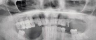
Cervical hemangioma in a child. In later stages it appears visually
On a note! The combination of pronounced symptoms often leads to the fact that the development of pathology can provoke coronary heart disease or stroke. First of all, this is due to possible pinching of the vertebral artery by the resulting tumor.
About radicular symptoms
In this case, we are talking about the appearance of sharp, shooting pains, reminiscent of an electric shock. They are provoked by any movement, shaking of the spine, that is, coughing, laughing, sneezing, straining in the toilet. This pain occurs when older people experience a shooting pain in the lower back, or lumbago.
With the long-term existence of radicular symptoms, a secondary, myofascial-tonic syndrome occurs. The deep-lying back muscles near the affected vertebra react to the surrounding inflammation, to the impacted fracture, with constant, chronic spasm. As a result of spasm, the muscle is deprived of the ability to promptly remove lactic acid, that is, waste products. She is deprived of nutrition from the arterial capillaries, which are also spasmed. This situation forms a vicious circle, and it can be broken either by eliminating the defective vertebra, or, if it is impossible for some reason to carry out restorative surgery, by prescribing central muscle relaxants and wearing a corset with limited mobility in the back, but this is not the best way out. provisions.
How often does radicular pain occur with hemangiomas? Very rarely, out of all diagnosed vertebral hemangiomas in all parts, disc collapse with the development of radicular symptoms occurs in 0.1% of cases. Therefore, the main “suppliers” of radicular symptoms can be considered protrusions and herniations of intervertebral discs.
FAQ
Does spinal hemangioma hurt?
Spinal hemangioma at an early stage of development rarely provokes the appearance of acute pain. Discomfort appears during physical activity and sudden movements, but quickly passes. Pain is typical for formations that reach 1 cm in diameter - they must be treated or removed promptly, otherwise the patient’s quality of life will decrease.
What contraindications for hemangioma should the patient be aware of?
The list of contraindications for spinal hemangioma includes:
- visiting saunas, solariums, prolonged exposure to the sun and other thermal effects;
- massage the affected area (provokes increased blood flow);
- manual therapy;
- physiotherapy;
- prolonged standing or sitting (more than 4 hours).
It is recommended to limit the intake of vitamins and immunostimulants, because such drugs can provoke the growth of tumors.
Is spinal hemangioma dangerous?
Hemangioma is dangerous if its course is uncontrolled. It can cause neurological disorders leading to a decrease in quality of life. There is a small chance of cells degenerating into cancer. The patient's condition must be monitored at all stages of treatment.
Where to go for diagnosis and treatment in Moscow?
If you suspect the development of a vertebral hemangioma in Moscow, you can contact JSC “Medicine” (clinic of Academician Roitberg), which has a convenient location in the Central Administrative District (Mayakovskaya metro station). Experienced staff will provide an individual approach, conduct an examination using high-quality equipment, differentiate the diagnosis and determine the best treatment regimen.
Diagnostics
Computer and magnetic resonance imaging reign supreme in the diagnosis of hemangiomas. Even the term “diagnosis of vertebral hemangiomas” is absurd, since they do not hurt and do not manifest themselves in any way, and therefore there is no point in looking for them. This can only be done during a scientific study on the incidence of vascular tumors of the vertebrae.
Therefore, almost 100% of the detection of all hemangiomas are accidental findings. They begin to be monitored, and with a reasonable approach, seeing a growing vascular tumor, when it reaches a certain size, and taking into account the risk factors and dangers outlined above, radical treatment is carried out, which makes the patient forget about the hemangioma. In some cases, hemangioma is initially suspected during an X-ray examination of the spine, but to accurately confirm the diagnosis, the patient is still sent for a tomography. There are no other diagnostic methods, just as there are no laboratory tests for hemangioma.
Treatment of patients with vertebral hemangioma
Since both stable and growing hemangioma are reliably hidden, as if inside a shell, inside a vertebra, then absolutely all conservative methods are completely ineffective, meaningless, and if they are offered to you, then this is nothing more than fraud.
Fraud includes attempts to “cure” hemangioma in such patients with the help of massage, acupuncture, osteopathy sessions, manual therapy, leeching, cauterization with wormwood cigars, electrical stimulation, electrophoresis sessions, and other techniques so beloved by our people. After the money is spent, the patient undergoes a second MRI. Hemangioma has not gone away! He comes to the doctor in anger, and the doctor with a charming smile informs the patient that “... you had a growing hemangioma, and we finally, using unique techniques, stopped its growth and achieved stabilization. Now you are in no danger, and you can forget about the operation”..... Comments, as they say, are unnecessary.
We must remember that a formation that is hidden inside a vertebra can neither be eliminated nor stop its growth without getting inside the vertebra itself. Fortunately, hemangioma can be treated with a simple, painless, and minimally invasive surgical method, without any incisions.
Percutaneous puncture vertebroplasty
Puncture vertebroplasty is a modern and very convenient method of removal. The goal is to eliminate the cavity inside the vertebra, filling it with special cement, which, when slightly heated, expands, completely destroys the tangle of blood vessels, and completely fills the entire space inside the vertebra.
Schematic representation of the procedure.
You don't need to make any cuts for this. It is enough to accurately determine the projection of the desired vertebra on the skin. Under X-ray control, to avoid mistakes, a needle is inserted into the vertebra under local anesthesia. After the needle falls into the internal cavity filled with a vascular tumor, a special biopolymer is fed inside, which is heated to a relatively high temperature, about 60 C°. This temperature is quite enough to completely destroy tumor cells, and the ability of the polymer to slightly expand during hardening makes it possible to guarantee that all voids are filled. Thus, the vertebra again becomes a single monolith.
In this case, a cementing substance consisting of:
- cement itself;
- radiopaque material (so that you can see the filling process directly under X-ray control “in real time”);
- antibacterial additive is approximately similar in density and mechanical characteristics to bone tissue, so that the vertebra continues to perform its function as before.
Cement.
Roughly speaking, this surgical intervention resembles filling a diseased tooth. No cuts, no blood, no long hospital stay. The next day, the patient can leave the hospital on his own, and local anesthesia allows percutaneous puncture vertebroplasty to be performed in patients with concomitant pathologies, including the elderly.
Contraindications for surgery
Taking advantage of the illiteracy of the population, many private clinics warm their hands well by doing the opposite - imposing unjustified surgical treatment. So, if they found a vertebral hemangioma on an MRI in a gullible, suggestible person, then they can make good money on this. Arguments such as “potential danger”, “risk of rapid, uncontrolled growth”, and “vertebral fracture in just a few weeks, or, in extreme cases, months” are used. Usually, urgent vertebroplasty is suggested, and the conversation ends with the words: “you are very lucky that you came to us, and we did the MRI on time.” Particularly arrogant “specialists” may even hint at the possibility of malignancy and “turning into cancer.” As we remember, this is impossible: hemangiomas of any location do not turn into “cancer”.
This is a very winning situation. Vertebroplasty is an operation with minimal risk for the patient, quite simple, but it can cost quite a lot of money. So, for the injection of bone cement into one vertebra you can easily charge 20 thousand rubles, and even more. Patients should know that this is not necessary at all, and they should not follow the lead of such doctors.
The contraindication to this small surgical intervention will be precisely the small size of the hemangioma and its stability. Therefore, if you are assured of the need for urgent vertebroplasty, then in any case, it is necessary to do a repeat tomography at least 6 months after the discovery of the tumor to make sure that the tumor is really growing and the potential danger.
A little history
It must be said that vertebroplasty is an extremely successful and beautiful way to eliminate a tumor and create a single bone block. Previously, before the introduction of this technique, sclerotherapy was used. In this case, a sclerosing substance, such as ethyl alcohol, was injected into the hemangioma. But the cavity remained: although the hemangioma itself died, the alcohol reduced the strength of the bone tissue, and as a result, a pathological fracture quite often occurred. In some cases, the tumor did not die completely, and a relapse occurred.
In addition to sclerotherapy, tumor embolization was also used, that is, an attempt to eliminate blood flow through the vessels. It was technically more difficult, and the matter could not be done with just a puncture. It required the skill of a vascular surgeon, which is not at all needed in modern vertebroplasty. But since the tumor itself remained, the death of the embolized vessels did not play a big role. The vessels actually died, but new vascular bundles were constantly being formed, because the pressure in the arterial section was maintained.
Etiology of the disease
To date, the etiology of the development of spinal hemangioma has not been fully studied. Previously, opinions regarding the origin of this type of tumor were divided into two points of view:
- the appearance of a tumor is the result of vasodilation;
- hemangioma is a true neoplasm.
In the 21st century, many research scientists believe that hemangioma is a benign tumor that appears as a consequence of genetic predisposition. This opinion is based on the fact that most of these formations develop at the site of embryonic clefts.
Forecast
As you may have guessed, in the vast majority of cases, a stable, small hemangioma does not affect either the quality of life or its duration. Before the widespread use of tomography, tens and even hundreds of generations lived perfectly well with hemangiomas, with calcification of the pineal gland in the brain, and with other random findings that nowadays cause stress and panic. It is not always necessary to take any emergency measures when you discover unfamiliar formations in your body.
Perhaps, if a “silent” vertebral hemangioma is detected, we can only advise you to keep your weight close to ideal, avoid heavy lifting, weightlifting, and contact, hard types of martial arts. Women can be advised to regularly perform densitometry after menopause, since in the presence of osteoporosis, a pathological fracture can occur long before the hemangioma reaches the theoretical strength limit in the region of 50-60% of the vertebral volume.
And if a compression fracture already occurs, then you will have to undergo extensive surgery. Yes, some vertebral compression fractures do not require surgical treatment; they heal on their own. But keep in mind that in this case there will be a vascular tumor between the bone fragments, and the hemangioma will be a factor of “non-union”, it will create tissue interposition between the fragments, and, lying freely, no longer covered by the vertebra, will create an additional risk of bleeding.
Therefore, the basis of the correct approach to the treatment of hemangiomas will be dynamic observation, taking into account additional risk factors.
Possible complications
Despite the fact that hemangioma does not grow quickly, in rare clinical cases the tumor, increasing in diameter, puts pressure on the patient’s bone elements. When a vertebra is damaged, not only is its integrity compromised, but its strength is also reduced, which is what causes pain.
Hemangioma may cause pain
In addition, the affected vertebrae become more vulnerable to fractures. But, in addition to pain, the risk of neurological disorders increases, including problems with urination and loss of sensation in the lower extremities. When it develops into an aggressive form, spinal hemangioma can pose a serious danger to the patient, so it is impossible to start the disease and ignore its first signs. This is the only way to protect yourself from painful complications.
Treatment of spinal hemangioma