Candidiasis or thrush
is a fungal, infectious disease caused by opportunistic fungi of the genus Candida. Microorganisms are present in most healthy people. A person may encounter them for the first time in the womb, during childbirth, or during breastfeeding. Candidiasis develops against the background of reduced immunity, when the body can no longer restrain the active proliferation of the fungus.
The infection affects the mucous membranes of the mouth and genitals. It can develop on smooth skin, nails, and in the intestines, as a type of dysbacteriosis. There is visceral (systemic) candidiasis that affects internal organs. Despite the fact that Candida fungi are present in many people, and the disease itself is considered a neglected disease, it must be treated even if there are no external manifestations.
Causes of candidiasis
- Frequent damage to the skin and mucous membranes, for example, due to illness, working with aggressive chemicals, dry skin, age-related changes.
- Prolonged exposure to water, humid and warm environments. This includes a climate that is not suitable for your skin type, as well as occupational hazards.
- Mechanical damage to the skin and mucous membranes: injection drug addiction, long-term surgical interventions, catheters, shunts, endotracheal tubes.
After entering the body, the fungus may not manifest itself for a long time, since its reproduction will be suppressed by the immune system. In some cases, natural defenses weaken, leading to candidiasis (thrush). Among the factors that contribute to the proliferation of Candida:
- Chronic, endocrine and immune system diseases (diabetes, HIV).
- Metabolic disorders or taking medications that disrupt the natural microflora (antibiotics, hormonal contraceptives).
- Unbalanced diet, lack of sleep, stress and depression.
The fungus can be found in raw meat, unpasteurized dairy products, and, in more rare cases, on fresh vegetables and fruits. You can become infected with candidiasis from animals: dogs, calves, poultry.
Treatment of candidiasis of the anus and rectum
Local therapy is supplemented by instillation of silver and antifungal agents into the anal canal. Also, ointments and creams containing antimycotics are applied to the skin of the perianal area and inside the rectum.
The same systemic drugs are used.
In case of systemic candidiasis of internal organs, treatment is carried out exclusively in hospital conditions by administering rather toxic, but most effective antifungal drugs.
Nail candidiasis requires long-term therapy with antifungals for up to 3-4 months, in combination with removal of the affected areas of the nail plates, local exposure to antifungal varnishes, solutions, laser and ultraviolet radiation. Often there is simultaneous damage to the skin of the periungual ridges and interdigital spaces, which also require treatment with creams and other local procedures under the supervision of a dermatovenerologist.
Risk factors for development
The likelihood of infection increases with casual sexual intercourse, uncontrolled use of antibiotics and drugs that disrupt the natural microflora. Foods with large amounts of sugar and carbohydrates create a favorable environment for fungal growth. Excessive sweating also leads to an exacerbation of candidiasis, so it is necessary to wear cotton underwear that allows the skin to breathe and moisture to evaporate.
At the same time, excessive cleanliness can also cause harm. We are talking about douching. It should not be used as a method of contraception, since it is not effective, and also as a means of hygiene, because it leads to the leaching of the protective flora. If you experience discomfort, you should first consult a doctor.
Treatment of candidiasis in men
Our clinic uses the following types of local treatment:
- Baths with antiseptics and antifungal drugs on the head of the penis.
- Wet-dry dressings for erosive and ulcerative lesions of the head and foreskin.
- Instillations into the urethra for candidal urethritis - 800 rubles.
- Total instillations for fungal infections of the posterior urethra and prostate. — 1000 rub.
- Ultraviolet irradiation of the skin of the scrotum and groin areas. — 800 rub.
- Electrophoresis of antifungal drugs into the lesion. -1200 rub.
- Laser irradiation of foci of chronic candidiasis. — 800 rub.
Classification of the disease
Based on the depth of damage, candidiasis is divided into superficial and systemic forms. The first appear on visible parts of the body: mucous membranes, skin and its appendages. The second, systemic, are combined forms of the disease affecting internal organs. Without proper treatment, systemic (visceral) candidiasis can lead to the development of candidal sepsis. Most often, the fungus affects:
- gastrointestinal tract (esophagus, stomach, intestines);
- urinary organs (bladder, urinary tract, kidneys);
- lower respiratory tract (trachea, bronchi, lungs).
The disease is also classified based on the rate of spread of infection and the presence of relapses. Candidiasis of the liver and spleen often takes a chronic form. The acute, generalized form is asymptomatic candidemia, candida septicemia, candida thrombophlebitis, myositis, arthritis, cerebral candidiasis and other forms.
Classification of candida lesions according to ICD-10
- Candidiasis.
- Candidal stomatitis.
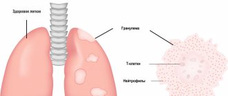
- Pulmonary candidiasis.
- Candidiasis of the skin and nails.
- Candidiasis of the vulva and vagina.
- Candidiasis of other urogenital locations.
- Candidal meningitis.
- Candidal endocarditis.
- Candidal septicemia.
- Candidiasis of other locations.
- Candidiasis, unspecified.
Officially, the diagnosis is made in accordance with ICD-10. In practical healthcare, such a classification is not entirely convenient, so they use the classification of deep candidiasis, which is a deeper list. When making a diagnosis, first indicate the name and form of the infection with a description of all localization points.
Skin treatment is carried out by a pediatrician in children and by a dermatologist in adults. Therapy is also carried out by doctors of other specializations, depending on the affected organ. For example, this could be a dentist, neurologist, gynecologist, pulmonologist.
Treatment of candidiasis in women
Basically, to the general treatment described above, vaginal treatments are added on the gynecological chair with antiseptic, antifungal solutions, during which fungal layers are completely removed from the surface of the vaginal mucosa and a high concentration of drugs is created, which has a detrimental effect on the remaining Candida cultures.
After the first treatment, 95% of women feel significant relief and a decrease in the severity of symptoms.
The cost of the procedure is 800 rubles.
In case of a chronic process, a course of such treatments is recommended, which, in combination with probiotics, not only removes fungi, but also restores the normal microflora of the vagina and the state of local immunity.
Physiotherapeutic treatment is also carried out with a vaginal laser, electrophoresis, and baths with an ozonized sodium chloride solution, which helps to either completely get rid of relapses of chronic vulvovaginal candidiasis or reduce its exacerbations.
In case of a chronic process, two-stage treatment is always recommended for all patients, and especially women.
The first stage is the destruction of the fungus.
The second stage is the restoration of normal microflora of the intestines and vagina.
Symptoms of candidiasis
The symptoms of the disease are directly related to the affected organ. Superficial forms have obvious signs, while systemic forms can be asymptomatic or be similar to other diseases. In the presence of chronic diseases (diabetes mellitus, HIV, AIDS, leukemia and other types of oncology), candidiasis may not be detected at all. Since its symptoms will be perceived by the patient as an unhealthy state due to the existing disease. To make matters worse, Candida susceptibility testing is not considered a routine procedure in many teaching hospitals and laboratories.
Candidiasis of the skin and nails
The infection is expressed in the form of allergic rashes (candidamycids), erythematous-squamous spots, urticarial and bullous type rashes. Nail pathologies are divided into:
- parochinia - purulent inflammation of the periungual and subungual spaces, absence of eponychium (skin at the base of the nail plate);
- onychia - purulent inflammation of the nail fold, deformation of the nail with discoloration and brittleness.
Candidal skin lesions may be accompanied by fever, inflammation and swelling of the lesions.
Candidiasis (thrush) of the oral mucosa
The fungus can affect both individual areas of the oral cavity and completely all mucous membranes: lips, gums, cheeks, palate, tongue, tonsils, uvula and pharynx. This is expressed in the presence of a white coating, compared to a curd mass. Swelling and redness may appear. There is often pain when swallowing, talking, or palpating. Saliva becomes viscous and bad breath appears.
Thrush may be accompanied by candidal cheilitis - damage to the lips and corners of the mouth. Painful cracks appear, covered with a white coating, and pieces of skin peel off from the lips. The disease is characterized by a long course with the possible addition of bacterial infections.
Intestinal candidiasis
Intestinal infection with fungi of the genus Candida can be an independent disease or develop as a result of damage to the oral cavity. Expressed as severe dysbacteriosis with the following symptoms:
- diarrhea;
- excessive gas formation;
- nagging pain in the rectum;
- an admixture of white flakes in the stool.
In most cases, symptoms are sluggish or completely absent. The disease is dangerous because the body does not receive enough vitamins and microelements, which is especially dangerous for a growing child.
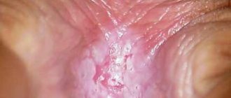
Candidiasis of the genitourinary system (urogenital candidiasis)
Unlike sexually transmitted diseases, candidiasis of the genitourinary system is often hidden and asymptomatic. The fungus is discovered during a planned infection or against the background of other diseases. Symptoms begin to appear against the background of declining immunity, ongoing pathological processes of microflora destabilization and re-infection. In women, candidiasis manifests itself as follows:
- itching and burning of the external genitalia;
- swelling and redness;
- white, cheesy discharge;
- pain during sexual intercourse.
In men, the symptoms are similar:
- itching and burning of the head of the penis;
- white plaque and discharge:
- pain during sexual intercourse and urination.
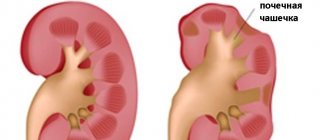
Fungal infections of the bladder, excretory tract and kidneys are characterized by frequent urination, including false urges, pain in the suprapubic region. Candidiasis can be accompanied by bacterial infections. Complications include: cystitis, pyelonephritis, necrosis of the papillae, abscesses, formation of mycelium in the renal pelvis. Without proper treatment, against the background of chronic diseases, there is a risk of developing kidney failure.
Complication
With timely treatment, candidiasis does not cause any particular harm to health. But the symptoms of candidiasis can cause a lot of discomfort. Long-term, it can lead to damage to other organs, most often the urethra, bladder and kidneys. In particularly severe cases, the progressive disease can affect the reproductive organs, leading to infertility in both men and women. But candidiasis poses the greatest danger to pregnant women, because... the risk of fetal damage is very high.
Diagnosis of candidiasis
The study is based on the isolation and identification of a species of fungus of the genus Candida. Currently, there are about 150 species, differing in morphological and biochemical properties. The most common fungus is C. albicans, accounting for up to 80% of cases of candidiasis of the digestive tract and up to 70% of genital infections. Before prescribing treatment, it is also necessary to determine the sensitivity of the isolated strain to antimycotics (antifungal medications): amphotericin B, voriconazole, itraconazole, fluconazole, flucytosine.
Microscopic diagnostics
1. Microscopy of a smear is taken from the area of the affected mucosa. Allows for comparative characterization of blastospores and pseudomycelia. During the study, fixed and native preparations that stain microorganisms are used. To increase the information content, pseudomycelia of cells are treated with dyes. The contrast in staining of microorganisms makes it easy to distinguish candida from other forms, including bacteria, under a microscope.
2. Bacterial culture allows you to identify the causative agent of infection and determine its concentration. The analysis is used to monitor the effectiveness of treatment, as well as to identify the sensitivity of candida fungi to various antimycotics.
Diagnostics by ELISA and PCP
1. Enzyme-linked immunosorbent assay (ELISA) is based on the determination of antibodies that are produced by the immune system in response to foreign substances in the blood. This technique allows you to identify the pathogen and the degree of its development, establishing whether the disease occurs in an acute or chronic stage.
2. Polymerase chain reaction (PCR) is a highly sensitive test that allows you to directly detect the infectious agent. Thanks to it, it is possible to differentiate Candida fungi with pseudomycelium from those that do not have it. These data are important for subsequent interpretation of results and deeper diagnosis.
Pathogenesis, clinical picture and diagnosis
The development of candidiasis of the digestive tract is characterized by the sequential passage of fungi through the following stages: adhesion, invasion, candidemia and visceral lesions. At the first stage, micromycetes adhere to the epithelial cells of any part of the mucous membrane. Subsequently, defects in the resistance system allow micromycetes to penetrate (invade) the mucous membrane and underlying tissues through transformation into pseudomycelium. Cytopenia is a decisive factor that allows invading fungi to reach the vascular wall, destroy it and circulate in the vascular bed (this stage is called candidemia). In the absence of adequate therapy, candidemia leads to the formation of foci of invasive candidiasis in the visceral organs, more often in the liver and spleen, lungs, and central nervous system.
At first glance, it is paradoxical, but the introduction of fungi of the genus Candida is often observed in areas of the mucous membrane represented by multilayer epithelium (oral cavity, esophagus), and much less often this occurs in relation to single-layer epithelium (stomach, intestines).
In practice, the clinician has to deal primarily with candidiasis, the frequency of which in healthy individuals reaches 25% in the oral cavity, in the intestines - up to 65–80% and significantly exceeds the frequency of the disease. Esophageal candidiasis, for example, occurs in general patients in 1–2% of cases, in patients with type 1 diabetes mellitus in 5–10%, and in patients with AIDS in 15–30% [2].
Symptoms of candidiasis are primarily characterized by the appearance of the so-called. thrush and irritation of the mucous membranes. Typical complaints are burning of the mucous membrane of the tongue and the oropharyngeal cavity, dys, odynophagia, retrosternal discomfort, but a latent course also occurs. Symptoms of esophageal candidiasis can disrupt the act of swallowing, which in turn leads to malnutrition and a significant decrease in quality of life.
The scope of examination for candidiasis of the digestive organs includes the study of anamnesis and clinical picture, assessment of routine clinical tests, endoscopic studies, mycological (cultural, morphological, serological) and immunological tests.
Endoscopic signs of candidiasis are hyperemia and contact vulnerability of the mucous membrane, as well as fibrinous deposits of various locations, configurations and sizes. The “standard” for diagnosing candidiasis of the mucous membranes is the detection of pseudomycelium of Candida spp. during morphological examination of biopsy specimens and scrapings.
In order to detect pseudomycelium, morphological mycological methods are used: cytological - with staining of smears according to Romanovsky-Giemsa, histological - with staining of biopsy specimens with the CHIC reaction.
Thus, taking into account the dimorphism of micromycetes Candida spp. is the key to the differential diagnosis between candidiasis and candidiasis. In modern conditions, the clinician must demand from the morphologist an accurate description of the morphological structures of the fungus, because the detection of individual yeast cells, as a rule, indicates candidiasis, and the detection of pseudomycelium allows confirming the diagnosis of candidiasis.
A disadvantage of morphological methods is their limited sensitivity for endoscopic biopsy. It is known that biopsy forceps make it possible to obtain a miniature fragment of tissue for study and the likelihood of detecting an informative sign with a single biopsy is insufficient.
The cultural mycological method is based on sowing biomaterials of mucous membranes on Sabouraud's medium. The advantage of this method is the possibility of species identification of fungi of the genus Candida and testing the culture for sensitivity to antimycotics. The relevance of such studies is due to
the fact that different species of Candida, in particular C. albicans, C. tropicalis, C. parapsilosis, C. krusei, C. glabrata, etc., have different sensitivities to modern antifungal drugs. Currently, modern laboratories have several diagnostic systems for determining the species of fungi and their sensitivity to antifungal drugs. Species identification and determination of sensitivity to antifungal drugs are based on the use of selective culture media, the study of the nature of sugar utilization by fungi and the enzyme profile of fungi, the dilution method, fluorescent hybridization, etc. (diagnostic systems Candiselect, Auxacolor, Fungiscreen, 60780 Fungitest, etc.). A cultural study of biomaterials of the mucous membranes with determination of the type of pathogen becomes strictly necessary in case of recurrent candidiasis or resistance to standard antimycotic therapy.
The disadvantage of the cultural method is the inability to differentiate candidiasis and candidiasis when studying materials from “open systems”, since mucous membranes can normally be contaminated with fungi of the genus Candida.
The sensitivity and specificity of serological tests for diagnosing esophageal candidiasis (enzyme-linked immunosorbent assay with Candida antigen, specific IgE level, Platelia latexagglutination test) have not yet reached the required accuracy, and their results should be assessed with some caution.
The management plan for patients with esophageal candidiasis must include diagnosis and correction of underlying diseases, other foci of candidal infection, rational antifungal therapy and immunocorrection.
In the clinic of the Scientific Research Institute of Medical Mycology of the St. Petersburg Medical Academy of Postgraduate Education, a patient with candidiasis must undergo a minimum amount of diagnostics of the alleged underlying disease, including: • clarification of the pharmacological history; • clinical blood test; • blood test for HIV infection; • blood test for glucose tolerance; • exclusion of the most common malignant tumors and, in connection with this, radiography of the lungs, fibrogastro, fibrocolonoscopy, additionally for men - ultrasound examination (ultrasound) of the prostate gland, additionally for women - ultrasound examination of the mammary glands and pelvis with consultation of a gynecologist.
Therapeutic measures
Treatment of mucosal candidiasis is based on the use of antifungal drugs. The general principle of action of all antifungal agents is inhibition of the biosynthesis of ergosterol in the cell wall of micromycetes. In general, antifungals used to treat candidiasis can be divided into three groups. The first is polyene antimycotics, which are practically non-resorbable when taken orally. These include amphotericin B, nystatin and natamycin. The second is azole antimycotics, which are relatively well absorbed when taken orally. These include: ketoconazole, fluconazole (most often prescribed for candidiasis), itraconazole, voriconazole, posaconazole. The third group is echinocandins: caspofungin, anidulafungin, micafungin.
The goal of treatment of candidiasis of the mucous membranes of the upper digestive tract is to eliminate symptoms and clinical laboratory signs of the disease, as well as to prevent relapses. A modern revision of the standards for the treatment of candidiasis of the mucous membranes of the upper digestive tract, published in the USA in 2009, contains fundamental changes and additions compared to a similar document of 2004 [3].
Most cases of oropharyngeal candidiasis and esophageal candidiasis are caused by C. albicans either as the only pathogen identified or as part of a mixed infection. However, cases of overt infection caused by species usually resistant to fluconazole, C. glabrata and C. krusei, are becoming increasingly common.
Numerous randomized prospective studies of the course of oropharyngeal candidiasis in patients with AIDS and cancer have been conducted in the USA and Europe; it was noted that the majority of patients respond to therapy with topical antimycotics. However, it has also been found that in patients with AIDS, relapses of candidiasis after therapy with locally acting antimycotics occur faster than after therapy with fluconazole, and resistance to antifungals can appear with all treatment regimens.
It is obvious that treatment with fluconazole and itraconazole in solution is more effective than treatment with ketoconazole and itraconazole capsules. For children aged 5 years and older, the recommended dose of itraconazole solution is 2.5 mg/kg twice daily, and the local effect of the solution may be as important as the systemic effect. In AIDS patients, posaconazole suspension is as effective as fluconazole therapy.
Recurrent candidiasis infection is typical for patients with immunodeficiency, especially for patients with AIDS. To prevent relapses of oropharyngeal candidiasis, long-term maintenance therapy with fluconazole is quite effective. It has been compared in efficacy to intermittent fluconazole in response to disease relapse, and long-term maintenance therapy has been shown to prevent relapse of candidiasis more effectively than intermittent therapy, but is associated with a greater risk of developing resistance.
At the same time, it has been shown that oral therapy with amphotericin B, nystatin and itraconazole capsules is less effective than treatment with fluconazole to prevent relapses of oropharyngeal candidiasis.
According to the recommendations, for the treatment of fluconazole-resistant candidiasis, an itraconazole solution should first be prescribed. It has been shown that at least 64, but not more than 80% of patients respond to such therapy. Posaconazole suspension is effective in approximately 74% of patients with recurrent oropharyngeal or esophageal candidiasis, and voriconazole may be effective in fluconazole-resistant candidiasis. Intravenously administered caspofungin, micafungin, and anidulafungin may be reasonable alternatives to triazole antimycotics. Oral or intravenous amphotericin B may also be effective in some patients.
Immunomodulation with the addition of granulocyte-macrophage colony-stimulating factor and interferon gamma preparations may be useful in recurrent oral candidiasis.
The simultaneous detection of oropharyngeal candidiasis and dysphagia or odynophagia may indicate that the patient has esophageal candidiasis. A trial of fluconazole treatment for such patients with suspected esophageal candidiasis may be a cost-effective alternative to endoscopic examination: most patients with esophageal candidiasis report resolution of complaints within 7 days of starting therapy. In the treatment of esophageal candidiasis, fluconazole is more effective than ketoconazole, itraconazole capsules and flucytosine, and itraconazole solution is comparable in effectiveness to fluconazole. Up to 80% of patients with fluconazole-resistant candidiasis respond to treatment with itraconazole solution. Voriconazole is as effective as fluconazole and is superior in the treatment of fluconazole-resistant candidiasis, although it is associated with more side effects.
The use of echinocandins is characterized by a higher frequency of relapses of candidiasis than after treatment with fluconazole. Fluconazole-resistant candidiasis responds to caspofungin, which has similar efficacy to micafungin and anidulafungin. In patients with AIDS, candidiasis usually becomes recurrent, and long-term maintenance therapy with fluconazole is effective in preventing relapses. In HIV-infected patients, the use of HAART (highly active antiretroviral therapy) is associated with a decrease in the degree of contamination of the mucous membranes with C. albicans fungi and a decrease in the incidence of oropharyngeal candidiasis. Thus, HAART should be used as adjunctive therapy (to the extent possible) in all HIV-infected patients with oropharyngeal candidiasis and esophageal candidiasis.
Chronic candidiasis of the skin and mucous membranes is a rare disease characterized by chronic persistent onychomycosis and damage to the mucous membranes caused by fungi of the genus Candida. Some of these patients suffer from thymoma or autoimmune polyendocrine syndrome type 1. These patients should be given fluconazole as initial therapy. When widespread skin and nail lesions are present, the response to antifungal therapy may be delayed and relapse almost always occurs. Thus, most patients require chronic maintenance antifungal therapy. The development of fluconazole-resistant candidiasis is common. Patients with this form of the disease should be treated with the same therapeutic regimens that are used for fluconazole resistance in AIDS patients.
Treatment of oropharyngeal candidiasis For the treatment of mild cases: irrigation with a solution of clotrimazole at a dose of 10 mg 5 times a day or a suspension of nystatin locally at a concentration of 100 thousand units/ml at a dose of 4-6 ml 4 times a day, or 1-2 lozenges with nystatin ( 200 thousand units each), prescribed 4 times a day for 7–14 days. Note that in the Russian Federation, dosage forms of nystatin in the form of suspensions or lozenges are not registered.
For the treatment of moderate to severe cases of the disease: oral fluconazole 100–200 mg (3 mg/kg) per day for 7–14 days.
For the treatment of fluconazole-resistant forms of the disease: either itraconazole solution at a dose of 200 mg/day or posaconazole suspension at a dose of 400 mg 400 mg twice daily for the first 3 days, and then 400 mg daily for up to 28 days. If these measures fail, voriconazole 200 mg twice daily or 1 ml of amphotericin B oral suspension given at a dose of 100 mg/ml 4 times daily is recommended. Please note that dosage forms of amphotericin B in the form of an oral suspension are not registered in the Russian Federation.
In case of refractory disease, echinocandins or intravenous amphotericin B at a dose of 0.3 mg/kg per day are also recommended. Chronic maintenance therapy is usually necessary for people with AIDS. In these cases, fluconazole is recommended at a dose of 100 mg 3 times a week. HAART is also recommended to prevent recurrence of infection.
For the treatment of oropharyngeal candidiasis associated with dentures, in addition to antifungal therapy, it is recommended to disinfect dentures.
Treatment of esophageal candidiasis
In all cases, treatment with systemically acting antimycotics is required. Local antifungal drugs (nystatin, natamycin, clotrimazole) are not effective. Oral fluconazole is recommended at a dose of 200–400 mg (3–6 mg/kg) per day for 14–21 days. For patients who cannot take oral medications, intravenous fluconazole 400 mg (6 mg/kg) per day or amphotericin B 0.3–0.7 mg/kg per day or echinocandins are recommended. It is acceptable to prescribe a diagnostic trial of antifungal therapy before performing an endoscopic examination (note that this recommendation is typical for the United States, where the cost of an endoscopic examination is significantly higher than the cost of a trial course of antifungal therapy).
For the treatment of fluconazole-resistant forms of the disease, the following are recommended: itraconazole solution at a dose of 200 mg/day, posaconazole suspension at a dose of 400 mg twice a day, or voriconazole 200 mg twice a day, prescribed intravenously or orally for a period of 14–21 days. Acceptable alternatives are micafungin 150 mg/day, or caspofungin 50 mg/day, or anidulafungin 200 mg/day, or amphotericin B 0.3–0.7 mg/kg/day.
For the treatment of recurrent esophageal candidiasis, fluconazole is recommended at a dose of 100–200 mg three times a week. HAART is recommended to prevent recurrence of infection in AIDS patients.
In general, the success of treatment of mucosal candidiasis depends not only on adequate antifungal therapy, but also on the success of correction of the underlying disease.
Interpretation of results
If there are clear signs of candidiasis (thrush), and during a laboratory test blastospores and pseudomycelia of the fungus were identified, the study ends here. A diagnosis is made and treatment begins.
If a microscopic examination gives a negative result, this does not indicate the absence of infection. The disease can occur in a latent chronic form. It is necessary to carry out a number of other tests, for example, microscopic examination of scrapings, determination of Candida DNA in scrapings, urine, and prostate secretions. Also prescribed:
- Clinical blood test.
- Test for HIV infection.
- Determination of trace element reserves in the body.
- Test for glucose and carbohydrate metabolism metabolites.
These laboratory tests can help identify conditions that may be causing the fungus to grow.
Candida in the blood, treatment
Sometimes patients come in who, as they believe, have a fungus in their blood.
If, indeed, as a result of culture or PCR analysis, which is extremely rare, Candida albicans or another type of it was found, then such a person needs urgent treatment in a hospital setting.
But more often than not, the analysis only shows the presence of antibodies to candida, which is not an emergency indication for treatment, especially if there are no clinical manifestations.
Such patients need to be examined in more detail using PCR and culture.
What happens if candidiasis is not treated or treated incorrectly?
In addition to the transition to a chronic form, a number of serious situations are possible that can lead to a significant loss of health of the body as a whole and individual organs and systems.
Advantages of taking tests at JSC "SZDCM"
- Own laboratory with the latest diagnostic equipment.
- Convenient location of terminals within transport accessibility from anywhere in the city.
- Qualified laboratory technicians and friendly staff.
- Fast analysis and several options for obtaining results. Choose the one that is most convenient for you.
Medical centers and laboratory terminals of the North-Western Center for Evidence-Based Medicine are located in St. Petersburg, Leningrad region, Veliky Novgorod, Okulovka, Kaliningrad and Pskov.
Analyzes
- Bacteriological study for opportunistic pathogenic flora (OPF)
- NC yeasts of the genus Candida: C.albicans, C.krusei, C.glabrata
- Study of the biocenosis of the urogenital tract in women (“Femoflor 13 - screening”)
- Candida albicans
- Mycoses: identification of clinically significant fungi with determination of sensitivity to antimycotic drugs (only for fungi of the genus Candida and Cryptococcus neoformans)
- Specific immunoglobulin E - Candida albicans
go to analyzes