Causes
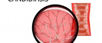
Candidiasis (thrush) affects not only external but also internal organs. The cause of the disease is yeast-like fungi that live in the body of every person.
- Many factors can provoke intensive reproduction of the fungus. For example, hypothermia, illness, stress, changes in hormonal levels (during pregnancy or taking hormonal medications).
- When taking antibiotics, along with pathogenic bacteria, the beneficial microflora of the intestine and vagina, which controls the growth and development of Candida fungi, also dies. The presence of a chronic disease that reduces the activity of the immune system (HIV, sexually transmitted diseases, infections) very often causes candidiasis.
- Candidiasis may be accompanied by endocrine diseases (diabetes, obesity, thyroid dysfunction).
- Finally, candidiasis can be caused by a hot climate or wearing uncomfortable tight or synthetic underwear.
The source of infection with candidiasis, as a rule, is the body’s own flora (autoinfection), but infection can occur from the outside. When causing a disease, the fungus does not change its properties - the body changes its properties (local protection decreases). Attaching to epithelial cells, the pathogenic fungus begins to parasitize them, penetrating deep into the tissues.
In the body's fight against candidiasis, a dynamic balance often arises when the fungus tries to penetrate deeper into the tissue, but cannot, and the body tries to reject it and also cannot. In this case, the process can last for years; a shift in balance in one direction or another will lead either to recovery or to an aggravation of the process.
Candidiasis occurs in several forms depending on certain characteristics.
- Carriage. A person is a carrier of the disease. There are no symptoms of candidiasis and there is no need to treat.
- Acute. Accompanied by itching, rashes, and discharge. Treatment must be comprehensive and high quality. Young children are most often susceptible to infection.
- Chronic. Symptoms subside and manifest; relapses are possible. It develops if it is not treated correctly with antibiotics for a long time, or if hormonal contraceptives are used.
This disease has several varieties, since it does not have an exact localization in the body:
- Urogenital candidiasis in women.
- male.
- Thrush of the lips
- On skin folds (armpits, area between the buttocks, inguinal folds).
- Gastrointestinal tract (stomach, esophagus, intestines, anus).
Magazine "Child's Health" 1(10) 2008
Chronic mucocutaneous candidiasis (CMC), or chronic generalized candidiasis, candido-endocrine syndrome, autoimmune polyendocrinopathy - candidiasis - ectodermal dystrophy, autoimmune polyglandular syndrome of the first type - a heterozygous group of diseases characterized by the persistence of a fungal infection of the skin, nails, mucous membranes caused by Candida fungi , rarely causing systemic disease or septicemia [5]. CCSC refers to primary combined immunodeficiency, which most often manifests itself in childhood [5]. Inheritance of the disease is autosomal recessive and autosomal dominant, sporadic cases are possible. More than 50% of patients have autosomal recessive inheritance and polyendocrinopathies [5]. Typical manifestations are mucocutaneous candidiasis, hypoparathyroidism, and chronic adrenal insufficiency. The presence of these signs is the basis for making a clinical diagnosis. The gene responsible for the development of this disease is located on the long arm of chromosome 21 (21q22.3), named AIRE and encodes a transcriptional regulatory protein [5, 6, 8]. The R257X mutation in the AIRE gene was detected with high frequency. In 26% of cases, the clinical picture is atypical, despite the presence of an identical R257X mutation [8]. Molecular genetic testing makes it possible to diagnose atypical cases of chronic mucocutaneous candidiasis.
The immunological characteristics of the CKS disease are most often vague and polymorphic. Most patients have a normal number of lymphocytes and their functional activity, but distinctive manifestations are the absence of in vitro T-cell proliferation to Candida antigen, decreased delayed-type hypersensitivity to Candida antigen, a defect in monocyte chemotaxis, and deficiency of the IgG2 and IgG4 subclasses [2–4].
According to the Register of Primary Immunodeficiencies of the Institute of Immunology of the FMBA, 31 patients with CCSC are being observed in Russia. Their clinical picture is heterogeneous. The dominant symptoms of the disease are determined by the presence of candidiasis of the mucous membranes (100%), skin (33%), onychomycosis (70%), multiple caries (58%), repeated bacterial infections of the respiratory tract (75%), skin (50%), ENT organs (25%), lymph nodes (12%), diarrhea (33%). Hyperplasia of the cervical and submandibular lymph nodes is detected (87%). Endocrinopathy often develops: hypoparathyroidism (46%), chronic adrenal insufficiency (29%), hypothyroidism (12%). Autoimmune diseases are noted: hemolytic anemia (8%), chronic aggressive hepatitis (12%), keratopathy (8%), alopecia (8%) [1].
The leading clinical marker of the disease - chronic candidiasis of the mucous membranes and skin - does not determine the severity of the disease and prognosis.
Bronchopulmonary infections are represented by chronic bronchitis, recurrent bronchitis, acute destructive pneumonia, and episodes of acute pneumonia. The severity of the clinical situation in patients is determined by a bacterial infection, and the prescription of antibiotics does not increase the manifestations of candidiasis [6]. The prognosis of the disease depends on the severity of polyendocrinopathy.
In the USA, cases of 3 men aged 21, 16 and 43 years with CKS were described, the onset of the disease in whom began in childhood with candidal lesions of the skin, nails, mucous membranes, refractory to antifungal therapy. The first patient developed an unusual, severe S. aureus septicemia at 3 months of age with cerebral and pulmonary abscesses leading to a pneumatocele requiring thoracic drainage and potent antibiotic therapy. The second patient developed multiple cerebral aneurysms caused by Candida infection at the age of 5 years. The third patient, 43 years old, had two episodes of pneumonia and pulmonary tuberculosis. During endoscopy, the patient was found to have epidermoid carcinoma in the esophagus. After 2 years, pulmonary metastases were revealed during radiotherapy and chemotherapy. All 3 patients were diagnosed with an autosomal dominant type of CCSC [1].
There are two patients with CCSC under our supervision: a girl and a boy.
In a girl (Fig. 1), currently 14 years old, the disease debuted at the age of 1 year with recurrent oral candidiasis, onychomycosis. At the age of 8, total alopecia, loss of eyebrows and eyelashes, and episodes of febrile fever 2-3 days a month for a year were noted. At the age of 9, during examination at the regional children's clinical hospital in Donetsk, congenital asplenia was discovered. At the age of 10, congenital ectodermal dystrophy of the cornea, keratitis, and congenital ectodermal dysplasia were diagnosed. From the age of 11, symptoms of adrenal insufficiency appeared: episodes of weakness, lethargy, decreased blood pressure; hypoglycemia and seizures. A low level of cortisol (32.5–76.9 nmol/l, normal 252–603 nmol/l), antibodies to thyroid peroxidase, and hypocalcemia were detected. The patient has been receiving cortico- and mineralocorticosteroids (prednisolone and cortinef) for replacement purposes since the age of 12, against which the symptoms of hypocorticism have regressed. The patient has somatogenic dwarfism, hypogonadism, and is retarded in physical development. Exacerbations of oral candidiasis - once or twice a month, for which he receives fluconazole.
The patient was examined at the Medical Genetic Research Center in Moscow and the City Center for Pediatric Immunology in Kyiv. A homozygous mutation R257X of the AIRE gene was detected.
The second patient is a 14-year-old boy. Onset of the disease at 3 years of age: alopecia, onychomycosis. From the age of 5 he was stunted. At the age of 11, total alopecia, loss of eyelashes and eyebrows, autoimmune thyroiditis, chronic autoimmune hepatitis, and chorioretinitis appeared. From the age of 12 years - transient hyperazotemia. From the age of 13, manifestations of chronic adrenal insufficiency were discovered: general weakness, increased fatigue, arterial hypotension, hyperpigmentation of the skin, for which the patient received prednisolone. Against the background of respiratory infections, episodes of acute adrenal insufficiency occurred repeatedly. The patient has a high titer of antibodies to thyroglobulin, antinuclear antibodies, decreased cortisol, hyperkalemia, hyponatremia, hyperazotemia, hypergammaglobulinemia. The child died at the age of 14 years due to pluriglandular insufficiency, which developed during an exacerbation of recurrent bronchitis.
Autopsy revealed autoimmune polyendocrinopathy: chronic thyroiditis with atrophy of thyroid tissue and hypothyroidism, adrenal atrophy, chronic adrenal insufficiency, nanism, hypogonadism, chronic hepatitis, alopecia, catarrhal-desquamative bronchitis, candidiasis of the gastrointestinal tract, dystrophic changes in the liver, kidneys, myocardium , pulmonary edema, edema of the membranes and substances of the brain. In the thymus there was a proliferation of adipose and connective tissue, in the thickness of which foci of accumulation of lymphocytes with single petrified Hassal bodies were observed. A small number of reduced lymphoid follicles, many of which without reproductive centers, were found in the spleen. In the mucous membrane of the esophagus and intestines there are a small number of lymphoid follicles.
The team of authors would like to thank O.E. for their cooperation and assistance in examining the patient with CCSC. Pashchenko, doctor of the Russian Federal Clinical Hospital of Moscow.
Symptoms of candidiasis
The disease is widespread. Pathogens of candidiasis have been found in the air, soil, vegetables, fruits, and confectionery products. Yeast-like fungi are found as saprophytes on healthy skin and mucous membranes.
Manifestations of candidiasis, and therefore symptoms and signs, depend on the location of the source of the disease.
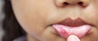
Candidiasis of the oral mucosa (oral candidiasis, infantile thrush) most often occurs in children; as a rule, they become infected from the mother through the birth canal. Symptoms:
- the mucous membranes of the cheeks, pharynx, tongue and gums become red,
- swelling appears,
- then pockets of white cheesy plaque appear on the oral mucosa.
With candidiasis of the skin and its appendages, the lesions are most often located in large folds:
- inguinal-femoral,
- intergluteal,
- armpits,
- under the mammary glands.
The skin in the interdigital folds may be affected, more often in children and adults suffering from serious illnesses - on the skin of the palms, feet, smooth skin of the torso and limbs. Lesions in large folds look like small 1-2 mm bubbles, which soon open to form erosions. Erosion increases in size and merges, forming large areas of damage.
Foci of candidiasis have an irregular shape, dark red color, and around the lesion there is a strip of exfoliating epidermis. Outside the folds, the lesions look like red spots with peeling in the center, and occasionally small blisters may appear around the lesion.
Vaginal candidiasis (candidiasis, thrush) is an infectious disease of the vaginal mucosa, which often spreads to the neck of the uterus and vulva. Almost every woman has encountered this disease, and some signs of candidiasis are constantly disturbing. Most often occurs in women of reproductive age, but can occur in girls
Intestinal candidiasis (dysbacteriosis) often accompanies vaginal candidiasis or develops in isolation. Typically, intestinal candidiasis appears after taking antibiotics or previous intestinal infections. Fungi of the genus Candida settle in the small intestine. Symptoms characteristic of this type of candidiasis: white curdled flakes are often found in the stool of a patient suffering from intestinal candidiasis.
Esophageal candidiasis is a disease that is very difficult to identify among all those available in the field of gastroenterology. The disease is characterized by a discrepancy between the severity of the disease, the level of damage and the condition of the patient himself.
Candidiasis of interdigital skin folds
Candidiasis of the interdigital folds is often an occupational disease and occurs in people employed in production using Candida spp. for industrial purposes, or those where there is a high probability of contamination by them (confectionery, canning, processing of vegetables and fruits, etc.), as well as for women who do a lot of housework without protective gloves, nurses.
Hand skin candidiasis
More often, the folds between the third and fourth fingers of the hands and the lateral surfaces of these fingers are affected, where the skin is macerated, whitish in color, and is easily torn off, exposing an eroded shiny surface.
Complication
With timely treatment, candidiasis does not cause any particular harm to health. But the symptoms of candidiasis can cause a lot of discomfort. Long-term, it can lead to damage to other organs, most often the urethra, bladder and kidneys. In particularly severe cases, the progressive disease can affect the reproductive organs, leading to infertility in both men and women. But candidiasis poses the greatest danger to pregnant women, because... the risk of fetal damage is very high.
Symptoms of scalp fungus
- Dryness and redness of the skin, severe itching, the appearance of small round plaques of a pinkish tint, profuse dandruff in the form of keratinized whitish or grayish scales.
- Painful appearance of hair - thinning, dryness, split ends, fragility of the hair shaft along the entire length. Partial or complete hair loss may occur in the affected areas.
- Increasing the intensity of the sebaceous glands, hair quickly loses its freshness and becomes oily at the roots within a few hours after washing.
- In severe forms of fungal infections, purulent inflammatory processes may begin due to blockage of the sebaceous ducts.
If left untreated, Pityrosporumovale bacteria multiply uncontrollably and damage to the skin of the face and neck may occur.
To pre-identify a scalp fungus, you can study photos of symptoms on the Internet. However, in any case, the course of treatment should be prescribed by an experienced dermatologist.
How is the disease diagnosed?
Visual methods for diagnosing candidiasis. Upon examination, inflammation of areas of the skin is revealed, limited to a border of exfoliating, macerated epidermis, and a whitish coating on the mucous membranes.
Laboratory diagnostics. Contrary to popular belief, the main method for diagnosing candidiasis is still microscopy of a smear from the affected areas of the mucosa. PCR (DNA diagnostics), which has become popular recently, is usually poorly suited for diagnosing candidiasis.
Laboratory diagnosis of the disease includes:
- microscopy of discharge smear
- cultural diagnostics (seeding)
- enzyme immunoassay (ELISA)
- polymerase chain reaction (PCR).
Causes of fungal skin infections
Skin, as an indicator of internal disorders, can signal various malfunctions in the functioning of internal systems. In particular, the following deviations from the norm can become incentives for intensive proliferation of fungi:
- imbalance of hormones of the reproductive system and thyroid gland;
- chronic overwork of the nervous system – regular stress and lack of sleep;
- disruptions in the digestive system and metabolic disorders;
- a decrease in general immunity due to past illnesses or as a result of drug treatment;
- vitamin deficiency and lack of vital microelements in the diet;
- increased sebaceous production of the skin.
Scalp fungus can develop not only as a result of diseases. The reason may be hereditary predisposition, improper hair care, the use of other people's personal hygiene products, etc.
Treatment of candidiasis
Treatment of candidiasis is aimed at eliminating factors that contribute to the occurrence of candidiasis. If the skin is affected, local treatment is carried out using an open method using antifungal ointments.
The attending physician prescribes systemic and local medications for this disease. Local agents are not absorbed into the blood - they act only on the mucous membrane affected by the Candida fungus. They stop the reproduction and growth of fungi, relieve discomfort and restore affected tissue.
Candidiasis of the skin outside the folds (smooth skin)
Candidiasis of the skin of the body develops, as a rule, secondarily, when the process spreads from the folds, mucous membranes or affected nails and periungual ridges, and is characterized by a wide variety of clinical manifestations.
Candidiasis of smooth skin
In the typical variant, the disease is characterized by the appearance of small blisters and pustules with a flaccid lining, which are highly resistant.
With atypical variants, for example with HIV infection (photo below), the manifestations of candidiasis can be completely unusual.
Facial candidiasis
Diagnostic methods
The treatment is carried out by a dentist-therapist. Diagnosis begins with an examination and a detailed survey: the doctor will find out what medications you have taken recently, and whether there are any chronic or infectious diseases. A cytological examination of plaque taken from the mucosa is mandatory. This is important because a buildup of non-fungal flora can easily be confused with a fungal infection.
The scraping is performed in the morning, on an empty stomach; there is no need to brush your teeth before the procedure. The day before, it is important to avoid eating foods rich in carbohydrates so as not to provoke the growth of pathogenic flora. Research allows not only to accurately determine the causative agent and type of Candida fungus, but also to find out the sensitivity of fungi to the main antifungal drugs. Based on the test results, the doctor will determine the fungus in the oral cavity and prescribe medication.
