If you think that dentists never tire of repeating the importance of preventive examinations and timely treatment of caries and gum inflammation just for the sake of advertising their profession, you are mistaken. Inattention to the health of your own teeth and vain fears of dental treatment can ultimately lead to complications, the treatment of which will be both difficult and expensive. A striking example of such a complication is dental granuloma - a pathological formation that occurs in the root part of the tooth.
In the early stages of its development, granuloma does not show itself with any noticeable symptoms, but as the tumor grows and becomes inflamed, severe pain in the tooth may appear, which cannot be relieved with tablets from the pharmacy. It is possible to remove a granuloma and save a diseased tooth only in dentistry and there is no need to delay contacting a doctor, because an untreated granuloma can lead to the development of serious complications, including osteomyelitis, general infections of the body and even cancer.
In this article we will tell you:
- What causes dental granuloma?
- Symptoms of dental granuloma;
- How is dental granuloma treated?
Also in the article you will find information on the prevention of dental granuloma and prices for treatment/removal of dental granuloma in Moscow.
Classification of granulomas and forms
In medicine, granulomas are classified according to several criteria: etiology, pathogenesis and morphology. Specific varieties are collected in a separate group. In the group according to the criterion of etiology there are types of established and unidentified etiology: infectious and non-infectious. The latter include dust granulomas, drug-type granulomas and neoplasms around foreign bodies. Pathogenesis group: immune and non-immune. The first type includes epithelioid cell granulomas. Non-immune ones occur due to toxic factors and acute infections. The morphological group is divided into two main groups: mature and epithelioid cell. According to the morphology of granulomas, there is the formation of a diffuse-type granulomatous infiltrate and the formation of tuberculoid-type granulomas.
Main types of granulomas
In medical practice, there are many types of neoplasms of this type. They can be single or multiple. It is not always possible to see the beginning of their formation, since pathogenic cells are located deep in the layers of the dermis. Granuloma in children and adults can appear and disappear without treatment. This should justify the need for gentle therapy in the future. Granulomas of the following type are most often diagnosed in children and adults:
- ring-shaped;
- pyogenic;
- tuberculosis.
Causes and development factors
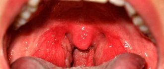
Granuloma annulare in children can appear for various reasons, which have not yet been fully established. The provoking factor is a focus of chronic infection in the form of rheumatism, tonsillitis. The presence of diabetes mellitus, allergic diseases, and metabolic pathologies increases the risk of development. In children, a lot depends on the immune system. If there is a failure, the development of the neoplasm will be started. Pyogenic granuloma in a child occurs due to a pyococcal infection. The reason for the development lies in skin injuries, due to which infection gets into the wounds. It is localized mainly on the face, legs, fingers and arms. Eosinophilic granuloma is rarely found in young children. The peak incidence occurs between 5 and 10 years. The causes of such granuloma in a child are ambiguous. The question still remains open. Scientists suggest that immunopathological processes are the basis.
Stages of granuloma development
From the moment of inception, the neoplasm goes through 4 stages. At the initial stage, there is an accumulation of cells with a tendency to phagocytosis. At the second stage, the process of their rapid growth begins. The third stage is characterized by the transformation of phagocytes into epithelial cells. At the last fourth stage, granuloma formation occurs directly.
Conservative (medicinal) treatment of dental granuloma
This technique is used only for those cases in which the development of granuloma has not yet progressed far. The essence of conservative therapy for tooth root granuloma will be the intake and use of various drugs - antibiotics, antiseptics, drugs that have an anti-inflammatory effect. All medications are prescribed by a dentist and the treatment process takes place under the strict supervision of a specialist. This treatment option for granuloma is effective with early diagnosis of the disease and, if you strictly follow the doctor’s instructions, the granuloma will resolve within 1-2 weeks.
In some cases, conservative treatment of granuloma is complemented by root canal therapy. Working with tooth canals for root granuloma is indicated:
- For constant toothaches, the intensity of which does not subside from taking painkillers;
- Swelling of the gums, spreading to the cheek and jaw;
- Treatment of tooth canals is clearly indicated when large granulomas are detected - from five millimeters.
Symptoms and signs
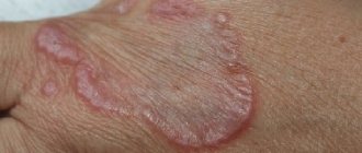
Granuloma annulare in children has different causes, but the symptoms do not change. Mainly affects the bones of the pelvis, skull, and spine. The course of the disease is slow. The symptom of eosinophilic granuloma is pain and swelling at the site of localization. There are cases where there is no discomfort. On the surface of the skin it appears as semicircular intradermal lesions. Granuloma annulare in children of a localized form is usually located on the hands and arms, feet and legs. It is diagnosed mainly before the age of 6 years. Ring-shaped lesions range in diameter from 2 to 5 cm. Giant sizes up to 10 cm are less common.
On the body, a pyogenic granuloma in a child does not exceed 2 cm in diameter. It has a thin surface and can bleed. The structure is fragile. Occurs quickly due to skin trauma. It does not turn pale when pressed. There is a rim of skin around the granuloma or it may be raised on a stalk. Rarely does a rash appear in the affected area. A characteristic symptom that children complain about is itching. Ring-shaped and pyogenic varieties occur in children more often than other neoplasms from the granulomatous group.
When to see a doctor
If papules of red-pink and bluish color appear on the skin, you should contact a dermatologist to confirm the diagnosis at JSC “Medicine” (clinic of Academician Roitberg) in the center of Moscow. Granuloma annulare in children causes and treatment depends on the degree of proliferation of foci and intensity. You will also need the help of a specialist in case of itching. Treating yourself or visiting a doctor is not a conflict of interest. Granuloma in a child may have characteristic causes, but the clinical manifestations can easily be confused with another serious disease. We are talking about lichen, which is sometimes mistakenly self-diagnosed based on external signs. There are a number of other diseases that stand apart, but can be mistaken for granuloma annulare: erythema raising, necrobiotic lesions, rheumatoid nodules.
What is dental granuloma, the reasons for the formation
Dental granuloma is a pathological neoplasm of periodontal tissues affected by inflammation, appearing in the upper part of the tooth root. Externally, the granuloma looks like a dense small nodule (up to 0.5 cm in diameter). If you do not urgently treat a dental granuloma, it will continue to develop and deform into a radicular cyst - a capsule that is filled not only with dead tissue cells, but also with pus and bacteria.
Some people believe that a cyst and a dental granuloma are the same thing. But these are different diseases. The tooth cyst is larger in size and does not show any symptoms longer. As granuloma develops, the tooth will become very painful.
What causes dental granuloma? There are a number of reasons that contribute to the occurrence of pathological formation at the apex of the tooth root:
1. Granuloma under the tooth can occur as a complication of caries, pulpitis, periodontitis, the treatment of which was not carried out on time. It is important to understand that no dental disease goes away on its own! If you don’t treat your teeth, they will continue to decay, and the inflammatory process will spread to the internal tissues of the tooth and to its root!
2. Poor quality of dental treatment can lead to the appearance of a granuloma on the root of a tooth. If mistakes were made during the treatment of caries or other dental diseases, for example, the tooth canals were poorly cleaned and treated, then in the future there is a high risk of various complications, including the formation of granulomas on the root of the tooth.
3. Severe tooth trauma, leading to infection penetration to the root.
These are the main reasons that can provoke the growth and development of granulomas on the roots of the tooth. If you do not want to face such an unpleasant disease, never delay dental treatment, which you can undergo without pain and stress in our dental clinic in Moscow - Firadent.
It is worth learning and remembering that granuloma can appear after tooth extraction. Most often this happens when the tooth was removed incorrectly or after the operation the patient did not follow the doctor’s recommendations on hygiene. Bacteria easily enter the open socket that remains after the tooth is extracted from the gums, and as a result of their active reproduction, an inflammatory process begins and a granuloma is formed.
Available diagnostic methods
At JSC “Medicine” (academician Roitberg’s clinic) near the Mayakovskaya metro station, you can comprehensively examine the health of adults and children. The latest equipment (ultrasound, MRI, CT) and advanced diagnostic methods, specially equipped rooms - everything is there to confirm or refute granuloma annulare in children. In the early stages of development, neoplasms cannot be detected. Symptoms appear when lesions appear on the surface of the epidermis. A diagnostic examination by a dermatologist is carried out when there are complaints of skin changes. Only after this do they turn to laboratory and histological analyzes of skin biopsies from lesions in case of suspicion of deep forms of lesions.
Clinical case
Based on Lam S. et al, Eosinophilic granuloma/Langerhans cell histiocytosis: Pediatric neurosurgery update. 2015
A 17-year-old young man was hospitalized due to an increasing scalp lesion over the past 6 weeks. The formation is painful on palpation and periodically bleeds due to ulceration, but no neurological deficit has been identified. CT and MRI revealed a large lesion in the frontal bone on the right, compressing the superior sagittal sinus. A total resection of the formation was performed, and the diagnosis of Langerhans cell histiocytosis of the skull bone was confirmed. At the outpatient stage, cytostatic therapy was carried out.
Figure 6. (a) CT examination without contrast - frontal scan (upper and middle part) and 3D reconstruction of the skull (lower part). (b) MRI scan. T1-weighted image in the coronal plane (top) and T2-weighted image in the sagittal plane
Sources
- Coppes-Zantinga A., Egeler RM The Langerhans cell histiocytosis X files revealed. Br J Haematol, 2002. - V. 116 - N. 1 - P. 3–9.
- Lam et al., Management of adult patients with Langerhans cell histiocytosis: recommendations from an expert panel on behalf of Euro-Histio-Net. Orphanet J Rare Dis, 2013 - V. 8. - N 72.
- Lam S., Reddy GD, Mayer R., Lin Y., Jea A. Eosinophilic granuloma/Langerhans cell histiocytosis: Pediatric neurosurgery update. Surg Neurol Int, 2015. - N. 6 (Suppl 17): S435 - S439.
- Langerhans' Cell Histiocytosis (Histiocytosis X). What is it? Harvard Medical School. Harvard Health Publishing. October, 2014. www.health.harvard.edu
- Sharma R., Singh R. et al. Langerhans cell histiocytosis (skeletal manifestations). Radiopaedia. https://radiopaedia.org/articles/langerhans-cell-histiocytosis-skeletal-manifestations-1
- Shea C. R, James W. D. et al. Langerhans Cell Histiocytosis. Medscape, 2021. https://emedicine.medscape.com/article/1100579‑overview
- Churilov L.P. Death on takeoff, or Who are you, Doctor Taratynov? Health is the basis of human potential: problems and ways to solve them, 2014. - T. 9 - No. 2 - P. 919–929.
- Yusupova L. A., Yunusova E. I., Garayeva Z. Sh., Mavlyutova G. I. Histiocytosis X. Practical Medicine, 2014 - T. 08 - No. 14.
Methods of treating the disease
Granuloma annulare in children requires restrained treatment. The characteristics of the body allow us to expect spontaneous resolution. According to statistics, in 75% of cases, granulomas regress within 2 years. A granuloma may reappear on the skin of a child in 40% of patients. It is important that recurrent tumors behave benignly. For painful symptoms, corticosteroid ointment with a pronounced anti-inflammatory and antiallergic effect is used. Another treatment method is superficial scarification. How to treat granuloma annulare in a child without harm to health is an important topic. The methods are based on the use of physiotherapeutic procedures: PUVA therapy, ultraviolet irradiation and phototherapy, pulsed laser. The treatment of pyogenic neoplasm is characterized by excessive proliferation of blood capillaries. With this diagnosis, it is recommended to be treated by excision, curettage, liquid nitrogen and electrocoagulation. Granuloma annulare in children is treated within a month. The need for manipulation and use of ointments is determined individually.
Complications of granuloma
If granuloma treatment is ignored, the following complications may occur:
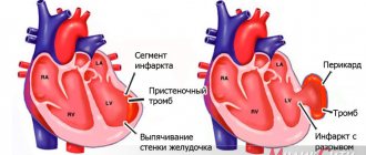
flux inflammation in the periosteal tissues; osteomyelitis is an inflammatory process in the jaw; cyst inflammatory neoplasm.
Since granuloma is a source of infection, complications can affect other body systems. So, the disease can be provoked by:
sepsis blood poisoning; sinusitis, inflammation of the maxillary sinuses; infectious myocarditis inflammation of the heart muscle; pyelonephritis is a kidney disease.
Prevention in children
Any granuloma in a child is treated only after consultation with a dermatologist. There are no special methods of prevention. Granuloma annulare in children has variable causes for the development of the disease. Taking into account the influence of immunity on the occurrence of formations, it is necessary to undergo timely treatment. It is necessary to take into account the infectious nature and metabolism in the body. Any disease should receive therapy to reduce risks. This is what prevention consists of – eliminating factors that can provoke the disease.
How to make an appointment with a dermatologist
You can make an appointment with a dermatologist using the special form “Make an appointment with a doctor” - just enter your data so that the administrator can call you back and coordinate the time of the visit. During the consultation, the doctor will examine the clinical picture, prescribe tests if necessary, and select treatment. You can make an appointment with a dermatologist using the number. JSC "Medicine" (clinic of academician Roitberg) has a convenient location at the address: 2nd Tverskoy-Yamskaya lane, building 10. Nearby there are metro stations: "Mayakovskaya", "Tverskaya", "Novoslobodskaya", "Belorusskaya", "Chekhovskaya" .