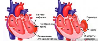
Shortness of breath is a protective reaction of the body aimed at normalizing the gas composition of the blood. According to the definition of clinicians and pathophysiologists, shortness of breath is a condition that is characterized by a severe subjective feeling of lack of air against the background of disturbances in the frequency, rhythm, depth of respiratory movements, and the duration of inhalation or exhalation. The Yusupov Hospital has created all the conditions for the treatment of patients who have inspiratory dyspnea:
- Chambers with European level of comfort;
- The latest diagnostic equipment, which allows you to determine the type and severity of shortness of breath, identify diseases that have led to impaired respiratory function;
- The use of modern drugs that are highly effective and have a minimal range of side effects is registered in the Russian Federation;
- Attentive attitude of the staff to the wishes of patients and their relatives.
Severe cases of diseases manifested by inspiratory dyspnea are discussed at a meeting of the Expert Council. Professors, doctors of medical sciences, and doctors of the highest category take part in its work. Patients of the therapy clinic are advised by pulmonologists and cardiologists. With severe inspiratory dyspnea, patients are transferred to the intensive care unit, which is equipped with expert-class breathing equipment.
Types and mechanisms of development of shortness of breath
According to the nature and form of external respiration disturbance, shortness of breath can be inspiratory, expiratory and mixed. In the presence of inspiratory dyspnea, the duration of the inhalation phase in relation to exhalation increases. Expiratory dyspnea is characterized by a predominance of exhalation over inhalation. With mixed shortness of breath against the background of an extended inhalation, the exhalation becomes even longer.
The development of inspiratory, expiratory and mixed dyspnea is ensured by the Hering-Breuer reflex. In its formation, an important role is played by highly sensitive, low-threshold mechanoreceptors of alveolar stretch. The impulses of these receptors are sent along the alpha fibers of the vagus nerve to the reticular formation of the brain stem and the respiratory center of the medulla oblongata, and then along the nerve pathways that go from the center to the respiratory muscles.
At the beginning of inspiration, when the alveoli are slightly filled with air and slightly stretched, low-frequency impulses are formed that are optimal for the inspiratory neurons of the respiratory center. Their activation leads to inhalation. As the degree of stretching of the alveoli increases, in cases where they are filled with air at the height of inspiration, a flow of impulses occurs from the mechanical alveolar stretch receptors and goes to the brain. They are not optimal for inspiratory neurons, which leads to interruption of the act of inhalation and the development of exhalation.
In cases of the development of limited disturbances in pulmonary ventilation due to pulmonary edema, pneumofibrosis, when the elasticity of the lung tissue decreases, the stretching of the alveoli under the influence of the physiological volume of inhaled air becomes significantly more difficult; from the mechanoreceptors of the stretching of the alveoli, low-frequency impulses enter the bulbar center for a long time. They stimulate inspiratory neurons for a long time, which leads to prolongation of the act of inspiration - the development of inspiratory dyspnea.
Inspiratory dyspnea in various pathologies
Reflex influences from the baroreceptors of the aorta and carotid sinuses are included in the mechanism of development of shortness of breath during blood loss, collapse, shock, and collapse. When blood pressure is below 70 mm Hg. Art. the flow of impulses that inhibit the inhalation center decreases.
If the oxygen tension in the blood decreases, the concentration of carbon dioxide increases, or the level of hydrogen ions increases, the flow of impulses from central and peripheral chemoreceptors to the bulbar respiratory center increases, inhalation is activated, and inspiratory dyspnea develops. This occurs in patients suffering from cardiac, respiratory, renal failure, anemia, as well as in acid-base balance disorders of various origins.
The cause of the feeling of insufficient breathing may be excessive stretching of the intercostal muscles during heavy physical work, narrowing of the upper respiratory tract, decreased elasticity of the lungs, when muscle proprioceptors are excited. From them, impulses travel to the higher parts of the brain. Stimulation of the respiratory center and inspiratory shortness of breath occurs with local disorders of cerebral circulation (spasm, thrombosis of cerebral vessels, embolism, development of destructive changes of allergic, infectious origin).
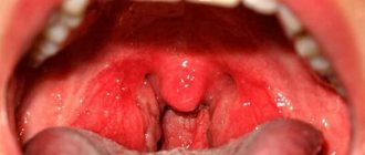
Inspiratory dyspnea can be a symptom of lung cancer, larynx, and carcinomatosis. Sometimes inspiratory dyspnea occurs in pregnant women due to the high position of the diaphragm. Inhalation becomes difficult when a foreign body enters the respiratory tract, laryngeal stenosis and swelling of the vocal cords, laryngitis.
Degrees and signs of inspiratory dyspnea
There are five degrees of severity of inspiratory dyspnea:
- Zero – there is no shortness of breath at rest, it occurs after physical exertion;
- Mild – inspiratory shortness of breath occurs after climbing stairs or walking quickly;
- Medium – breathing is difficult during normal walking, which forces a person to slow down and stop to rest;
- Severe – a person cannot walk more than 100 m without stopping;
- Very severe - bothers the patient in a state of physical and emotional rest.
The main symptom of inspiratory dyspnea is difficulty breathing during inspiration. A person sometimes hears whistling or wheezing when breathing. With severe inspiratory dyspnea, these noises can be heard by people around.
Since inspiratory dyspnea is not an independent pathology, but a symptom of the disease, the patient is concerned about other manifestations of the disease:
- When foreign objects enter the respiratory tract, heaviness in the chest, pain at the location of the foreign body, coughing, and suffocation develop;
- In patients suffering from pleurisy and other diseases in which there is a loss of elasticity of the respiratory tract, cough, chest pain, and pale skin occur;
- With diseases of the cardiovascular system, the patient experiences heart rhythm disturbances, pain in the chest while walking or after emotional stress;
- In patients with lung cancer, shortness of breath and a mild but prolonged cough become the first signs of pathology;
- With pulmonary tuberculosis, patients complain of difficulty breathing, coughing, night sweats, and low-grade body temperature.
To find out the cause of the development of inspiratory dyspnea, contact the doctors at the Yusupov Hospital.
Differential diagnosis of dyspnea in clinical practice
Mechanisms of dyspnea
Shortness of breath is a manifestation of a discrepancy between the increased demands of gas exchange and the load performed by the respiratory muscles.
Dyspnea can occur during normal gas exchange, but also in the presence of pathology of the respiratory muscles [3]. Shortness of breath is a subjective sensation that is difficult to measure objectively. At the same time, it is possible to determine indirect signs of shortness of breath: changes in frequency (tachypnea), dependence on position (orthopnea, platypnea) and disruption of the respiratory cycle (pathological types of breathing). There are also ways to objectify the patient’s sensations - various scales and questionnaires that are provided to the patient to assess the severity of shortness of breath. However, such instruments are used more in research than in clinical practice. The respiratory regulation system consists of an efferent (directing impulses to the respiratory muscles), an afferent (directing impulses from the receptors to the brain) link and the breathing center itself, which processes information. A disruption in each of these links can lead to shortness of breath. Thus, if external respiration is impaired, the work of the respiratory muscles increases, and with pathology of the respiratory muscles, greater efforts are required. It is believed that impulses from motor efferent neurons, in parallel with the respiratory muscles, are sent to the sensory cortex, which leads to the sensation of shortness of breath. Sensory afferent impulses are enhanced by activation of chemoreceptors by hypoxia, hypercapnia and acidosis. This also results in a feeling of shortness of breath. The same mechanism is activated in response to bronchospasm, increased pulmonary artery pressure, and even hyperinflation. Finally, in severe anxiety disorders, shortness of breath is a consequence of misinterpretation of signals from the afferent circuit, as well as increased breathing rates that exceed physiological needs [4–6].
Pathophysiological causes of shortness of breath are usually divided into pulmonary and extrapulmonary. The first includes the occurrence of shortness of breath in obstructive diseases (bronchial obstruction, stenosis of the upper respiratory tract), restrictive lung pathology (infiltrative processes, pulmonary fibrosis, after resection of a lung lobe) and vascular diseases of the lungs (PE, pulmonary hypertension, intrapulmonary shunts). Extrapulmonary mechanisms include extrapulmonary restriction (morbid obesity, diaphragmatic paralysis, neuromuscular diseases, severe kyphoscoliosis), cardiovascular diseases (systolic and diastolic myocardial dysfunction, valvular defects) and other causes, including anemia, severe acidosis of any origin, third trimester pregnancy and respiratory regulation disorders (panic attack with hyperventilation syndrome, alveolar hyperventilation) (Tables 1, 2).
Shortness of breath due to lung diseases
In clinical practice, shortness of breath most often occurs in diseases with the development of obstructive disorders of the external respiration function, i.e., with increased resistance to the flow of inhalation or exhalation. With restrictive respiratory disorders due to infiltration, fibrosis or edema, the compliance of the lungs decreases. Detection of severe broncho-obstructive syndrome usually does not cause significant difficulties due to the typical medical history and characteristic auscultatory pattern (dry wheezing, including during forced exhalation). At the same time, with a combination of obstructive and restrictive lung lesions, the auscultatory picture can be quite poor. The same is true for mild persistent bronchial asthma in trained individuals. The key to identifying bronchial obstruction as the cause of shortness of breath in such patients is to conduct a pulmonary function test (always using a bronchodilator) and body plethysmography in unclear cases.
Dyspnea due to chronic pulmonary pathology is characterized by aggravation during exercise, as well as a lack of connection between shortness of breath and body position. The exception is orthopnea in patients with severe exacerbation of bronchial asthma or chronic obstructive pulmonary disease (COPD), platypnea (increased shortness of breath in an upright position due to the development of hypoxemia) in patients with the presence of intrapulmonary shunts with blood discharge from right to left (with vascular malformations, portopulmonary hypertension ), as well as after pneumonectomy. It is believed that the development of platypnea may be associated with the opening of the oval window after pneumonectomy. In a standing position, the flow of blood from right to left increases, which leads to shortness of breath [7].
Shortness of breath due to heart disease
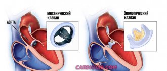
Dyspnea is one of the main manifestations of left ventricular failure, both systolic and diastolic. Shortness of breath is associated with increased pressure in the chambers of the heart and, as a consequence, pulmonary venous hypertension. Hypervolemia, another manifestation of heart failure, plays a significant role in increasing the pressure in the chambers. Dyspnea in heart failure increases with exertion, and in case of decompensation, at rest. In the latter case, shortness of breath increases in the supine position (orthopnea), including after falling asleep (cardiac asthma). Decompensated left-sided heart failure is characterized by signs of hypervolemia (moist rales in the lungs, pleural effusions, bulging of the external jugular vein, edema). In some cases, with decompensation due to swelling of the bronchial wall, bronchial obstruction with characteristic manifestations may develop (wheezing, dry rales, changes in pulmonary function tests). The presence of signs of fluid overload and known heart pathology (history of myocardial infarction, valve defects, long-term history of arterial hypertension, atrial fibrillation) allows diagnosing heart failure as the cause of shortness of breath without much difficulty. It is much more difficult to identify heart failure as the cause of dyspnea in the absence of signs of volume overload, which is especially true in diastolic heart failure. In this situation, determining the level of brain-type natriuretic peptide (BNP) may be useful.
The concentration of MNUP increases in parallel with the increase in overload of the ventricular myocardium (right or left) with volume or pressure, i.e., the filling pressure of the chambers. Values of BNP (BNP) more than 400 pg/ml, and its n-terminal precursor (NT-proBNP) - more than 1600 mg/dl - indicate a cardiac cause of shortness of breath. BNP values less than 100 pg/ml, and NT-proBNP values less than 300 pg/ml are likely to exclude it. On the other hand, MNUP also reflects an increase in pressure in the right chambers, thus its content in the blood can increase with pulmonary hypertension, PE and cor pulmonale. In morbidly obese patients, especially women, BNP levels, on the contrary, may be significantly reduced even in the presence of heart failure [8].
A difficult clinical task is the differential diagnosis between shortness of breath in heart failure with preserved ejection fraction, without yet signs of severe fluid overload, and shortness of breath as an equivalent of angina pectoris. One gets the impression that the latter is overdiagnosed in domestic clinical practice. Key to the differential diagnosis in this case are the characteristics of dyspnea (longer in heart failure), the results of stress testing, and the response to loop diuretic therapy. It should be noted that nitrates reduce shortness of breath in both cases. Therefore, in these patients, a positive response to nitroglycerin cannot be considered as a differential diagnostic sign.
Another cause of short attacks of shortness of breath may be heart rhythm disturbances, for example, frequent ventricular extrasystole, especially of the bigeminy or trigeminy type, with an initially low pulse and short paroxysms of atrial fibrillation. Rhythm disturbances are not always detected when recording a standard 12-lead ECG. Daily Holter ECG monitoring may be required to clarify the nature of rhythm disturbances and their correspondence in time to symptoms.
Another cause of short-term episodes of shortness of breath may be pulmonary arterial hypertension (primary, as part of systemic connective tissue diseases), which is characterized by “crises” - increases in pressure in the pulmonary vessels, accompanied by shortness of breath.
Despite this, in most cases, the differential diagnosis of “cardiac” and “pulmonary” dyspnea does not cause great difficulties. The exception is patients with concomitant diseases of the heart and lungs, in whom it is necessary to identify the prevailing mechanism.
Other causes of shortness of breath
Shortness of breath during moderate exertion is quite common in anemia and thyrotoxicosis, conditions with high cardiac output. In this case, the severity of shortness of breath depends on the initial state of the cardiovascular system.
Dyspnea and tachypnea, even at rest, accompany metabolic acidosis of any origin. In clinical practice, most often this is diabetic ketoacidosis, acidosis due to renal failure (including renal tubular acidosis with hyperkalemia in patients with diabetic nephropathy and a moderate decrease in filtration during spironolactone therapy), as well as acidosis due to poisoning with salicylates and antifreeze. An increase in progesterone concentrations, characteristic of the third trimester of pregnancy, also contributes to the development of shortness of breath with light exertion.
Dyspnea during exertion is also caused by diseases that cause extrapulmonary restrictive disorders, including severe kyphoscoliosis, pleural effusion, significant thickening of the pleura and pathology of the diaphragm.
Finally, shortness of breath as part of hyperventilation syndrome is a common manifestation of anxiety disorders and a number of neuroses and neurosis-like conditions, in which clinical manifestations can be quite pronounced.
Clinical approach to a patient with complaints of shortness of breath
When analyzing complaints and medical history, special attention should be paid to the description of the patient’s feeling of shortness of breath, the speed of its development and the effect on the severity of shortness of breath of changes in body position, the addition of infections and changes in external factors, such as temperature and humidity. The range of diseases leading to the sudden onset of shortness of breath and its gradual development varies. Moreover, a sharp increase in long-term shortness of breath may indicate both the progression of the main process and the addition of a second disease. Among the diseases leading to the sudden development of severe shortness of breath, the most common in clinical practice are pneumonia, decompensated or acute heart failure (including with the development of silent myocardial infarction of the status asthmaticus type), pulmonary embolism, broncho-obstructive syndrome (exacerbation of bronchial asthma or COPD ), pneumothorax (including spontaneous), foreign body aspiration, hyperventilation syndrome and metabolic acidosis (most often ketoacidosis) [9]. Most of these diseases, with a typical clinical picture, do not cause significant difficulties for diagnosis, with the exception of PE, in which most often the only symptoms are shortness of breath, tachycardia, chest pain and decreased oxygen saturation at rest. It should be noted that cyanosis and hemoptysis occur in a minority of patients with PE [10]. The same applies to the classic ECG change Q1S3T3 (the most common ECG change in PE is nonspecific ST–T changes along the anterior wall of the left ventricle) [11]. Most diseases that lead to the development of severe shortness of breath require hospitalization and inpatient treatment.
In outpatient practice, we more often encounter cases of chronic shortness of breath, when the differential diagnosis is made between cardiac, pulmonary, cardiopulmonary and “non-cardiac and non-pulmonary” causes of shortness of breath. The occurrence of shortness of breath in a horizontal position is most typical for heart failure, but also occurs in bronchial asthma associated with gastroesophageal reflux and morbid obesity. Night attacks of shortness of breath and suffocation suggest the presence of heart failure or bronchial asthma. When collecting anamnesis, it is necessary to pay special attention to cardiovascular risk factors and the patient’s professional path (Fig. 1).
Shortness of breath when talking indicates a significant decrease in the vital capacity of the lungs (with pulmonary edema, late stages of interstitial diseases) or hyperstimulation of the respiratory center (panic attack, acidosis). The participation of accessory muscles during breathing indicates severe bronchial obstruction and/or a significant decrease in lung elasticity. A careful examination may reveal signs of certain diseases associated with shortness of breath. Thus, swelling of the neck veins in a sitting position indicates an increase in pressure in the right atrium, i.e., the presence of right ventricular heart failure. Thickening of the nail phalanges like Hippocrates' fingers may indicate the presence of interstitial lung diseases as a cause of shortness of breath; Raynaud's syndrome is associated with pulmonary hypertension in systemic scleroderma and other systemic connective tissue diseases. Paradoxical movement of the abdominal wall (inward movement during inspiration while lying down) indicates damage to the diaphragm, usually bilateral.
In many cases, a thorough analysis of complaints, anamnesis and examination of the patient is sufficient to make a diagnosis. If the cause of shortness of breath is not clear, the next step is chest x-ray (CH), which can identify cardiomegaly as a common manifestation of heart failure, as well as infiltrative changes in the lungs, hyperinflation as a manifestation of broncho-obstructive diseases and pleural effusion. Most patients also need to undergo electrocardiography and pulmonary function testing if ventilation disorders are suspected. In many cases, the definition of MNLP, as discussed above, provides significant assistance. Among other causes of chronic shortness of breath in clinical practice, the most common are anemia, thyrotoxicosis, obesity or exercise, chest pathology and neuromuscular diseases [9]. Therefore, performing a clinical blood test as well as TSH can provide the information necessary to make a diagnosis.
In the absence of a clear clinical picture, as well as the presence of concomitant diseases of the heart and lungs, it is necessary to conduct a stress test with gas analysis and spiroergometry. This technique allows you to determine indicators of pulmonary gas exchange during exercise: oxygen consumption, carbon dioxide production, as well as minute pulmonary ventilation. Since in lung diseases, exercise tolerance is limited by disturbances in respiratory mechanics (obstructive or restrictive), shortness of breath occurs as a result of achieving maximum voluntary ventilation (MVV). The difference between MVV and VEmax measured at peak load is called respiratory reserve and is normally 50–80% of MVV. In patients with chronic lung diseases, VEmax during exercise approaches MVV to a much greater extent. This means that exercise tolerance in such patients has “pulmonary limits”, respiratory reserve <50%.
In cardiac disease, shortness of breath occurs due to decreased contractile reserve, so the limitation of peak oxygen uptake (VO2peak) and ventilatory threshold (VT) are caused by inadequate transport of oxygen to the periphery, and the ventilatory reserve remains normal (> 50%). There are other respiratory parameters for differentiating dyspnea, each of which has more or less good sensitivity and specificity. Through a comprehensive analysis of these parameters, spiroergometry allows one to draw conclusions about the factors limiting physical performance [12, 13].
Shortness of breath is a common complaint that prompts medical attention. The use of a stepwise approach, based on the analysis of complaints, the clinical picture and the use of additional methods in individual cases, makes it possible to identify the cause of shortness of breath in most patients at the outpatient level.
Literature
- Elliott MW, Adams L, Cockcroft A et al. The language of breathlessness. Use of verbal descriptors by patients with cardiopulmonary disease // Am. Rev. Respirat. Disease. 1991. Vol. 144. P. 826–832.
- Chuchalin A.G. Dyspnea: pathophysiological and clinical aspects // Pulmonology: scientific and practical journal. 2004. No. 5. P. 6–16.
- Tobin MJ Dyspnea. Pathophysiologic basis, clinical presentation, and management // Arch. Intern. Med. 1990. Vol. 150. P. 1604–1613.
- Banzett RB, Pedersen SH, Schwartzstein RM, Lansing RW The affective dimension of laboratory dyspnea: air hunger is more unpleasant than work/effort // Am. J. Respira. Crit. Care Med. 2008. Vol. 177(12). P. 1384–1390.
- American thoracic society consensus. Dyspnea. Mechanisms, assessment, and management: a consensus statement. American Thoracic Society // Am. J. Respira. Crit. Care Med. 1999. Vol. 159(1). P. 321–340.
- Mahler D. A., Harver A., Lentine T. et al. Descriptors of breathlessness in cardiorespiratory diseases // Am. J. Respira. Crit. Care Med. 1996. Vol. 154(5). P. 1357–1363.
- Amao E., Val E., Michel F. Platypnea-orthodeoxia syndrome // Rev. Clin. Esp. 2013. Vol. 213(2). P. 120–121.
- Morrison LK, Harrison A, Krishnaswamy P et al. Utility of a rapid B-natriuretic peptide assay in differentiating congestive heart failure from lung disease in patients presenting with dyspnea // J. Am. Coll. Cardiol. 2002. Vol. 39(2). P. 202–209.
- Ponka D., Kirlew M. Top 10 differential diagnoses in family medicine: Dyspnea // Can. Fam. Physician. 2007. Vol. 53(8). P.1333.
- Worsley DF, Alavi A. Comprehensive analysis of the results of the PIOPED Study. Prospective Investigation of Pulmonary Embolism Diagnosis Study // J. Nucl. Med. 1995. Vol. 36(12). P. 2380–2387.
- Rodger M., Makropoulos D., Turek M. et al. Diagnostic value of the electrocardiogram in suspected pulmonary embolism // Am. J. Cardiol. 2000. Vol. 86(7). P. 807–9. A10
- Toma N., Bicescu G., Dragoi R. et al. Cardiopulmonary exercise testing in differential diagnosis of dyspnea // Maedica (Buchar). 2010. Vol. 5(3). P. 214–218.
- Arena R., Sietsema KE Cardiopulmonary Exercise Testing in the Clinical Evaluation of Patients With Heart and Lung Disease // Circulation. 2011. Vol.123. P. 668–680.
Diagnostic tests for shortness of breath
Pulmonologists conduct a general examination of patients who are concerned about inspiratory shortness of breath, count the frequency of respiratory movements and heart contractions, and measure blood pressure. During the physical examination, the chest is palpated and percussed, the lower boundaries and excursion of the lungs are determined.
Then the following studies are carried out:
- Blood gas analysis;
- Electrocardiography (to assess the condition of the heart);
- Spirometry (a study of the function of external respiration, which includes the measurement of volume and velocity parameters).
If a neoplasm is suspected, bronchoscopy and biopsy are performed, followed by histological examination of the chest. The main method for diagnosing respiratory pathologies in which inspiratory dyspnea develops is radiography. Important information about the condition of the lungs is obtained using computed tomography or magnetic resonance imaging. Doctors at the Yusupov Hospital take a differentiated approach to the choice of examination methods for each patient with inspiratory dyspnea.
Diagnostic features
First of all, it is necessary to exclude (or confirm) the presence of somatic pathologies. They start with the simplest studies: general clinical and biochemical analysis of blood and urine, ultrasound of the abdominal organs, heart, thyroid gland, ECG, chest x-ray. Research is also needed for latent infections, markers of autoimmune and allergic diseases, diabetes, and endocrine pathologies. According to indications, it is recommended to do a tomography of the lungs, angiography of the coronary vessels, and a lipid profile.
The absence of pathological changes indicates the neurological nature of expiratory dyspnea. The exact form of the neurotic disorder and its severity are assessed using psychosocial testing.
Treatment of inspiratory dyspnea
Treatment of patients with inspiratory dyspnea is aimed at eliminating the underlying disease that led to its development. If the patient has viscous sputum, which makes it difficult to inhale, doctors prescribe inhalations with drugs that dilate the bronchi and relieve bronchospasm, thinning the sputum. Antihistamines reduce the body's sensitivity to allergens. An effective method of treating inspiratory dyspnea is oxygen therapy. Many patients benefit from breathing exercises.
If inspiratory dyspnea is caused by a foreign body entering the respiratory tract, doctors remove it using special techniques that cause coughing. If the measures taken are ineffective, bronchoscopy is performed. For patients with shortness of breath due to the presence of a malignant neoplasm, oncologists at the Yusupov Hospital perform surgical interventions, a course of chemotherapy and radiation treatment.
For cardiovascular diseases, medications are prescribed to improve myocardial function. Patients are advised to stop smoking, control body weight, and eat right. To treat arterial hypertension, cardiologists individually select antihypertensive drugs to control blood pressure.
The main causes of expiratory dyspnea
If a patient comes to the clinic complaining of difficulty breathing, the first thing to do is rule out the most obvious cause of this condition—a foreign body. If we are talking about children or patients with severe mental development disorders, these can be small parts from construction toys, toys, decorations, interior items, etc.; in adults, these are bones from fish, berries, etc.
In addition, expiratory shortness of breath can be a consequence of the following pathologies:
- coronary heart disease, heart failure, myocardial malformations, structural defects of the heart;
- toxic damage to the myocardium by alcohol, narcotic substances, and certain medications;
- severe arrhythmia and hypertension, which are accompanied by myocardial hypertrophy;
- infectious lesions of the heart muscle;
- hypertrophy of the thyroid gland;
- obstructive pathologies of the bronchopulmonary system;
- asthma;
- lung cancer;
- viral, bacterial or fungal pneumonia;
- pulmonary fibrosis;
- various forms of anemia.
The expiratory type of shortness of breath often occurs in people with obesity, metabolic disorders, and diabetes. But such a syndrome is one of the typical manifestations of anxiety and phobic disorders, neurasthenia. The etiology of such diseases is associated either with a severe psychotraumatic situation or with prolonged stress caused by:
- accident (fire, robbery, attack) or global catastrophe (flood, earthquake, etc.);
- dismissal;
- a previous illness diagnosed either in the patient himself or in one of his relatives;
- problems with children: poor academic performance, truancy, running away from home, expulsion from university, addiction to alcohol, drugs, connections with an antisocial company;
- loss of property, money;
- divorce, betrayal of a spouse, constant family troubles;
- severe fear in early childhood, to which the parents were unable to react in a timely and adequate manner and calm the child.
Emergency care for inspiratory dyspnea
If severe inspiratory dyspnea occurs, it is necessary to calm the patient and provide access to clean air (open the window, untie the tie, unbutton the collar, remove clothing that restricts breathing movements). The patient is asked to sit down. If you feel better, you can drink a glass of warm tea with honey.
If the patient uses inhalers, they must be used in the dose prescribed by the doctor. You should not take any medications on your own. If a person does not feel better after 10-15 minutes, you need to call an ambulance and call the specialists at the contact center of the Yusupov Hospital. Before the patient arrives, doctors will prepare everything necessary to provide specialized medical care.
First aid rules
If a person experiences expiratory shortness of breath that is caused by unknown causes, the first thing to do is call a medical team. Before the ambulance arrives, you can help ease the victim’s breathing. To do this, you need to open all the windows to allow fresh air into the room. If there are any objects on the patient’s body that obstruct breathing, they must be removed. You can additionally turn on the fan and direct it towards the person. It's good if you can use an oxygen mask.
Sometimes shortness of breath develops in people due to nervous tension and severe stress. In this case, you need to invite the person to sit down, drink water and calm down. The technique of counting to 10 with your eyes closed helps.
When shortness of breath occurs against the background of an allergic reaction, it is necessary to eliminate the patient’s contact with the allergen and offer him antihistamines.
You should not put the patient to bed; it is better to give his body a semi-sitting position. This way, blood will flow better from the lungs and heart, which will allow you to quickly relieve an attack of shortness of breath and suffocation.
Sometimes steaming your feet in a bowl of hot water can help you cope with shortness of breath. This measure is especially effective for patients with cardiac asthma.
If there is a humidifier in the room, you can turn it on. This will make the viscous mucus thinner and remove it from the lungs faster.
Prevention of inspiratory dyspnea
In order to prevent inspiratory dyspnea, the following preventive measures must be observed:
- Select adequate physical activity that does not cause breathing difficulties; if inspiratory shortness of breath occurs while walking, you should stop and rest;
- Do not drink large amounts of coffee, which increases blood pressure, breathing rate and heart rate;
- Give up bad habits (smoking and alcohol abuse);
- Sleep with the window open or in a well-ventilated room;
- Do not stay in a stuffy room for a long time.
To avoid inspiratory shortness of breath at night, you should not eat large amounts of food during dinner. After emotional stress, you need to drink soothing tea or take sedative herbal medicines. If inspiratory shortness of breath occurs during pregnancy, you should be examined by your gynecologist. If you experience shortness of breath, call the Yusupov Hospital contact center.