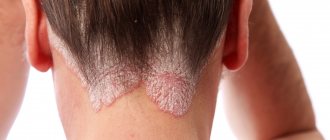
Where is increased fat content possible?
The content of the article
Problem areas:
- upper back;
- facial part, especially the chin, forehead and cheeks;
- cervical and thoracic parts of the body;
- genitals in both sexes;
- peripapillary zone.
These are the most common and most common, but there may be others. The only places on the body where acne cannot appear are the palms and feet. Due to the too thick and rough layer of the epidermis, incapable of this kind of inflammation.
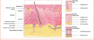
Leather
Skin is the largest human organ by area. The skin forms the outer covering that separates internal organs and tissues from the environment.
The skin consists of the epidermis (from the Greek epi - above and derma - skin) - the outer layer, and the dermis (the skin itself) - the inner connective tissue layer. Below the skin is the hypodermis (Greek hypo - down), represented by adipose tissue.
Epidermis
The epidermis of the skin is represented by stratified keratinizing epithelium. In the epidermis there are (from bottom to top) 5 layers: basal, spinous, granular, shiny and horny. In the basal layer, cells rapidly divide by mitosis; as cells move to the surface, they die and become keratinized. Keratinization is associated with the accumulation of a special substance by cells - keratin.
The horny (uppermost) layer of the epidermis is completely renewed in 7-11 days. Thanks to this renewal, the epidermis is very resistant to mechanical and chemical factors, is a barrier to microbes - bacteria, and is impenetrable to water.
In the basal layer there are melanocytes (from the Greek melanos - black) - cells that accumulate the black pigment - melanin. The synthesis of this pigment increases with prolonged exposure to the sun, which is the reason for the appearance of “tanning” on the skin.
In fact, tanning represents the body's protective reaction to the harmful effects of ultraviolet rays, which prevents them from passing through the skin into internal tissues and organs.
Dermis
Under the epidermis is the dermis (the skin itself), in which you can find sweat and sebaceous glands, as well as hair follicles (lat. folliculus - pouch). The dermis contains blood and lymphatic vessels, nerves, and muscle fibers.
The dermis has two layers:
- Papillary
- Reticulate
Formed by loose connective tissue in the form of papillae, protruding into the lower layers of the epidermis. It is the papillary layer that determines the unique pattern of human skin. Blood and lymphatic vessels and nerve endings are located here.
Formed by dense fibrous connective tissue. Structural proteins - collagen and elastin (along with hyaluronic acid) - give this layer (and the skin in general) strength and elasticity. Sweat and sebaceous glands and hair follicles are localized in the reticular layer.
We begin to study the appendages of the skin: sebaceous, sweat glands, hair and nails. The term appendages in no way downplays the significance of these formations; it only emphasizes that all of them are a derivative (formed from) the epidermis of the skin.
Sweat glands are tubular exocrine glands whose ducts open into pores on the surface of the skin. A secretion is secreted - sweat, which contains water, urea, uric acid, and salts. Sweat glands are found almost throughout the surface of the skin.
Functions of sweat glands:
- Excretory - removes urea and uric acid from the body
- Participation in water and salt metabolism - water and salts are released with sweat to maintain homeostasis
- Thermoregulation - when sweat evaporates, the skin cools, getting rid of excess heat
The sebaceous glands are located, unlike the sweat glands, more superficially. Their excretory ducts can open both into the hair follicle and onto the surface of the skin. The secret of the sebaceous glands is sebum, which prevents the development of microbes on the skin, prevents the skin from drying out, softens its surface and is a lubricant for the skin appendages - hair.
Hair is a derivative of the epidermis, consisting of a root and a shaft. The hair root ends in a hair follicle, into which the hair papilla with vessels and nerves enters from below. Hair growth occurs due to cell division of the hair follicle. Outside, the hair root is surrounded by a hair follicle, to which the levator pili muscle is attached.
The duct of the sebaceous gland opens into the hair funnel - the place where the hair root passes into the shaft. The rod consists of medulla and cortex, represented by keratinized cells. With old age, the amount of pigment in keratinized cells (scales) decreases, and the number of gas bubbles increases, which is the cause of graying of hair.
Human hair, compared to many other animals, is tiny and cannot serve as thermal insulation. Eyelashes, eyebrows, nose and ear hairs perform a protective function. Eyebrows serve to prevent sweat, an irritant, from getting into the eyes.
Nails are derivatives of the epidermis, which are convex horny plates located in the nail bed. The nail bed consists of germinal epithelium and connective tissue, rich in nerve endings and blood vessels. Nail growth occurs due to the division of germ epithelial cells.
In the lower part, the nail bed is surrounded by a dense leathery cushion - the cuticle, which protects the growth zone of the nail from bacteria and foreign particles. The function of the nail is to protect the sensitive part of the finger from mechanical damage and create support for it.
Skin is an organ of thermoregulation
You already know that due to the evaporation of sweat, the skin can cool, thereby performing a thermoregulatory function. However, this is not the only mechanism of thermoregulation. The skin contains networks of blood vessels.
During the heat, blood vessels dilate, blood fills them - heat transfer increases, thus the body gives off excess heat to the environment.
During cold weather, the blood vessels narrow, there is less blood in them (heat transfer decreases), it rushes to the internal organs (liver) so that the body can maintain the optimal temperature for as long as possible.
Skin is the organ of touch
The skin contains nerve endings (receptors) that perceive various stimuli: cold, heat, pressure, pain. Cold receptors are located at the surface of the skin, while thermal receptors are located in the dermis (the skin itself). Pain is perceived through free nerve endings.
Skin is the site of vitamin D synthesis
The skin is actively involved in the synthesis of vitamin D. It contains a substance that is a precursor to vitamin D - ergosterol, which is converted into vitamin D under ultraviolet rays (which is why it is useful to be in the sun).
In children, with a lack of solar radiation (insolation), rickets can develop - softening of bone tissue, since vitamin D is involved in the absorption of calcium.
Skin functions
At the end of the article, we will collect all the functions of the skin together into a system to make it easier to memorize. So, the functions of the skin:
- Protective
- Immune
- Thermoregulatory
- excretory
- Blood depot
- Receptor
- Participation in metabolism
Protects internal organs and tissues from mechanical damage, covered with sebum, which prevents the development of pathogenic microorganisms.
When foreign substances (antigens) enter the skin, they are recognized, destroyed and removed. Inflammation of the skin is called dermatitis (from ancient Greek δέρμα, δέρματος - skin + lat. itis - inflammation).
Thermoregulation is carried out by sweat glands, blood vessels and subcutaneous fat, which insulates internal organs and tissues.
Thanks to the work of the sweat glands, uric acid and urea, metabolic by-products, are removed from the body.
When the skin vessels are filled, up to 1 liter of blood can be deposited in them.
The skin contains temperature, cold, pain, and pressure receptors. All of them provide the tactile function of the skin.
Due to the work of the sweat glands, the skin takes part in water-salt metabolism, and due to the formation of vitamin D during insolation (sun exposure).
Diseases
The branch of medicine that studies the skin is called dermatology. A severe hereditary skin disease is known - ichthyosis (Greek "ichthys" - fish). It is characterized by a violation of keratinization of the skin: scales are formed that resemble fish scales. Sometimes keratinization is so severe that it is incompatible with life.
© Bellevich Yuri Sergeevich 2018-2021
This article was written by Yuri Sergeevich Bellevich and is his intellectual property. Copying, distribution (including by copying to other sites and resources on the Internet) or any other use of information and objects without the prior consent of the copyright holder is punishable by law. To obtain article materials and permission to use them, please contact Yuri Bellevich
.
Causes
- Dry skin;
- Hormonal changes in the whole body;
- Infection;
- Too much sebum production.
In addition to these reasons, there is another factor that was recently established by scientists - heredity.
Let's look at each reason in more detail.
Dry skin
One of the most common reasons. Due to dryness, the skin exfoliates and its particles clog the pores, which causes inflammation. It is very important to consider several points that cause excessive dry skin:
- Age. Over the years, the skin gradually loses many of its important properties, which include moisture retention.
- Environment. Dry and cold air, low humidity, and so on.
- Shower with hot water. Leads to the destruction of the protective layer of the skin.
- Allergic diseases.
- Hygiene products. Using regular soap on sensitive areas of the skin: facial skin, intimate areas, can cause dryness and cause acne.
Hormonal background
The reason lies in the incorrect proportion of sex hormones in the blood. Androgens (male hormones) play a very important role here. If their level is exceeded, then excess secretion begins from the sebaceous and sweat glands, which is the reason for the appearance of acne. You can also isolate progesterone, a hormone secreted in pregnant women. Leads to clogging of pores and, as a result, sebaceous inflammation appears.
Lack of estrogen can also cause excessive oily skin.
What does the work of the sebaceous glands depend on?
The amount of secretion produced by the sebaceous glands is not the same in all people. With dry skin, the production of sebocytes is reduced, and oily skin is characterized by their increased reproduction. Contrary to popular belief, only 10 percent of acne cases in adults are associated with hormonal changes in the body. For example, they appear with ovarian cysts and disappear after treatment of the underlying disease. A similar effect can be observed in adolescents during puberty - with an increase in the production of sex hormones in the body. True, experts often attribute the appearance of teenage acne to increased emotionality and psychological instability characteristic of this age. The main reason
for the appearance of acne is the increased sensitivity of the sebaceous glands themselves.
On their surface there are receptors sensitive to male sex hormones - androgens. Androgens are released not only in the male body, but also in the female body. If the number of these receptors or their sensitivity to hormones increases, the activity of sebocyte division in the sebaceous gland increases. The sensitivity of the receptors is determined genetically
. That is why contacting an endocrinologist with acne problems and attempting hormonal treatment often does not bring results.
Sebum production
There are 3 factors that can affect the amount of sebum production. Androgen, or rather its excess in the blood, is one of the reasons. Rarely, but still there is a special disease called “seborrhea”. And of course, a very common cause in the 21st century is poor nutrition.
Here is a list of products that promote the active secretion of sebum:
- carbonated drinks;
- alcoholic drinks;
- products in the production of which dyes were used;
- caffeinated drinks (black tea, coffee;
- sweet and flour;
- spicy, smoked food.
How to prevent it?
Despite the fact that the level of gland secretion is genetically determined, the appearance of acne can be reduced by reducing risk factors
. First of all, by reducing the intake of substances into the body that provoke an increase in the secretion of the sebaceous glands. The main ones are: - large amounts of spicy and salty foods, - added sugar and chocolate, - nicotine and other toxic substances from tobacco smoke. - alcohol. This is another reason why teenagers should avoid drinking and smoking. But the hygiene factor in the prevention of acne is far from leading. Bacteria will remain on the skin in any case, no matter how thoroughly a person washes his face.
Treatment and prevention
It would be best and safest to contact a competent doctor - an endocrinologist, so that further treatment takes place under his strict supervision. You will have to start with a healthy diet that limits the consumption of foods that are harmful to good skin condition. And it won’t hurt to start special cosmetic skin care.
ONLINE REGISTRATION at the DIANA clinic
You can sign up by calling the toll-free phone number 8-800-707-15-60 or filling out the contact form. In this case, we will contact you ourselves.
How to fight?
One of the most common methods of dealing with acne is squeezing it out. Beauty salons even provide deep facial cleansing services, which supposedly helps get rid of acne forever. However, such procedures do not affect any of the factors in the formation of acne. The improvement is only temporary
and does not affect the further development of the disease.
Some cosmetic products allow you to gently remove the keratin plug from the sebaceous gland duct, improving the outflow of secretions and preventing the proliferation of bacteria. There are also products with an antibacterial effect that continue to act even when inflammation has already begun in the sebaceous gland. A qualified dermatologist will select products
suitable for your specific skin type and severity of symptoms. But if these measures are not enough, then the treatment of acne requires serious medical intervention and the use of special medications.
Solving problems with an endocrinologist
If there is an imbalance in the hormonal system, then an endocrinologist will eliminate it. I, Georgy Nikitich Romanov, am an endocrinologist with more than 20 years of experience in the specialty. If you have very oily facial skin, and conventional methods do not help to eliminate this problem, then the best solution would be to contact me for help. I will take a detailed history, prescribe tests based on what I see, evaluate the results, and determine effective treatment. Women of different ages approached me with this problem, and we always found the best solution to this problem.
I provide face-to-face consultations at a medical clinic in Gomel, as well as online consultations. You can sign up for an online consultation in one of the suggested sources: Viber, Telegram, Instagram, WhatsApp, Skype, Vkontakte.
My experience and knowledge work for you to solve skin problems and eliminate existing discomfort.
Let's sum it up
There can be many reasons for the occurrence of increased oiliness in the dermis, but the solutions to all problems are basically the same. Therefore, it is very important to figure out how to reduce or remove, get rid of excess oily skin on the face, what to do when the chosen gentle products no longer help, and what you can count on. Modern cosmetology offers many different options for influencing the dermis, but not all of them are equally safe and effective, and many problems can be completely solved with high-quality home care.
Recommendations
- Lovasi, Marianne; Szegedy, Andrea; Zouboulis, Christos C.; Toroczyk, Daniel (October 17, 2021). "The immunobiology of the sebaceous glands is controlled by sebum lipids." Dermato-endocrinology
.
Informa UK Limited. 9
(1):e1375636. Doi:10.1080/19381980.2017.1375636. ISSN 1938-1980. PMC 5821166. PMID 29484100. - ^ a b
James, William D.;
Berger, Timothy; Alston, Dirk M. (2006). Andrews Skin Diseases: Clinical Dermatology
. Saunders Elsevier. paragraph 7. ISBN 978-0-7216-2921-6. - ^ a b c d
Young, Barbra;
Lowe, James S; Stevens, Alan; Heath, John W; Deakin, Philip J (March 2006). Wither's Functional Histology
(5th ed.). Elsevier Health Sciences. pp. 175–178. ISBN 978-0-443-06850-8. - ^ a b c d f f gram
Smith, K. R.;
Thibouteau, D. M. (2007). “Series of thematic reviews: Skin lipids. Sebaceous gland lipids: friend or foe? Journal of Lipid Research
.
49
(2): 271–281. doi:10.1194/ml. R700015-JLR200. PMID 17975220. - ^ a b
Thibouteau, D. (July 2004).
“Regulation of the human sebaceous glands.” Journal of Investigative Dermatology
.
123
(1):1–12. Doi:10.1111/j.1523-1747.2004.t01-2-.x. PMID 15191536. - ^ a b
Thody, A.J.;
Schuster, S. (1989). "Control and function of the sebaceous glands." Physiological Reviews
.
69
(2):383–416. Doi:10.1152/Physrev.1989.69.2.383. PMID 2648418. - ^ a b
Hanukoglu I., Boggula V.R., Vaknin H., Sharma S., Kleiman T., Hanukoglu A. (January 2021).
"Expression of epithelial sodium channel (ENaC) and CFTR in human epidermis and epidermal appendages". Histochemistry and Cell Biology
.
147
(6):733–748. Doi:10.1007/s00418-016-1535-3. PMID 28130590. S2CID 8504408. - Dellman's Textbook of Veterinary Histology
(405 pages), Jo Ann Koers Eurrell, Brian L. Frappier, 2006, p.29, weblink: Books-Google-RTOC. - ^ a b
Zubulis CC (2004).
"Acne and sebaceous gland function." Dermatology clinics
.
22
(5): 360–366. doi:10.1016/j.clindermatol.2004.03.004. PMID 15556719. - Porter A. M. (2001). “Why do we have apocrine and sebaceous glands?” JR Soc Med
.
94
(5):236–7. Doi:10.1177/014107680109400509. PMC 1281456. PMID 11385091. - Victor Eroshchenko, DiFiore Atlas of Histology with Functional Correlations
, Lippincott Williams & Wilkins, 10th edition, 2005 p. 41 years old - Dorlanda (2012). Dorland's Illustrated Medical Dictionary
(32nd ed.). Elsevier Saunders. item 866. ISBN 978-0-19-856878-0. - ^ a b
Cheng JB, Russell DW (September 2004).
"Mammalian wax biosynthesis II: expression cloning of a wax synthase cDNA encoding a member of the acyltransferase enzyme family" (PDF). Journal of Biological Chemistry
.
279
(36):37798–807. Doi:10.1074/jbc.M406226200. PMC 2743083. PMID 15220349. - Webster, Guy F.; Anthony W. Rawlings (2007). Acne and their treatment
.
Fundamental and clinical dermatology. 40
. CRC Press. p. 311. ISBN 978-0-8247-2971-4. - ^ a b
Draelos, Zoe Diana (2005).
Hair Care: An Illustrated Dermatological Guide
. London; New York: Taylor and Francis. paragraph 26. ISBN 978-1-84184-194-6. - Sweeney TM (December 1968). "The influence of estrogen and androgen on the renewal time of the sebaceous glands." Journal of Investigative Dermatology
.
53
(1): 8–10. Doi:10.1038/jid.1969.100. PMID 5793140. - Amory JC, Anawalt BD, Matsumoto AM, Page ST, Bremner WJ, Wang S, Swerdlow RS, Clark RW (June 2008). "Effects of 5alpha reductase inhibition by dutasteride and finasteride on bone mineral density, serum lipoproteins, hemoglobin, prostate specific antigen and sexual function in healthy young men." J. Urol
.
179
(6):2333–8. Doi:10.1016/j.juro.2008.01.145. PMC 2684818. PMID 18423697. - Wilkinson, P.F. Millington, R. (1983). Leather
(Digital print version - ed.). Cambridge (UK) [etc.]: Cambridge University Press. paragraph 151. ISBN 978-0-521-24122-9. - Monika-Hildegard Schmid-Wendtner; Corting Schmid-Wendtner (2007). Ph and skin care
. ABW Wissenschaftsverlag. P. 31–. ISBN 978-3-936072-64-8. Retrieved June 19, 2012. - Zlotogorsky A (1987). "Skin surface pH distribution on the forehead and cheek of adults." Arch.
Dermatol. Res .
279
(6):398–401. Doi:10.1007/bf00412626. PMID 3674963. S2CID 3065931. - Schmid MH, Korting HC (1995). "The Skin Acid Mantle Concept: Its Relevance to Skin Cleanser Selection" (PDF). Dermatology
.
191
(4):276–80. Doi:10.1159/000246568. PMID 8573921. Archived from the original (PDF) on March 1, 2011. - Yoon, S. W. (2010). "The role of sebum secretion in the pathogenesis of acne: facts and controversies." Dermatology clinics
.
28
(1): 8–11. doi:10.1016/j.clindermatol.2009.03.011. PMID 20082943. - “Series of thematic reviews: Skin lipids. Antimicrobial lipids on the skin surface." May 10, 2011
- Thiele, Jens J.; Weber, Stefan W.; Packer, Lester (1999). “The secretion of the sebaceous glands is the main physiological route for the delivery of vitamin E to the skin.” Journal of Investigative Dermatology
.
113
(6):1006–1010. doi:10.1046/j.1523-1747.1999.00794.x. PMID 10594744. - Zouboulis, Christos C.; Baron, Jens Malthe; Boehm, Markus; Kippenberger, Stefan; Kurzen, Hjalmar; Reichrath, Jörg; Tilitz, Anya (2008). "Borders of biology and pathology of the sebaceous glands." Experimental Dermatology
.
17
(6):542–551. Doi:10.1111/j.1600-0625.2008.00725.x. PMID 18474083. - Doucet, Sebastian; Susignan, Robert; Sago, Paul; Schaal, Benoit (2009). Hausberger, Martina (ed.). "Secretion from the areolar (Montgomery) glands in lactating women causes a selective unconditioned response in newborns." PLOS ONE
.
4
(10):e7579. doi:10.1371/journal.pone.0007579. PMC 2761488. PMID 19851461.CS1 maint: ref = harv (communication) - McCully, J.P.; Shine On, WE (March 2004). "The lipid layer of tears: depends on the function of the meibomian glands." Experimental studies of the eye
.
78
(3): 361–5. Doi:10.1016/s0014-4835 (03) 00203-3. PMID 15106913. - Dorlanda (2012). Dorland's Illustrated Medical Dictionary
(32nd ed.). Elsevier Saunders. item 781. ISBN 978-0-19-856878-0. - Dorlanda (2012). Dorland's Illustrated Medical Dictionary
(32nd ed.). Elsevier Saunders. item 802. ISBN 978-0-19-856878-0. - Roeser, R.J.; Ballachanda, B. B. (December 1997). "Physiology, pathophysiology and anthropology/epidemiology of human ear canal secretions." Journal of the American Academy of Audiology
.
8
(6): 391–400. PMID 9433685. - ^ a b
Britton, editors Nicky R. College, Brian R. Walker, Stuart H. Ralston;
illustrated by Robert (2010). Davidson's Principles and Practice of Medicine
(21st ed.). Edinburgh: Churchill Livingston/Elsevier. pp. 1267–1268. ISBN 978-0-7020-3085-7. - Farrell L. N., Strauss J. S., Stranieri A. M. (December 1980). “Treatment of severe cystic acne with 13-cis-retinoic acid. Evaluation of sebum production and clinical response in a multi-dose trial." Journal of the American Academy of Dermatology
.
3
(6): 602–11. Doi:10.1016/S0190-9622(80)80074-0. PMID 6451637. - Zhao E, Peng Y, Wang XL, Wu LP, Wang M, Yang HL, Xiao XH (2011). "Demodex-associated facial dermatosis: a case-control study." J Zhejiang Univ Sci B
.
12
(12): 1008–15. Doi:10.1631/jzus.B1100179. PMC 3232434. PMID 22135150. - James, William D.; Berger, Timothy G. (2006). Andrews' Cutaneous Diseases: Clinical Dermatology
. Saunders Elsevier. item 662. ISBN 978-0-7216-2921-6. - Dessinioti, C.; Katsambas, A. (2013). "Seborrheic dermatitis: etiology, risk factors and treatment: facts and controversies." Klin dermatol
.
31
(4): 343–51. doi:10.1016/j.clindermatol.2013.01.001. PMID 23806151. - ^ a b
Rapini, Ronald P.;
Bologna, Jean L.; Iorizzo, Joseph L. (2007). Dermatology: 2-volume set
. St. Louis: Mosby. ISBN 978-1-4160-2999-1. - Nelson BR, Hamlett CR, Gillard M, Raylan D, Johnson TM (July 1995). "Sebaceous carcinoma." Jam. Academician Dermatol. 33
(1): 1–15, test 16–8. Doi:10.1016/0190-9622(95)90001-2. PMID 7601925. - Neville BW, Damm JD, Allen CA, Bouquot JE (2002). Pathology of the oral cavity and maxillofacial region
(2nd ed.). Philadelphia: W. B. Saunders. paragraph 31. ISBN 978-0-7216-9003-2. - Kovic O, Hale E (2005). "Nevus sebaceous." Online Journal of Dermatology
.
11
(4): 16. PMID 16403388. - Harper, Douglas. "Sebaceous". Etymology online
. Retrieved April 5, 2014. - ^ a b c d
Astruc, Jean (1746).
A general and detailed treatise on all childhood diseases
. J. Nourse. p.. Sebaceous glands. - Rosenthal, Stanley A; Furnari, Domenica (1958). "Slide agglutination as a preliminary test in the laboratory diagnosis of Candida Albicans1." Journal of Investigative Dermatology
.
31
(5): 251–253. Doi:10.1038/jid.1958.50. PMID 13598929. - Dobson, G. E. (1878). Catalog of bats in the collection of the British Museum. Order of trustees.
- Gutierrez, Mercedes; Aoki, Agustin (1973). "Fine structure of the glandular gland of the free-tailed bat." Tadarida
brasiliensis."
Journal of Morphology
.
141
(3):293–305. Doi:10.1002/jmor.1051410305. PMID 4753444. - Heideman, P. D., Erickson, K. R., & Bowles, J. B. (1990). Notes on the reproductive biology, glands and roosting habits of Molossus sinaloae (Chiroptera, Molossidae). Zeitschrift für Säugetierkunde, 55 (5), 303-307.
- Lars Mecklenburg; Monika Linek; Desmond J. Tobin (September 15, 2009). Alopecia disorders in pets
. John Wiley and Sons. P. 269–. ISBN 978-0-8138-1934-1.