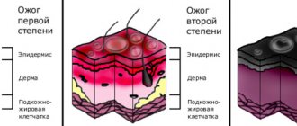
Skin structure
The skin consists of 3 layers: epidermis, dermis and subcutaneous fat (SAT) (Fig. 1). The epidermis is the thinnest of them and is a stratified keratinizing epithelium. The dermis is the middle layer of the skin. Mainly composed of fibrils of the structural protein collagen. PFA contains fat cells - adipocytes. The thickness of these layers can vary significantly depending on the anatomical location.
Fig.1. Skin structure
Skin thermoregulation
The skin takes part in heat regulation. It protects the body from overheating and cooling, helps maintain a constant body temperature. About 80% of the heat generated in the body is released through the skin. The skin is the main organ of thermoregulation. Through the skin, the body releases heat to the environment through heat conduction, heat radiation, and a significant part through the evaporation of sweat. If, for example, you hold a wet hand in the air, a feeling of cold appears because the evaporation of water cools the hand.
Heat transfer through the evaporation of sweat occurs in different ways: with the evaporation of sweat, the formation of which depends on the amount of blood passing through the skin vessels; as a result of radiation of heat into the surrounding air; due to the transfer of heat to objects in direct contact with the body, such as clothing; due to the mixing of air currents, in which the warm air surrounding the surface of the body is replaced by cooler air.
Heat transfer through the evaporation of sweat occurs continuously in the form of perspiration, imperceptible to us. During the day, the skin secretes twice as much water through sweat as the lungs do during the same time. In hot weather, the body loses more heat because the blood vessels dilate, the blood flow in them accelerates, and the amount of blood flowing near the surface of our body increases, where it cools. In cold weather, the skin vessels narrow, the skin turns pale, and sweat production decreases sharply, resulting in reduced heat transfer.
Epidermis
Rice.
2. Epidermis The epidermis is a constantly exfoliating epithelial layer of the skin, which consists mainly of 2 types of cells - keratinocytes and dendritic cells. Melanocytes, Langerhans cells, Merkel cells, and intraepidermal T lymphocytes are present in small quantities in the epidermis. Structurally, the epidermis is divided into 5 layers: basal, spinous, granular, shiny and horny, differing in the position and degree of differentiation of keratinocytes, the main cell population of the epidermis (Fig. 2).
Keratinization. As keratinocytes differentiate and move from the basal layer to the stratum corneum, their keratinization (keratinization) occurs - a process that begins with the phase of keratin synthesis by keratinocytes and ends with their cellular degradation. Keratin serves as a building block for intermediate filaments. Bundles of these filaments, reaching the cytoplasmic membrane, form desmosomes necessary for the formation of strong contacts between neighboring cells. Further, as epithelial differentiation progresses, epidermal cells enter the degradation phase. The nuclei and cytoplasmic organelles are destroyed and disappear, metabolism stops, and cell apoptosis occurs when it is completely keratinized (turns into horny scales).
The basal layer of the epidermis consists of a single row of mitotically active keratinocytes, which divide on average every 24 hours and give rise to new cells in the overlying epidermal layers. They are activated only in special cases, such as when a wound occurs. Next, a new cell, a keratinocyte, is pushed into the spinous layer, where it spends up to 2 weeks, gradually approaching the granular layer. The movement of the cell to the stratum corneum takes another 14 days. Thus, the lifespan of a keratinocyte is about 28 days.
It should be noted that not all cells of the basal layer divide at the same rate as keratinocytes. Epidermal stem cells under normal conditions form a long-lived population with a slow proliferation cycle.
The spinous layer of the epidermis consists of 5-10 layers of keratinocytes, differing in shape, structure and intracellular content, which is determined by the position of the cell. Thus, closer to the basal layer, the cells have a polyhedral shape and a round nucleus, but as the cells approach the granular layer, they become larger, acquire a flatter shape, and lamellar granules appear in them, containing various hydrolytic enzymes in abundance. Cells intensively synthesize keratin filaments, which, gathering into intermediate filaments, remain unconnected on the nuclear side, but participate in the formation of multiple desmosomes on the membrane side, forming connections with neighboring cells. The presence of a large number of desmosomes gives this layer a spiny appearance, for which it received the name “spinous.”
The granular layer of the epidermis is composed of still living keratinocytes, distinguished by their flattened shape and a large number of keratohyalin granules. The latter are responsible for the synthesis and modification of proteins involved in keratinization. The granular layer is the most keratogenic layer of the epidermis. In addition to keratohyalin granules, keratinocytes of this layer contain large quantities of lysosomal granules. Their enzymes break down cellular organelles during the transition of the keratinocyte to the phase of terminal differentiation and subsequent apoptosis. The thickness of the granular layer can vary; its value, proportional to the thickness of the overlying stratum corneum, is maximum in the skin of the palms and soles of the feet.
The shiny layer of the epidermis (so named for its special shine when viewing skin preparations on a light microscope) is thin, consists of flat keratinocytes, in which the nuclei and organelles are completely destroyed. The cells are filled with eleidin, an intermediate form of keratin. It is well developed only in some parts of the body - on the palms and soles.
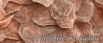
The stratum corneum of the epidermis is composed of corneocytes (dead, terminally differentiated keratinocytes) with a high protein content. The cells are surrounded by a waterproof lipid matrix, the components of which contain compounds necessary for exfoliation of the stratum corneum (Fig. 3). The physical and biochemical properties of cells in the stratum corneum vary depending on the position of the cell within the layer, directing the exfoliation process outward. For example, cells in the middle layers of the stratum corneum have stronger water-binding properties due to the high concentration of free amino acids in their cytoplasm.
Rice. 3. Schematic representation of the stratum corneum with the underlying granular layer of the epidermis.
Regulation of proliferation and differentiation of epidermal keratinocytes. Being a continuously renewing tissue, the epidermis contains a relatively constant number of cells and regulates all interactions and contacts between them: adhesion between keratinocytes, interaction between keratinocytes and migrating cells, adhesion with the basement membrane and underlying dermis, the process of terminal differentiation into corneocytes. The main mechanism for regulating homeostasis in the epidermis is supported by a number of signaling molecules - hormones, growth factors and cytokines. In addition, epidermal morphogenesis and differentiation are partially regulated by the underlying dermis, which plays a critical role in maintaining postnatal skin structure and function.
Protective function of the skin
| Skin function | |
| Receptor | Tactile: sensation of touch; Temperature: perception of cold and hot; Protective:
|
| excretory | During the day, 0.5 liters of water, salt, and lactic acid are released through the skin |
| Thermoregulatory | More than 80% of heat is lost through the surface of the skin |
| Participation in blood circulation | At the same time, the skin contains up to 1 liter of blood |
| Participates in mineral metabolism | Produces vitamin D and melanin |
The skin is equipped with a large number of different sensory nerves and corresponding nerve endings (receptors), due to which it functions as a complex sensory organ. Skin receptors perceive various irritations from the external environment and signal them to the cerebral cortex. That is why we are able to feel touch and feel objects, feel heat and cold, experience pressure and pain. Skin receptors make it possible to obtain ideas about the objects around us, their size, shape, the nature of their surface, temperature.
Dermis
The dermis is a complex, loose connective tissue consisting of individual fibers, cells, a network of blood vessels and nerve endings, as well as epidermal outgrowths surrounding the hair follicles and sebaceous glands. The cellular elements of the dermis are represented by fibroblasts, macrophages and mast cells. Lymphocytes, leukocytes, and other cells are capable of migrating into the dermis in response to various stimuli.
The dermis, making up the main volume of the skin, performs primarily trophic and supporting functions, providing the skin with such mechanical properties as plasticity, elasticity and strength, which it needs to protect the internal organs of the body from mechanical damage. The dermis also retains water, participates in thermoregulation and contains mechanoreceptors. Finally, its interaction with the epidermis supports the normal functioning of these layers of skin.
In the dermis there is no such directed and structured process of cell differentiation as in the epidermis, however, it also exhibits a clear structural organization of elements depending on the depth of their occurrence. Both the cells and the extracellular matrix of the dermis also undergo constant renewal and remodeling.
The extracellular matrix (ECM) of the dermis, or intercellular substance, which consists of various proteins (mainly collagen, elastin), glycosaminoglycans, the most famous of which is hyaluronic acid, and proteoglycans (fibronectin, laminin, decorin, versican, fibrillin). All these substances are secreted by dermal fibroblasts. The ECM is not a random accumulation of all components, but a complexly organized network, the composition and architectonics of which determine such biomechanical properties of the skin as rigidity, extensibility and elasticity. Epidermal keratinocytes, which are closely connected to each other, are attached to the ECM proteins. They form a dense protective layer of the skin. The structure of the ECM is also capable of exerting a regulatory effect on the cells immersed in it. Regulation can be either direct or indirect. In the first case, ECM proteins and glycosaminoglycans directly interact with cell receptors and initiate specific signal transduction pathways in them. Indirect regulation is carried out through the action of cytokines and growth factors retained in the cells of the ECM network and released at a certain moment to interact with cell receptors. The ECM structural network is subject to remodeling by enzymes from the matrix metalloproteinase (MMP) family. In particular, MMP-1 and MMP-13 initiate the degradation of collagen types I and III. The density of the ECM network of the dermis is uneven - in the papillary layer it is looser, in the reticular layer it is much denser, both due to the closer arrangement of the fibers of structural proteins and due to the increase in the diameter of these fibers.
Collagen is one of the main components of the ECM of the dermis. Synthesized by fibroblasts. The process of its biosynthesis is complex and multi-stage, as a result of which the fibroblast secretes procollagen into the extracellular space, consisting of three polypeptide α-chains folded into one triple helix. Procollagen monomers are then enzymatically assembled into extended fibrillar structures of various types. In total, there are at least 15 types of collagen in the skin; in the dermis, types I, III and V of this protein are most abundant: 88, 10 and 2%, respectively. Type IV collagen is localized in the basement membrane zone, and type VII collagen, secreted by keratinocytes, plays the role of an adapter protein for attaching ECM fibrils to the basement membrane (Fig. 4). Fibers of structural collagens of types I, III and V serve as a framework to which other ECM proteins are attached, in particular collagens of types XII and XIV. It is believed that these minor collagens, as well as small proteoglycans (decorin, fibromodulin and lumican), regulate the formation of structural collagen fibers, their diameter and the density of the formed network. The interaction of oligomeric and polymeric collagen complexes with other proteins, ECM polysaccharides, various growth factors and cytokines leads to the formation of a special network with certain biological activity, stability and biophysical characteristics important for the normal functioning of the skin. In the papillary layer of the dermis, collagen fibers are located loosely and more freely, while its reticular layer contains larger strands of collagen fibers.
Rice. 4. Schematic representation of skin layers and distribution of different types of collagens.
Collagen is constantly renewed, degrading under the action of proteolytic enzymes collagenases and being replaced by newly synthesized fibers. This protein makes up 70% of the skin's dry weight. It is the collagen fibers that “withstand the blow” under mechanical influence on it.
Elastin forms another network of fibers in the dermis, giving the skin such qualities as firmness and elasticity. Compared to collagen, elastin fibers are less rigid and curl around collagen fibers. It is with elastin fibers that proteins such as fibulins and fibrillins bind, to which, in turn, latent TGF-β-binding protein (LTBP) binds. Dissociation of this complex results in the release and activation of TGF-β, the most potent of all growth factors. It controls the expression, deposition and distribution of collagens and other matrix proteins in the skin. Thus, the intact network of elastin fibers serves as a depot for TGF-β.
Hyaluronic acid (HA) is a linear polysaccharide consisting of repeating dimers of D-glucuronic acid and N-acetylglucosamine. The number of dimers in the polymer varies, which leads to the formation of HA molecules of different molecular weights and lengths - 1x10⁵-107 Da (2-25 μm), which, accordingly, have different biological effects.
HA is a highly hydrophilic substance that affects the movement and distribution of water in the dermal matrix. Thanks to this property, our skin, normally and in youth, has high turgor and resistance to mechanical pressure.
HA easily forms secondary hydrogen bonds both within one molecule and between neighboring molecules. In the first case, they ensure the formation of relatively rigid spiral structures. In the second, association with other HA molecules and nonspecific interaction with cell membranes occurs, which leads to the formation of a network of polysaccharide polymers with fibroblasts included in it. Shorter molecules of proteoglycans (versican, lumican, decorin, etc.) “sit” on a long HA molecule, like on a thread, forming aggregates of enormous sizes. Extended in all directions, they create a scaffold, contributing to the stabilization of the ECM protein network and fixing fibroblasts in a specific matrix environment. Taken together, all these properties of HA endow the matrix with certain chemical characteristics - viscosity, density of “cells” and stability. However, the ECM network is a dynamic structure that depends on the state of the organism. For example, under conditions of inflammation, HA aggregates with proteoglycans dissociate, and the formation of new aggregates between newly synthesized HA molecules (renewed every 3 days) and proteoglycans is blocked. This leads to a change in the spatial structure of the matrix: the size of its cells increases, the distribution of all fibers changes, the structure becomes looser, the cells change their shape and functional activity. All this affects the condition of the skin, leading to a decrease in its tone.
In addition to regulating water balance and stabilizing the ECM, HA plays an important regulatory role in maintaining epidermal and dermal homeostasis. HA actively regulates dynamic processes in the epidermis, including keratinocyte proliferation and differentiation, oxidative stress and inflammatory response, maintenance of the epidermal barrier and wound healing. In the dermis, HA also regulates fibroblast activity and collagen synthesis. By remodeling the matrix, HA controls the functioning of cells in the matrix, influencing their availability to various growth factors and changing their functional activity. Cell migration and the immune response in tissue depend on the action of GC. Thus, changes in the distribution, organization, molecular weight and metabolism of GCs have significant physiological consequences.
Fibroblasts are the main type of cellular elements of the dermis. It is these cells that are responsible for the production of HA, collagen, elastin, fibronectin and many other intercellular matrix proteins necessary for the formation of connective tissue. Fibroblasts in different layers of the dermis differ both morphologically and functionally. Not only the amount of collagen they synthesize depends on the depth of their location in the dermis, but also the ratio of the types of this collagen, for example types I and III, as well as the synthesis of collagenase: fibroblasts in the deeper layers of the dermis produce less of it. In general, fibroblasts are very plastic cells, capable of changing their functions and physiological response and even differentiating into another cell type depending on the stimulus received. The role of the latter can be played by signaling molecules synthesized by neighboring cells and the restructuring of the surrounding ECM.
The structure of human skin and its main functions
Skin is one of the types of tissue of the human body, which has a number of inherent properties such as: elasticity, strength, some porosity, waterproofness, sensitivity and antibacteriality.
The skin protects the body from the negative effects of the environment (from excess solar radiation, for example), it is capable of secreting fat and producing odorous substances, selectively absorbing some chemicals useful to the body and rejecting others. In addition, the skin has such an important function as regeneration, which gives it the ability to independently recover from damage.
On average, there are about 5 million hairs on the surface of the skin of an adult, with about 200 receptors and 100 pores located on each square centimeter.
Which layers of skin are affected by cosmetics?
In accordance with the legislation of most countries, cosmetic products can only affect the outer layers of the skin; their deep penetration and impact on the living layers of the skin is unacceptable. That is, cosmetics should have exclusively external use, which is its fundamental difference from medications.
It should be noted here that there is no barrier in the lower part of the epidermis that could effectively prevent the penetration of cosmetics into the lymphatic and blood vessels, and, consequently, the distribution of the substances they contain throughout the human body. The possibility of effective exchange between the dermis and epidermis has been proven experimentally; accordingly, the risk of substances crossing the transepidermal barrier is quite high.
What substances are able to overcome the transepidermal barrier and enter the dermis?
It was experimentally established that substances such as caffeine, nicotine, hyaluronic acid, essential oils, vitamin E (it accumulates at the border of the dermis and epidermis). Nanoparticles contained in sunscreens penetrate deep into the skin, as well as liposomes, which easily penetrate into the deep layers of the skin and deliver the necessary nutrients there.
The structure of human skin
The versatility and multifunctionality of the skin is based on the characteristics of its structure. Our skin consists of 3 important layers, each of which performs its own function:
|
Epidermis The epidermis is the outer layer of skin, which consists of keratinized (dead) skin cells. In layers of the epidermis distant from the surface, the cells are living. They actively divide and make a slow movement to the surface, where over time they are replaced by keratinized ones, and then peel off.
The epidermis is an effective barrier to water and aqueous solutions . Fat-soluble substances penetrate the skin through the epidermis much better. This is due to the fact that cell membranes themselves contain a significant amount of fats and fat-soluble substances dissolve in them naturally.
Almost all epidermal cells produce keratin , which is why they are called keratinocytes. These keratinocytes are in constant motion, as a result of which the keratinocyte loses its own nucleus and main organelles, turning into a kind of flat “bag” filled with keratin - a corneocyte. Corneocytes form the stratum corneum of dead epidermal cells and look like flat scales. They are responsible for the barrier function of the epidermis. Corneocytes are held together by a double layer of lipids (ceramides), a kind of plastic “cement.”
The basal layer of the skin contains melanocytes, which produce melanin . Melanin is a pigment that gives our skin color and reduces the effects of external radiation. So, thanks to its presence, the skin completely blocks infrared rays and is partially capable of blocking ultraviolet rays. Often, the formation of pigment spots on the skin will depend on the condition of the basement membrane. The epidermis also contains Langerhans cells, which protect the body from microbes and foreign bodies .
The thickness of the epidermis and the rate of its renewal As a rule, the thickness of the epidermis layer varies from 0.07 to 0.12 millimeters (this is the usual thickness of a sheet of paper or plastic film). Especially rough skin can be much thicker - up to 2 mm. (on the soles of your feet, for example). The process of skin cell renewal is continuous. During our lifetime, we get rid of approximately 18 kg of dead skin, while losing about 10 billion cells every day.
The rate of complete renewal of the outer layer of the epidermis slows down with age. So, if in childhood and adolescence this process takes 21-28 days, then already at 25 years old 35-45, and after 50 years 56-72 days.
The process of division and advancement of mature skin cells is not only slower, but also heterogeneous in different areas, which also affects the aesthetic appearance of the skin. If dead skin cells become layered, the cell division process occurs more slowly, leading to faster skin aging. In addition, the layer of dead cells makes it difficult for oxygen and nutrients to penetrate the skin. Therefore, the older a person is, the more often peelings are required.
Dermis The dermis is the inner layer of the skin, which also contains functional skin glands, thanks to which excess salts and moisture are removed from the body: sweat and sebaceous glands, producing sweat and sebum, respectively.
Sebum protects the skin from the aggressive effects of the external environment and gives the skin such properties as bactericidal (due to the acidic environment it creates and then has a detrimental effect on microorganisms) and water resistance. Sweat glands, in addition, participate in the natural thermoregulation of the body.
Hypodermis The hypodermis is a subcutaneous fat layer that protects us from both excess cold and excess heat, and in addition significantly mitigates damage from impacts.
Subcutaneous fatty tissue is capable of storing fat-soluble vitamins such as A, E, F, and K. An important function of the hypodermis is that it serves as the basis for the outer layers of the skin. Skin whose hypodermis layer is weakly expressed has more folds and wrinkles and is subject to faster aging. Another important function of the hypodermis is the hormone-producing function. Adipose tissue is not only capable of accumulating estrogens, but also stimulating their synthesis.
The hypodermis layer also contains leptin, a unique hormone responsible for the feeling of fullness during meals. Thanks to it, the body is able to regulate our appetite and, as a direct consequence, the amount of fat in the subcutaneous layer.
Subcutaneous fat
Subcutaneous fat, or hypodermis, is the lowest layer of skin, located under the dermis. It consists of fat lobules separated from each other by connective tissue septa containing collagen and penetrated by large vessels. The main cells of fat lobules are adipocytes, the number of which varies in different areas of the body. Currently, PFA is considered not only as an energy depot, but also as an endocrine organ, the adipocytes of which are involved in the production of a number of hormones (leptin, adiponectin, resistin), cytokines and mediators that affect metabolism, insulin sensitivity, reproductive and immune functional activity. systems
Skin appendages - nails and hair
All human fingers have nails on the back side of the terminal phalanges - thin and transparent keratinized plates. These are adnexal formations of the skin that develop from the epidermis. The nails are curved according to the surface of the nail phalanx of the finger. Most of the nail - its body - ends with a free edge and goes into the thinnest part of the nail - the root, which protrudes deep into the fold of skin. At the point where the body of the nail transitions into the root, under the skin ridge, a semi-lunar-shaped lighter part of the nail protrudes - the place of its growth. The entire nail is located in the nail bed. It is a skin formation and is tightly connected to the tuberosity of the nail phalanx using bundles of connective tissue fibers. There are no glands in the nail bed area, but there are a large number of sensory nerve endings.
The structure of the nail is similar to the structure of the epidermis. It distinguishes between the germinal and horny layers. The first corresponds to the germ layer, the proliferation of cells of which determines the growth of the nail. In a living person, the nail grows continuously and quite quickly - up to 4 mm per month, especially on the fingers. The substance of most of the nail consists of the stratum corneum. The nail plate is devoid of blood vessels and nerves. The pink color of the nail depends on the subungual vessels, which are visible through the transparent stratum corneum. When stagnation occurs due to changes in the color of the blood, the color of the nails also changes (cyanosis).
Skin appendages also include filamentous formations - hairs that cover a significant part of the surface of the skin. Only in some places are they completely absent (palms and palmar surfaces of the fingers, soles and plantar surfaces of the toes, dorsal surfaces of the terminal phalanges of all fingers). All hair is divided into several types according to its appearance. Long hair includes the hair of the head, beard and mustache. Short, bristly hair includes hair from the eyebrows, eyelashes, as well as hair located near the opening of the nose and the external auditory canal. There are also thin, woolly hairs (fluff) that are found on the body, limbs and face.
Hair is located obliquely to the surface of the skin. The root begins as a thickening, a hair follicle. The free part of each hair that protrudes above the skin is called its shaft. Each hair has a special straightening muscle attached to it. The hair has different colors - from pale yellow to black. Their color depends on the presence of pigment in the cortical layer, the intensity of the color depends on the amount of the latter. With the loss of pigment, graying of the hair occurs, which is intensified by the penetration of air between the cells of the cortex. Hair in a living person grows continuously. Hair serves as a protective device: eyelashes, eyebrows protect the eyes. These bristly hairs not only mechanically trap large dust particles, but also act as sensitive “antennas.” Even a light touch to them causes irritation of the sensitive nerve endings that are densely entwined at the hair root.