What is the brain, encephalon, its meaning?
The brain is the anterior part of the central nervous system. Brain
located in the cranial cavity, it interacts with the human body (men, women) with the external environment, integrating the functioning of all body systems.
The brain has the ability to assimilate, organize, store, and retrieve information about past experience. The brain is the material substrate of higher nervous activity. Phylogenetically, the brain is the anterior end of the neural tube. Ontogenetically, the brain is a derivative of the cerebral vesicles, from which parts of the brain are formed: the telencephalon, the diencephalon, the midbrain, the hindbrain, which is represented by formations such as the pons, cerebellum, and medulla oblongata. The cavities of the brain vesicles develop into the ventricles of the brain.
Human brain
August 25, 2014 Neurology
The human nervous system is represented by the brain, located in the cranial cavity; the spinal cord, located in the spinal cavity, and a branched system of nerves that extend from the brain (cranial nerves) and innervate the organs of the head; a system of nerves that branch from the spinal cord and innervate the arms, legs, torso, and internal organs. The brain and spinal cord represent the central nervous system, and the nerve system represents the peripheral nervous system.
All formations of the nervous system consist of many neurons (cells of the nervous system) and their processes, through which nerve impulses are transmitted in ascending and descending directions due to the diverse connections that exist between neurons.
Despite the fact that different neurons perform different functions and have differences in structure, they all have a body, a receptive structure, and a process, a dendrite, a conducting structure.
According to their functional characteristics, neurons are divided into motor - executive, and sensitive - perceiving, as well as interneurons that interact between them.
The nerve cell performs two main functions: 1) processing of incoming information, transmission of nerve impulses, and 2) biosynthetic, aimed at maintaining its vital functions.
This is the schematic diagram of the structure of a neuron.
This is what the human brain looks like.
This is a complex structure, consisting of many different formations that are in close interaction; carrying out conducting, analyzing, regulating and coordinating functions. All body movements, human feelings, the work of internal organs, his mind, intellect, memory, consciousness, sleep, wakefulness, everything is controlled by the brain. The human brain can be compared to a complex computer with embedded programs that are constantly modified throughout a person’s life.
Schematically, the brain can be divided into lobes: frontal, occipital, temporal, parietal; cerebellum, brain stem. The lobes of the brain are covered with a cortex, which is a collection of highly differentiated neurons that carry out higher integrative activities.
In the frontal lobes there are centers for the regulation of voluntary movements, when damaged, weakness develops in the arms, legs on one side, or only the arms or legs. In the frontal lobes there are also rotations of the eyes and head, when damaged, deviation of the eyes and head occurs towards the pathological focus. The frontal lobes also contain centers for coordination of movements, which, when damaged, cause disturbances in standing and walking. And finally, when the frontal lobe cortex is damaged, behavioral and mental disorders develop.
The parietal lobes are responsible for a person’s ability to recognize objects by touch, the ability to perform complex goal-directed actions, the ability to decipher written characters and the ability to write.
The temporal lobes contain auditory, gustatory and olfactory centers, centers for understanding and reproducing speech, and centers for coordination of movements.
The visual lobes contain the centers for the perception of visual images and visual memory. The cerebellum is one of the main coordinating centers.
In the brain stem there are centers for the regulation of life-supporting organ systems, respiratory, cardiovascular, intermediate centers for the regulation of cranial nerves, the pathways of the motor and sensory systems.
In the brainstem, in its tegmentum, are located the nuclei of the cranial nerves, the bodies of nerve cells responsible for the innervation of the organs of the head and face, providing the functions of the gustatory, auditory, visual, vestibular and olfactory analyzers.
The cranial nerves of the caudal group are distinguished: 1) Accessory nerve, 11th pair, innervates the muscle that turns the head to the side. 2) Hypoglossal nerve, 12th pair, innervating the tongue. 3) Glossopharyngeal nerve, 9th pair, innervating the pharyngeal muscles, tongue, palate, middle ear, salivary glands. 4) Vagus nerve, 10th pair, innervating the muscles of the pharynx, soft palate, larynx, smooth muscles of the bronchi, trachea, esophagus, stomach, intestines.
Next, the cranial nerves of the cerebellopontine angle are distinguished: 1) Facial nerve, 7th pair, innervating the facial muscles. 2) Vestibulocochlear nerve, 8th pair, innervating the inner ear. 3) Trigeminal nerve, 3rd pair, innervating the skin of the face, jaws, and masticatory muscles. Next comes a group of oculomotor nerves: 3, 4, 6 pairs.
And finally, the optic nerve, 2nd pair, innervating the retina, and the olfactory nerve, 1st pair, innervating the nasal mucosa.
Brain structure
The cerebrum, or telencephalon, is represented by two hemispheres, which are connected to each other by the corpus callosum, corpus callosum. It consists of nerve fibers running transversely from one hemisphere to the other. The corpus callosum ensures the unity of functioning of both hemispheres. When the corpus callosum is cut, each hemisphere of the brain begins to function independently of each other. Under the corpus callosum is the fornix, fornix. Anterior to the pillars is the anterior commissure, comissura anterior. Between the anterior part of the columns of the fornix and the knee of the corpus callosum there is a thin vertical plate of brain tissue - the transparent septum. Between the plates of the septum there is a slit-like cavity that does not have an ependymal lining. A number of authors call it the 5th ventricle.
The surface of the hemispheres is covered with a layer of gray matter - this is the cerebral cortex. Underneath it is the white matter and subcortical nuclei: striopallidal system, extrapyramidal system.
If you make a horizontal section of the brain through the cerebral hemispheres at the level of the thalamus and subthalamic nuclei, you can see the following formations: pineal body, superior colliculus, thalamus, frenulum, posterior limb of the internal capsule, globus pallidus of the lenticular nucleus, putamen of the lenticular nucleus, lateral sulcus, fence , anterior part of the internal capsule, head of the caudate nucleus, column of the fornix, anterior horn of the lateral ventricle, genu of the corpus callosum, septum pellucidum, interthalamic commissure, lentiform nucleus, external capsule, extreme capsule, convolutions of the insula, lateral sulcus, internal capsule, subthalamic nucleus, tail caudate nucleus, lateral geniculate nucleus, red nucleus, gray matter of the superior colliculus, cerebellar vermis.
Structure of brain tissue
The brain is a very complex and very fragile structure, so nature has created a triple system of brain protection. As a result, the brain is covered in three protective layers.
This is, firstly, a soft choroid that fuses with the brain and fills its entire space.
Secondly, this is the arachnoid membrane, which fuses with the soft one and connects with the hard one.
And thirdly, it is a hard shell that fuses with the periosteum of the skull and connects with the arachnoid.
The structure of the shells can be schematically represented as follows:
The terms “epidural space”, “subdural space”,
The “subarachnoid space,” which you can read on the diagram, refers to the space between the layers of the brain. CSF channels are channels through which cerebrospinal fluid circulates. Sometimes it is also called cerebral fluid.
Liquor is generated from blood plasma, penetrates into the space under the membranes and “goes” to the lymph nodes. Cerebrospinal fluid nourishes nerve cells, maintains stability of intracranial pressure, protects the brain from concussions and removes harmful metabolic products.
The brain itself consists of gray and white matter. Gray matter is on the outside, forming the cerebral cortex, white matter is on the inside. The gray matter layer can range from 1.3 to 4.5 millimeters, with the layer being thicker at the front of the brain.
The gray matter contains neurons and glia, the supporting cells of the central nervous system. It is the number of neural connections that determines the effectiveness of a person’s thinking, the ability to compare new information with existing information, etc.
The number of neurons in the brain is in the billions. Thus, there is evidence that the adult male brain contains on average 86.1 billion neurons +/- 8.1 billion [F. Azevedo et al., 2009]. And some scientists believe that the number of neurons in the human brain can reach 100 billion [R. Hodson, 2019].
Neurons have processes called axons and dendrites. The function of axons is the propagation of nerve impulses, the function of dendrites is the receipt of nerve impulses. The relationship between axons and dendrites can be schematically represented as follows:
Glial cells (glia) are responsible for the coordinated functioning of neurons, providing neurons with nutrients. The cerebral cortex plays a leading role in higher nervous activity, ensures communication between brain cells, and corrects deviations in the functioning of human systems and organs.
The white matter of the brain consists primarily of axons, which are covered with myelin. Hence the name “white matter” because myelin is white in color. Please note that the so-called unmyelinated axons, which are not covered with myelin, are concentrated in the gray matter.
White matter connects different areas of gray matter, where nerve cells are located, to each other, and provides the transfer of impulses between neurons. The myelin coating in this case functions as a signal accelerator.
The brain contains 12 pairs of nerves, each of which provides a specific function:
- 1 pair – olfactory.
- 2nd pair – visual.
- 3 pairs – oculomotor.
- 4th pair – trochlear (innervate the muscles that rotate the eyeballs outward and downward).
- 5 pair - trigeminal (responsible for the sensitivity of the facial muscles).
- 6th pair – abductors (innervate the muscles responsible for abduction of the eyeball).
- 7th pair – facial (innervates facial muscles).
- 8th pair – vestibulocochlear (responsible for transmitting auditory signals and impulses from the inner ear).
- 9th pair – glossopharyngeal.
- 10th pair – vagus (innervates internal organs).
- 11th pair – accessory (innervate the muscles responsible for turning the head).
- 12 pair - sublingual.
In general, the functions of the nerves are clear from their names, so explanations in parentheses are provided only where the names are not very informative. You can see the location of the nerves in the brain in the following picture:
The cerebral cortex is divided into lobes, the location and functions of which we discussed above, and is dotted with convolutions, due to which the surface area of the cortex can reach 2-2.5 square meters. The more convolutions, the more gray matter is “fitted” in the brain and the more neural connections can potentially be formed, and the higher the brain's performance. The appearance of a large number of convolutions is a product of evolution, as a result of which complex cognitive structures can develop without increasing the volume of the cranium.
So, we have examined in the most general simplified form how the human brain works, its structure and the structure of the tissues of which it consists. This, in general, is enough to understand the general patterns of brain function and what thought processes actually depend on. You can develop thinking techniques more deeply by studying our Cognitive Science program.
We wish that your brain always functions flawlessly and never fails you. And in conclusion, we suggest you take a short test on the topic of the article :
We also recommend reading:
- Storytelling
- 3 tasks for quick brain training
- Books about development and for development
- Neuroplasticity
- 10 simple ways to keep your brain sharp
- Brain structure
- Creativity and logic: the myth of functional asymmetry of the cerebral hemispheres
- Intelligence and its development: several recommendations
- “Are you out of your mind?” or 32 exercises for brain development
- 5 neurolifehacks: how to improve brain productivity
- Brain digest
Key words:1Cognitive science
Brain of a newborn, children, child, human: structure, anatomy
The brain of a newborn baby is shorter and wider than that of school-age children and adults. It is devoid of all tertiary and a number of secondary grooves. By the end of the child’s first year of life, the brain increases in size by 2–2.6 times. By 3 years it increases 3 times. From birth to adulthood, brain weight increases 4 times, and body weight increases 21 times.
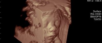
The mass of the right hemisphere is most often greater than the mass of the left hemisphere. After birth, the parietal and frontal lobes develop most intensively. And because of this, the overall configuration of the brain changes. Unlike the adult brain, in a newborn the neurons of different layers are located closely next to each other, because of this, the radial striation of the cortex may be absent. Single neurons may be located in the subcortical white matter. In the substantia nigra of the brain stem, neurons do not yet have the pigment melanin, which usually appears by 3–4 years of age. Up to 3–6 months of extrauterine life, the outer embryonic layer, which is called “Obersteiner’s layer,” remains in the cerebellar cortex. Obersteiner's layer consists of medulloblasts and spongioblasts. The surface of the inferior olives of the fetal medulla oblongata is smooth. After the birth of a child, the olives acquire elevations and then noticeably increase in size with age. Almost always in newborns, immature cellular elements are found in the subependymal parts of the ventricular system of the lateral ventricles, the presence of which erroneously resembles manifestations of Virchow's local encephalitis. Immature cells are located in the subependymal layer diffusely or in the form of separate foci. Sometimes they can be traced along the blood vessels over a significant extent of the white matter. By 3–6 months of a child’s life, these cells gradually disappear. The presence of a large number of immature cells in the subependymal parts of the ventricular system is an additional morphological sign of fetal prematurity.
Who needs to undergo an MRI of the brain?
Most often, a neurologist refers you to magnetic resonance imaging. Indications for research may include:
- regular dizziness and headaches of unspecified nature;
- decreased hearing or vision in the absence of an explanation from an ENT or ophthalmologist;
- frequent fainting;
- head injuries;
- infectious diseases of the brain;
- stenosis (narrowing of blood vessels);
- aneurysms (dilation of blood vessels);
- epilepsy;
- cerebrovascular accident;
- other neurological diseases.
Although brain tomography is a completely safe procedure, there are still contraindications. First of all, they are due to the presence of metal elements that cannot be removed during the procedure:
- braces;
- pacemakers;
- prostheses;
- staples on vessels;
- foreign objects;
- implants.
If there are fixed metal parts and devices, MRI cannot be performed.
Overweight patients may be limited by the diameter of the tomograph capsule.
It is not recommended to undergo an MRI during the first trimester of pregnancy, however, if there are medical indications, the diagnosis will be carried out. At a later date, the examination will not harm either the mother or the child. Breastfeeding women also do not have to worry: magnetic waves do not affect either lactation or the quality of milk.
How does a child's brain mass change with age?
If you monitor how the mass of a child’s brain changes depending on age, you will notice the following picture. If the child’s age is from 3 to 8 days, body length is 49 – 50 cm, then the brain weight will be 336 grams. At 1 month, the child’s height is 52 cm, brain weight is 360 grams. At 3 months, the child’s height is 56 cm, brain weight is 520 grams. At 6 months, with a height of 62 cm, GM weight is 670 g. At 9 months, with a height of 67 cm, GM weight is 760 g. At 1 year of age, the child’s height is 73 cm, brain weight is 960 g. At 1.5 years, with a height of 79 cm, the weight of the GM is 1045 g. At 2 years old, with a height of 85 cm, the weight of the GM is 1070 g. At 3 years old, the child’s height is 89 cm, and the brain weight is 1150 g. At 5 years old, height is 106 cm, weight is 1240 g. At 10 years old, height 132 cm, brain weight 1300 g. At 12 years old, height 145 cm, brain weight 1370 grams.
Physiology, brain function
Physiologically, all the work of the brain is built on the principles of hierarchy, integrity, systematicity and plasticity. These are the principles of functioning that carry out all conditioned and unconditioned reflexes. They contribute to the flow of conscious mental activity of a person. The principle of hierarchy is that phylogenetically younger parts of the brain carry out higher-order control, complementing but not replacing the function of phylogenetically more ancient parts. As a result of this, the body’s capabilities in more subtle differentiation of each stimulus by each analyzer are expanded, and a more adequate perception of the overall picture of the world is achieved based on the correlation of the results of the activities of many analyzers.
The highest form of expression of the hierarchical principle is the process of corticalization of functions. The principle of hierarchy is combined with the principles of integrity and systematicity, which consist in the fact that the brain functions as a single whole with the entire nervous system, while receiving afferent impulses, carries out its analysis and synthesis, forms a flow of efferent impulses that determine the adequate activity of all peripheral organs . As a result, a stable system is formed that provides continuous information communication: center - periphery - environment - periphery - center. Plasticity refers to the functional variability of nerve centers, which is clearly manifested in the process of compensation for impaired brain functions.
Irradiation of excitation plays an important role in the normal functioning of the brain. The feedback mechanism consists of closing the input and output of the same element or system. The dominant mechanism regulates the relationships between nerve centers.
Diseases, disorders, brain lesions
Diseases, disorders, and brain lesions are varied. In further articles, we will focus on pathologies such as brain tumor, brain cyst (including arachnoid, retrocerebellar, liquor), trauma, concussion or bruise of the brain, brain cancer, hydrocephalus (hydropsis), vascular atherosclerosis, aneurysm, encephalopathy, demyelination, ischemia, ischemic or hemorrhagic stroke, heart attack, atrophy, spasm or vasoconstriction, glioblastoma, meningioma, dysfunction, dystonia, diffuse changes, hypoxia (oxygen starvation), encephalitis, inflammation, vascular diseases, atrophic changes. The clinical picture of such diseases depends on the type of pathology.
What does a brain MRI show?
Having received 3D images of the brain in different projections, the doctor has the opportunity to examine each area in detail. This allows us to draw conclusions about:
- processes occurring in brain tissue;
- condition of blood vessels;
- the presence, location, shape and size of neoplasms, hematomas or blood clots;
- the presence, stage and localization of other brain pathologies.
Thanks to MRI, it is possible to visualize the brain in different projections, which significantly increases the information content of the examination
If the results are not accurate enough or additional visualization of a specific area is required, an MRI of the brain with contrast may be ordered. The introduction of a contrast agent makes it possible to obtain a more accurate idea of the structure of brain tissue or pathological formation and to clarify its boundaries.
Brain treatment in Saratov, Russia
Sarklinik provides treatment for a number of diseases, diseases of the central and peripheral nervous system in Saratov, in Russia for children, adolescents, adults, boys, girls, guys, girls, men, women, brain treatment in Saratov . Hardware and non-hardware treatment methods make it possible to restore the functioning of the human nervous system.
Sign up for a consultation. There are contraindications. Specialist consultation is required.
Text: ® SARCLINIC | Sarclinic.com \ Sarlcinic.ru Photo: © pixologic / Photobank Photogenica / photogenica.ru The people depicted in the photo are models, do not suffer from the diseases described and/or all coincidences are excluded.
Related posts:
I'm afraid to speak, logophobia: treatment of logophobia for stuttering, the child is afraid to speak
Agnosia: visual, auditory agnosia, treatment of agnosia
Depersonalization: causes, symptoms, treatment of depersonalization in Saratov, Russia
Neurologist neuropathologist difference, what is different, what treats what diseases
Kogan syndrome: treatment, how to treat Kogan, oculomotor apraxia