After removal of the lens nucleus, the lens masses must be completely removed by aspiration.
Aspiration is the removal of air or liquid from a body cavity. In ophthalmology, this term refers to the suction of lens masses during cataract surgery.
The lens masses subject to aspiration are adjacent to the posterior chamber or are mobile. The moving masses are usually always soft and are located in the anterior chamber in front of the iris. The adjacent masses are "fluffy" or "layered". In most cases, they are soft enough to be easily suctioned. But in some situations they have a dense consistency, so significant aspiration force is required to remove them.
About aspiration
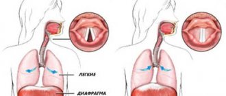
Aspiration is when food or liquid enters the airway instead of the esophagus. The esophagus is a tubular organ that carries food and liquids from the mouth to the stomach. Aspiration can happen when you eat or drink, or while receiving feeding tubes. It is also possible with vomiting or heartburn.
You may be at risk for aspiration if you have trouble swallowing. This occurs because food or liquid can get stuck in the back of the throat and enter the airways. Aspiration can cause pneumonia, respiratory infections (nose, throat, or lung infections), and other health problems.
Signs of aspiration
Signs of aspiration include:
- cough;
- suffocation;
- gagging;
- sore throat;
- vomiting
You and your caregiver should watch for these signs before, during, and after eating or drinking food or drinking a tube.
If you have any of these signs, stop eating, drinking, or receiving feeding tubes. Call your healthcare provider immediately.
to come back to the beginning
In what cases is aspiration used?
Cataracts can be not only acquired, but also a congenital disease. Children often suffer from it. As mentioned above, the only effective treatment is replacing the lens with an IOL. Children's cataracts are generally very mild. They can be easily removed using the extracapsular method of aspiration and washing out using a special instrument through an incision of only 3 millimeters.
When performing extracapsular extraction on a child's eye, doctors make a small incision and cut through the anterior capsule of the lens, removing the lens masses. The optimal combination of surgical treatment of cataracts in children is extraction and aspiration.
Aspiration is necessary because remnants of the lens masses may be located in the posterior or anterior chamber of the eye or in the cavity of the vitreous eye, causing inflammatory processes. The complexity of inflammatory processes depends on the size of the lens fragments. Mostly small in volume and relatively soft material is resorbed within 7-21 days after surgery. However, dense fragments of the lens can increase intraocular pressure for a long time and cause inflammatory reactions.
Removal of lens fragments is necessary in a number of cases:
- Against the background of anti-inflammatory therapy, inflammation occurs;
- Fragments of the lens are located near the pupil, reducing visual acuity;
- There are large fragments of the lens in the eye.
Prevention of aspiration
Follow these guidelines to prevent aspiration when eating or drinking:
- Avoid distractions, such as talking on the phone or watching TV, while eating or drinking.
- Cut food into small pieces that fit entirely in your mouth. Always chew food thoroughly before swallowing.
- Eat and drink slowly.
- If possible, sit up straight when eating or drinking.
- If you eat or drink on the bed, use a wedge-shaped pillow to prop yourself up. You can order this type of pillow online or buy it at your local surgical supply store.
- Remain in a sitting position (minimum 45 degrees) for at least 1 hour after consuming food or drinks (see Figure 1).
Figure 1. Sitting position at a 45 degree angle
- If possible, always keep the head of the bed elevated by using a wedge pillow.
Follow these guidelines to prevent aspiration while receiving tube feedings:
- If possible, sit up straight while receiving feeding tubes.
- If you are tube feeding on a bed, use a wedge pillow to prop yourself up. You can order this type of pillow online or buy it at your local surgical supply store.
- Remain in a sitting position (at least 45 degrees) for at least 1 hour after finishing the tube feeding (see Figure 1).
- If possible, always keep the head of the bed elevated by using a wedge pillow.
to come back to the beginning
Vacuum aspiration after medical abortion
Medical abortion and vacuum aspiration are minimally invasive methods of terminating pregnancy. However, the medicinal version is more effective for up to 4 weeks, and the vacuum version is more effective for up to 5 weeks.
In approximately 3% of cases, pharmaceutical abortion ends unsuccessfully - the fertilized egg remains inside. In this case, in order not to injure the uterus with surgical instruments, vacuum aspiration is used.
This must be done because the fetus exposed to abortifacient drugs is damaged. There is a high risk that the child will be born very sick and even non-viable.
If time permits, then vacuum cleaning is the best way out, especially for nulliparous women. Vacuum aspiration in this case also, as a rule, preserves the woman’s ability to become a mother in the future. Restoring health after it occurs faster than after a surgical abortion.
Maintaining a rhythm of eating
To avoid aspiration, it is important to take your time when eating. To avoid eating more than you can digest, follow these guidelines:
- If you are bolus feeding tube feedings, do not give more than 360 milliliters (ml) of formula at a time. Give each serving of food over at least 15 minutes.
- If you are gravity fed, do not give more than 480 ml of formula at a time. Give each serving of food over at least 30 minutes.
- If you are feeding through a tube inserted into the small intestine (duodenum or jejunum), do not give more than 150 ml of formula at a time.
If you have questions, call a clinical dietitian dietitian at 212-639-7312 or a qualified nutritionist at 212-639-6984.
to come back to the beginning
Irrigation-aspiration process
The operation to remove lens masses is performed using a special instrument, in particular a peristaltic pump. This process must meet a number of mandatory requirements:
- Progressiveness. The entire removal process must be carried out phase by phase. At each stage, a limited area of mass is removed.
- Gradualism. Lens masses must be removed gradually; for this, the instrument is programmed to a low vacuum value.
- Safety. The operation must be carried out with utmost care to avoid the possibility of damage to the structure of the eye. Even before the operation begins, the masses are separated from the posterior capsule. This is necessary to minimize the risk of damage to the ciliary ligament and to prevent vitreous prolapse.
- Efficiency. The more manipulations are performed in the anterior chamber, and the longer they last, the higher the likelihood of pupil constriction. In this regard, aspiration is carried out promptly and without delay.
- Efficiency. The instrument must remove any lens masses, regardless of their size and density. To do this, set the correct power of the aspirator and select the desired tip hole.
- Complete aspiration. In surgery, it is not allowed for even small lens masses to remain after cataract surgery. After aspiration, the masses should be completely removed to avoid the need for repeat surgery.
When to Contact Your Healthcare Provider
Contact your healthcare provider if you develop any of the following symptoms:
- any signs of aspiration, such as coughing or gagging;
- temperature 100.4°F (38°C) or higher;
- breathing problems;
- whistling when breathing;
- pain when breathing;
- cough with mucus.
If you have trouble breathing or any other emergency, call 911 or go to the nearest emergency room immediately.
to come back to the beginning
How to Prepare for Vacuum Aspiration
Removing a fetus using a vacuum syringe is as serious a procedure as any surgical intervention.
Therefore, preparation must be taken with full responsibility. The gynecologist will definitely refer the woman to studies such as:
- general blood test, general urinalysis;
- coagulogram;
- establishing accurate information about blood group and rhesus;
- blood for syphilis, HIV, gonorrhea, hepatitis, and other internal infections.
The gynecologist will also examine the patient in a gynecological chair, take smears from the vagina and cervix for microbiological examination and culture. An electrocardiogram may be required, as well as a thyroid hormone test.
This should be done for those who have problems with the heart or endocrine system.
The woman should also visit an ultrasound diagnostic room, where a specialist will verify the presence of pregnancy, set the exact timing, and make sure that there are no inflammatory processes in the pelvic organs or ectopic pregnancy. It is also important to determine the site of attachment of the fetus. All studies are aimed at excluding possible pathologies and contraindications.
On the day of surgery, you should take a light breakfast, do not pass it on. We need to take care of the way back home. If a woman is prone to fainting, she may need to arrange for someone to meet her and accompany her home.
It is also worth taking mental preparation seriously. Termination of pregnancy, as a rule. accompanied by the woman's mental suffering. Therefore, it is necessary to weigh everything and make a decision. And if this decision is final, tune in to a successful outcome.
Which doctor performs vacuum aspiration?
Vacuum cleaning is performed by a gynecologist. It is within his competence to establish pregnancy, determine indications and contraindications for any type of abortion, and help to rehabilitate after an artificial termination of pregnancy. He also performs abortions provided he has completed additional training.
Our clinic employs a gynecologist who has certificates for all types of services. He has gained extensive experience and an impeccable reputation.
Diagnosis of diseases using vacuum aspiration
Some diseases require analysis of a fragment of the endometrium of the uterus. For example, if endometrial hyperplasia is suspected, it is necessary to obtain material for analysis. Since mechanical intervention in the uterus is very traumatic, in this case a vacuum is used. This procedure is called aspiration biopsy. Through vacuum aspiration, the doctor obtains a fragment of the endometrium, while the operation itself does not require anesthesia for the patient; local anesthesia with a lidocaine solution is often sufficient, as well as taking conventional antispasmodics.
In our clinic, patients can receive vacuum aspiration services using the latest modern devices. Competent doctors provide a full range of services, from vacuum abortion to aspiration biopsy. Only here women can be sure that the medical services provided are performed at the highest level.
Why is vacuum aspiration dangerous?
The vacuum method of abortion is one of the most reliable and gentle. However, there is no safe abortion. And this method is fraught with a number of complications. The dangers are:
- disruption of hormonal levels, leading to serious consequences in the form of weight changes, tumor formation, metabolic disorders, and so on;
- psychological trauma that risks developing into depression;
- intrauterine bleeding;
- pain in the lower abdomen;
- inflammatory processes in the pelvic organs;
- incomplete abortion, when particles of the fertilized egg remain;
- menstrual disorder;
- infection;
- air embolism, when air enters the blood vessels and the woman dies (this is extremely rare);
- perforation of the uterine wall while measuring its depth with a probe;
- damage to the cervix with the need for suturing;
- Infertility is a rare complication, but can also occur due to improperly performed surgery.
Complications can occur early, that is, immediately after surgery. And also late - they are revealed months later in the form of hormonal problems, disruption of the menstrual cycle, and infertility.
Ultrasound after vacuum aspiration
After any method of abortion, it is recommended to undergo an ultrasound to ensure a successful result.
A vacuum mini-abortion is performed under the control of an ultrasound machine. But even in this case, after two weeks you need to do a repeat ultrasound examination.
This is done to ensure that:
- the entire fertilized egg has been removed;
- there is no internal bleeding and no delay in the removal of blood to the outside;
- no inflammatory processes, formation of polyps;
- the epidermis at the site of fetal separation heals well.
If the operation was performed without the control of an ultrasound machine, then an ultrasound examination is prescribed 2-3 days after the vacuum abortion.
A pelvic ultrasound after an abortion also gives an idea of the health of the organs in this area.
What anesthesia is used for vacuum aspiration?
Vacuum cleaning, although a minimally invasive procedure, still requires anesthesia, as it involves pain. The anesthetic agents in our clinic are modern, more gentle and do not cause allergic reactions.
What types of anesthesia are there:
- Local anesthesia is most often used. It completely removes pain, but the woman is conscious, understands what is happening, and answers questions.
- The half-sleep method is also used, when an anesthetic drug is injected intravenously, reducing the ability to perceive reality. In this case, the woman is almost in a sleeping position, does not understand what is happening to her, does not feel pain, but can hear distant sounds. She breathes on her own and quickly recovers from anesthesia.
- At the woman's request, general anesthesia can be used. It requires an equipped operating room and the presence of an anesthesiologist. The woman is completely asleep, the artificial respiration machine supports vital systems. The woman doesn’t remember anything, but wakes up in the ward.
There are many drugs for anesthesia. The doctor chooses the one that is safest for a particular woman, taking into account her physiology and health status.


