Fungal infections (mycoses) are diseases that are caused by different types of microscopic fungi.
Mycoses of the skin, mucous membranes, and nails are the most common among all human fungal diseases. Systemic mycoses (affecting several body systems) are predominantly found in people with immunodeficiency. Most often these are HIV-infected patients, patients after organ transplantation, cancer patients, etc.
The main causative agents of skin mycoses: trichophyton fungi Trichophyton rubrum, Trichophyton mentagrophytes, var. interdigitale, Epidermophyton floccosum and candida Candida.
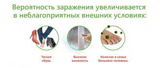
Infection with fungi occurs through direct contact with a patient, through shoes, clothing, when using general hygiene products (washcloths, manicure tools), when visiting gyms, baths, saunas, and swimming pools. The main factor of infection is the presence of abrasions and cracks in the skin, which occur with increased sweating, dryness, and poor circulation in the extremities. People with diabetes, immune disorders, blood diseases; Those taking antibiotics, glucocorticosteroids, and cytostatics for a long time are especially susceptible to the development of mycoses.
Most often, microscopic fungi cause damage to the skin of the feet and large skin folds (axillary, groin and others).
Mycosis: causes and treatment
Mycosis is a fungal infectious disease. Its causative agents are pathogenic and opportunistic microorganisms. Fungal diseases are very common these days; Potentially dangerous places include public beaches, swimming pools, bathhouses, and gyms . The fungus enters the human body through the respiratory system , microcracks in the skin and through mucous membranes.
Mycosis is a highly contagious disease - it can spread quite quickly not only among people, but also among animals.
Here are the reasons
that may contribute to the development of
fungal diseases
: • Weak immunity (promotes the engraftment of the fungus and the development of symptoms). • Frequent use of antibiotics (reduces immunity). • Varicose veins. • Diabetes mellitus (poorly healing wounds and cracks are the gateway for fungal penetration). • Abrasions, ulcers. • Narrow interdigital space (increased sweating in this area is a favorable environment for the development of fungus). • Increased body humidity, sweating. • Contact with an infected person or animal. • Using someone else's shoes, toiletries, towels. • Non-sterile manicure/pedicure instruments.
The signs of a fungal infection are well known:
with mycosis of the feet (dermatomycosis) - itching, redness, peeling of the skin.
With nail fungus (onychomycosis) - a change in the color of the nail (yellow, brown, gray or black) and its structure, which, under the influence of the disease, becomes loose and layered, thins or thickens. However, onychomycosis
is easily confused with other disorders. For example, with neurotic lesions of the nails or psoriasis, in which the nail becomes like a thimble (into a small hole) or turns yellow. Orthopedic problems, including lumbosacral osteochondrosis, disorders of peripheral innervation and blood supply, as well as vascular diseases, thyroid gland and other endocrine pathologies, greatly change the appearance of the nail. In addition, today a blurred picture of the disease is often encountered, which leads to late referral to specialists and ultimately to the development of advanced forms of fungal disease. Typically, there are two stages of toenail fungus, and it is easy to miss at the initial stage.
The first signs of fungus:
• small white dots on the nails; • changing the shape of the nail; • appearance of white stripes; • nail fragility; • change in surface, it can become hard and ridged; • The skin around the affected nail often turns red and peels.
In the advanced stage, the color of the nail plate changes, it becomes rougher and thicker, and the nail begins to crumble. Gradually, the fungus can spread to neighboring nails.
Tips on hygiene, prevention and treatment
Since a combination of mycosis of the skin and nails often develops, treating these processes separately is ineffective.
required :
a cream for the affected skin of the feet, a deeply penetrating solution and/or varnish for the treatment of nails.
If you request varnishes/solutions for the treatment of mycoses or if you have complaints about fungal infections of the nail plate, we recommend Mikoderil solution
or
Onihelp varnish
Exol
kit for removing fungal infections from the nail plate and its further restoration.
an antifungal cream (Terbized) or gel (Lamifungin)
is required . And don’t forget that you need to prepare for long-term treatment.
NB! External remedies can be effective only if no more than ⅓ of the nail plate and no more than 3 nails are affected by the fungus. To combat an advanced infection, long-term oral use of systemic antimycotics, which are prescribed by a doctor based on test results, will be required.
To maintain foot hygiene, broad-spectrum antiseptics ( MestaMidin-sens solution)
/
MykoStop
spray ) are effective preventative agents. Such preparations are also necessary when using varnishes and solutions, because when filing the nail plate, fungal spores can get onto the surrounding skin.
One of the reasons for the occurrence of mycoses may be reduced immunity. To maintain the body's defense system in the spring-summer period, it is recommended to take antioxidant vitamins C, D, Zn, Se ( VerrumVit
*,
Selenium WTF
*) or immunostimulants (
Ingaron
) and immunoadaptogens (
Trekrezan
). This will have a beneficial effect on susceptibility to any kind of infections and diseases.
Consequences of the disease
- not only severe itching, but also an unsatisfactory appearance of the nail plate.
Therapeutic and prophylactic varnishes will help mask these manifestations, and sedatives will help to cope with the nervousness and irritability that accompany the disease ( Passnovel*/Passiflora plus*
).
Among other things, fungi are allergens and produce mycotoxins that harm almost all body systems. They contribute to the development and complicate the course of bronchial asthma, urticaria, atopic dermatitis, allergic rhinitis, nasal polyps, and conjunctivitis. Therefore, it would be useful to offer sorbents in the form of tablets ( Normasorb
*,
Nor Masorb-Lacto
*) or powders (
Sorboxan
*,
Liquid Charcoal
*) for oral administration.
If hygiene rules are not followed, relapses of the disease may develop. To ensure dry feet, if we are talking about skin and toenail fungus
(after all, a humid and warm environment is an excellent environment for fungal growth),
DryDry
,
Salton
, and
Scholl deodorants are suitable.
And we definitely recommend treating shoes where fungal spores can remain for a long time.
For this we offer antiseptics (MestaMidin-sens)
or special sprays (
MikoStop
). To avoid contracting fungal infections, you should adhere to generally accepted rules of hygiene and avoid contact of spores with mucous membranes.
THERE ARE CONTRAINDICATIONS, YOU MUST READ THE INSTRUCTIONS OR CONSULT WITH A SPECIALIST. Dietary supplement NOT A MEDICINE.
What types of fungal infections are there?
There are a fairly large number of fungi that can cause skin lesions in humans.
The main differences between them:
- Favorite localization locations. Some fungi only affect the skin. Others are usually localized on the scalp and cause damage to the skin and hair. There are fungi that affect the nails (a group of diseases - onychomycosis).
- Seasonality. Different seasons are favorable for different types of fungi.
- Elements that appear on the skin during a disease.
Currently, there are five main groups of dermatomycosis:
- Keratomycosis - the fungus affects only the top layer of skin (epidermis).
- Actually, dermatomycosis is a disease in which the fungus affects the skin, hair, and nails.
- Candidiasis is a group of fungal diseases caused by yeast-like fungi Candida. They can affect not only the skin, but also mucous membranes. Typically, candidiasis develops with a significant decrease in immunity and long-term use of certain medications.
- Deep mycoses are diseases in which the fungus affects internal organs and bones.
- Pseudomycoses are diseases that superficially resemble mycoses, but are caused by other pathogens.
conclusions
Superficial fungal infections are most often caused by dermatophytes from the genera Trichophyton, Epidermophyton and Microsporum. These organisms metabolize keratin and cause a number of pathological clinical manifestations, including tinea pedis, dermatophytal dermatitis, dermatophytosis, etc. Based on clinical findings, the diagnosis of cutaneous dermatophyte infection can be strongly suspected. To confirm the diagnosis, potassium hydroxide should be used. Most dermatophyte infections can be managed with topical treatment. Examples of effective topical antifungal agents include azoles, allylamines, ciclopirox, butenafine, and tolnaftate. Oral antifungal therapy is used for widespread infections or infections that are resistant to topical therapy. Nystatin is not effective against dermatophyte infections.
List of literature / References
- Havlickova B, Czaika VA, Friedrich M. Epidemiological trends in skin mycoses worldwide // Mycoses. 2008 Sep;51 Suppl 4:2-15. doi: 10.1111/j.1439-0507.2008.01606.x. Erratum in: Mycoses. 2009 Jan;52(1):95. PMID: 18783559.
- Havlickova B, Czaika VA, Friedrich M. Epidemiological trends in skin mycoses worldwide // Mycoses 2008; 51 Suppl 4:2.
- Seebacher C, Bouchara JP, Mignon B. Updates on the epidemiology of dermatophyte infections // Mycopathologia 2008; 166:335.
- Ameen M. Epidemiology of superficial fungal infections // Clin Dermatol 2010; 28:197.
- El-Gohary M, van Zuuren EJ, Fedorowicz Z, et al. Topical antifungal treatments for tinea cruris and tinea corporis // Cochrane Database Syst Rev 2014; :CD009992.
- Alston SJ, Cohen BA, Braun M. Persistent and recurrent tinea corporis in children treated with combination antifungal/ corticosteroid agents // Pediatrics 2003; 111:201.
- Greenberg HL, Shwayder TA, Bieszk N, Fivenson DP. Clotrimazole/betamethasone diproprionate: a review of costs and complications in the treatment of common cutaneous fungal infections // Pediatr Dermatol 2002; 19:78.
- Rosen T, Elewski BE. Failure of clotrimazole-betamethasone dipropionate cream in treatment of Microsporum canis infections // J Am Acad Dermatol 1995; 32:1050.
- Hawkins DM, Smidt AC. Superficial fungal infections in children // Pediatr Clin North Am 2014; 61:443.
- Crawford F, Hollis S. Topical treatments for fungal infections of the skin and nails of the foot // Cochrane Database Syst Rev 2007; :CD001434.
- Korting HC, Tietz HJ, Brautigam M, et al. One week terbinafine 1% cream (Lamisil) once daily is effective in the treatment of interdigital tinea pedis: a vehicle controlled study. LAS-INT-06 Study Group // Med Mycol 2001; 39:335.
- Gupta AK, Cooper EA. Update in antifungal therapy of dermatophytosis // Mycopathologia 2008; 166:353.
- Bell-Syer SE, Khan SM, Torgerson DJ. Oral treatments for fungal infections of the skin of the foot // Cochrane Database Syst Rev 2012; 10:CD003584.
- Adams BB. Tinea corporis gladiatorum // J Am Acad Dermatol 2002; 47:286.
- van Zuuren EJ, Fedorowicz Z, El-Gohary M. Evidence-based topical treatments for tinea cruris and tinea corporis: a summary of a Cochrane systematic review // Br J Dermatol 2015; 172:616.6.
- Bourlond A, Lachapelle JM, Aussems J, et al. Double-blind comparison of itraconazole with griseofulvin in the treatment of tinea corporis and tinea cruris // Int J Dermatol 1989; 28:410.
- Cole GW, Stricklin G. A comparison of a new oral antifungal, terbinafine, with griseofulvin as therapy for tinea corporis // Arch Dermatol 1989; 125:1537.
- Panagiotidou D, Kousidou T, Chaidemenos G, et al. A comparison of itraconazole and griseofulvin in the treatment of tinea corporis and tinea cruris: a double-blind study // J Int Med Res 1992; 20:392.
- Faergemann J, Mörk NJ, Haglund A, Odegård T. A multicentre (double-blind) comparative study to assess the safety and efficacy of fluconazole and griseofulvin in the treatment of tinea corporis and tinea cruris // Br J Dermatol 1997; 136:575.
- Elewski BE, Hughey LC, Sobera JO. Fungal diseases. In: Dermatology, 3rd ed, Bolognia JL, Jorizzo JL, Schaffer JV (Eds), Elsevier Limited, Philadelphia; London 2012. Vol 2, p.1251.
- Voravutinon V. Oral treatment of tinea corporis and tinea cruris with terbinafine and griseofulvin: a randomized double blind comparative study // J Med Assoc Thai 1993; 76:388.
- Farag A, Taha M, Halim S. One-week therapy with oral terbinafine in cases of tinea cruris/corporis // Br J Dermatol 1994; 131:684.
- Smith KJ, Neafie RC, Skelton HG 3rd, et al. Majocchi's granuloma // J Cutan Pathol 1991; 18:28.
- Gill M, Sachdeva B, Gill PS, et al. Majocchi's granuloma of the face in an immunocompetent patient // J Dermatol 2007; 34:702.
- Cho HR, Lee MH, Haw CR. Majocchi's granuloma of the scrotum // Mycoses 2007; 50:520.
- Tse KC, Yeung CK, Tang S, et al. Majocchi's granuloma and posttransplant lymphoproliferative disease in a renal transplant recipient // Am J Kidney Dis 2001; 38:E38.
- Kim ST, Baek JW, Kim TK, et al. Majocchi's granuloma in a woman with iatrogenic Cushing's syndrome // J Dermatol 2008; 35:789.
- Akiba H, Motoki Y, Satoh M, et al. Recalcitrant trichophytic granuloma associated with NK-cell deficiency in a SLE patient treated with corticosteroid // Eur J Dermatol 2001; 11:58.
- Ilkit M, Durdu M, Karakaş M. Majocchi's granuloma: a symptom complex caused by fungal pathogens // Med Mycol 2012; 50:449.
- Novick NL, Tapia L, Bottone EJ. Invasive trichophyton rubrum infection in an immunocompromised host. Case report and review of the literature // Am J Med 1987; 82:321.
- Feng WW, Chen HC, Chen HC. Majocchi's granuloma in a 3-year-old boy // Pediatr Infect Dis J 2006; 25:658.
- Gupta AK, Prussick R, Sibbald RG, Knowles SR. Terbinafine in the treatment of Majocchi's granuloma // Int J Dermatol 1995; 34:489.
- McMichael A, Sanchez DG, Kelly P. Folliculitis and the follicular occlusion tetrad. In: Dermatology, 2nd ed, Bolognia JL, Jorizzo JL, Rapini RP (Eds), Elsevier Limited, St. Louis 2008.
- Gupta AK, Groen K, Woestenborghs R, De Doncker P. Itraconazole pulse therapy is effective in the treatment of Majocchi's granuloma: a clinical and pharmacokinetic evaluation and implications for possible effectiveness in tinea capitis // Clin Exp Dermatol 1998; 23:103.
- Burg M, Jaekel D, Kiss E, Kliem V. Majocchi's granuloma after kidney transplantation // Exp Clin Transplant 2006; 4:518.
- Liao YH, Chu SH, Hsiao GH, et al. Majocchi's granuloma caused by Trichophyton tonsurans in a cardiac transplant recipient // Br J Dermatol 1999; 140:1194.
- Bonifaz A, Vázquez-González D. Tinea imbricata in the Americas // Curr Opin Infect Dis 2011; 24:106.
- Cheng N, Rucker Wright D, Cohen BA. Dermatophytid in tinea capitis: rarely reported common phenomenon with clinical implications // Pediatrics 2011; 128:e453.
- Romano C, Rubegni P, Ghilardi A, Fimiani M. A case of bullous tinea pedis with dermatophytid reaction caused by Trichophyton violaceum // Mycoses 2006; 49:249.
- Al Aboud K, Al Hawsawi K, Alfadley A. Tinea incognito on the hand causing a facial dermatophytid reaction // Acta Derm Venereol 2003; 83:59.
- Veien NK, Hattel T, Laurberg G. Plantar Trichophyton rubrum infections may cause dermatophytids on the hands // Acta Derm Venereol 1994; 74:403.
What are the signs of skin fungus? When should you visit a dermatologist?
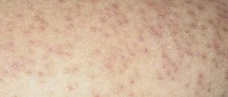
Fungal skin infection can be suspected if the following signs are present:
- peeling;
- increased skin dryness;
- redness, swelling - signs of inflammation;
- pain, itching;
- bubbles;
- cracks.
If these symptoms do not go away on their own, then you should see a dermatologist for timely treatment.