Amebiasis - symptoms and treatment
Amoebiasis (Amoebic Dysentery) is an acute and chronic disease that is caused by pathogenic strains of dysenteric amoeba. Penetrating into the body, they lead to ulcerative damage to the intestines, which is accompanied by moderate intoxication, stool disorders, weight loss and sometimes the appearance of abscesses in the liver, intestines, lungs and brain. The disease can last a long time, sometimes leading to death, especially in weakened patients and in the absence of medical care.
Pathogen
Taxonomy:
- domain - eukaryotes;
- branch - amoeba-like;
- type - Evosea;
- class - Archamoebae;
- family - Entamoebidae;
- genus - Entamoeba (entamoeba);
- species - dysentery amoeba (Entamoeba histolytica).
The Russian scientist F. A. Lesh was the first to describe the pathogen and prove its pathogenicity in 1875. At least 22 strains have now been isolated, of which 9 are pathogenic, and the remaining 13 are most likely not pathogenic for humans.
Changing during life, amoebas go through two stages of development:
- the vegetative, or active, stage in the form of a trophozoite ;
- a resting stage, or "dormant" stage, in the form of a cyst .
The vegetative stage divides amoebas according to function and structure into large vegetative, luminal and tissue. All of them, suddenly entering the external environment, die within 30 minutes.
Large vegetative trophozoites (forma magna) reach 20-60 microns. This form of amoebas moves due to forward thrusts. Contains the nucleus and cytoplasm - the main part of the cell. The cytoplasm is divided into a glassy transparent mass and an inner liquid layer with digestive vacuoles in which red blood cells captured by the amoeba are digested. In acute amoebiasis, large trophozoites are detected in fresh feces - “warm feces”. Capable of breaking down protein molecules. They have surface pectins, with the help of which amoebas attach to the intestinal mucosa.
The luminal vegetative trophozoites (forma minuta) are much smaller than the large form: they reach 15-20 microns. Such trophozoites are inactive. They have one core, which is not visible without coloring. Their cytoplasm contains small vacuoles, but without erytrophytes. Luminal trophozoites live in the upper part of the large intestine, feed on bacteria, reproduce, and do not cause obvious harm. They are detected in the feces of acute amoebiasis at the beginning of recovery, in chronic amoebiasis, as well as in amoeba carriers, but to identify them it is necessary to carry out deep intestinal lavages or examine the final portions of feces after taking a saline laxative. In the lower part of the large intestine, with a gradual deterioration of conditions, for example, lack of fluid, disruption of the bacterial flora, taking medications or changes in the pH of the environment, they usually transform into a cyst form, which is gradually released into the environment. If the body's defenses are weakened, luminal trophozoites can transform into a larger vegetative form and become aggressive.
Tissue vegetative trophozoites are formed from the luminal form, but are similar in appearance to the vegetative one. They reach 20-25 microns, are mobile, parasitize, penetrating the mucous membrane of the colon, and infect the intestines. They are detected only in acute amoebiasis in the affected organs; they are found extremely rarely in liquid feces - during the disintegration of intestinal ulcers.
Cysts form from the luminal form in the lower parts of the colon. They have a round shape and reach 8-15 microns. They are immobile, covered with a dense shell of chitin, and contain chromatoid bodies (RNA with protein) and glycogen. Depending on the maturity of the cysts, they contain from 1 to 4 nuclei. They are more often detected in patients with chronic amoebiasis and parasite carriers. They are very stable in the external environment: they remain in feces at room temperature for up to 2 weeks or longer, when frozen to −21°C - no more than 3 months, in water - up to 8 months. When heated to 100°C and boiled, they quickly die. Standard household disinfectants with chlorine have almost no effect on cysts. Soap-cresol preparations, a solution of sublimate 1:1000 and a 3% solution of carbolic acid can affect them [1][3][7][10].
Epidemiology
Amoebiasis is a widespread disease, mainly found in the countries of Southern and Western Africa, Central and South America, as well as in India, China and Korea. In Russia, isolated cases are mainly recorded, mainly in the southern regions, the Caucasus and the Far East. Although recently the incidence in our country has been increasing. This may be due to the influx of migrants from border regions and the development of tourism.
On average, about 50 million cases of amoebiasis are registered annually in the world, of which about 100 thousand end in death. In terms of the number of deaths, this disease ranks third among parasitic diseases. Approximately 90% of cases of the disease are intestinal amoebiasis, the remaining forms are extraintestinal.
The source of infection is a person (patient or carrier). With its feces, amoebas in the form of cysts enter the environment.
The transmission mechanism is fecal-oral. Includes water, food, household contact and sexual (oral-anal) transmission routes. Infection can occur when any substance (water, food, dirt, fingers) that comes into contact with the feces of an infected person or contains some part of it enters the mouth. Mechanical spreaders and carriers can be cockroaches and flies. In extremely rare cases, homosexuals and patients with secondary immunodeficiency may become infected when amoebas enter directly into the wound.
A person with amoebiasis runs the risk of transmitting the infection to family members, but this risk is low, especially if the person follows the rules of personal hygiene: thoroughly washes his hands after using the restroom and before preparing food.
Situations that increase the risk of infection:
- visiting tropical countries with low levels of sanitation;
- interaction with immigrants from tropical countries with poor sanitary conditions;
- neglect of personal hygiene rules;
- consumption of raw, undisinfected water from open sources and the water supply system;
- homosexual contacts;
- presence of mental disorders.
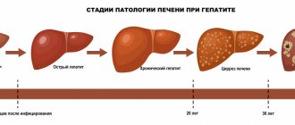
Immunity after an illness is unstable and weakly expressed, repeated infections are possible. Even a large number of antibodies formed, for example in amoebic liver abscesses, do not protect against the progression of amebiasis [1][2][5][6].
Causes of amoebiasis
The causative agent of amebiasis is a single-celled organism Entamoeba histolytica. It has 2 stages of development - vegetative and cyst (resting phase). The microorganism is excreted in the feces of the patient or carrier and enters the environment.
The development of the disease is provoked by additional causes of amebiasis. Risk group:
- staying in warm countries with unsanitary conditions;
- failure to comply with personal hygiene rules in a closed group;
- immunodeficiency or severe concomitant diseases.
Treatment of amoebiasis
The most effective medications for killing pathogens are metronidazole and tinidazole. They are prescribed in a course of 3 to 8 days.
Treatment of amebiasis includes additional antimicrobial drugs (intestopan, tetracyclines), medications to eliminate diarrhea, bloating, enterosorbents, and vitamins. When abscesses form, surgical treatment is necessary.
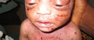
For the cutaneous form, metronidazole in the form of a cream is additionally prescribed.
With timely treatment, the prognosis for this disease is favorable. Late-diagnosed liver or lung abscesses are life-threatening.
Prevention
A vaccine against the pathogen has not been created. Prevention of amebiasis includes the following measures:
- hospitalization and isolation of patients, their treatment until the pathogens disappear in the feces;
- identification and treatment of disease carriers;
- a ban on persons who have suffered amoebiasis from working in public catering establishments;
- careful adherence to personal hygiene, drinking only boiled water, especially when staying in countries with hot climates;
- during tourist trips to dangerous areas, do not eat vegetables and fruits that have already been washed by someone, wash them thoroughly before eating, use a straw to drink from water bottles, do not drink iced or carbonated drinks, do not eat milk, cheese and other unpasteurized products , do not buy food on the street.
Self-medication of amoebiasis is unacceptable. When the first signs of illness appear, you should consult a doctor.
Treatment of amebiasis at the Mama Papa Ya clinic
The network of family clinics “Mama Papa Ya” provides medical services for suspected amoebiasis. If you experience loose stools or pain in the right half of the abdomen, especially after returning from a vacation from warm countries, we recommend contacting a pediatrician, therapist or gastroenterologist.
Advantages of our clinic:
- a network of branches is located in Moscow and other cities;
- quick appointment with a specialist without queues;
- good training of doctors and extensive experience to suggest the correct diagnosis clinically;
- confirmation of the diagnosis is carried out through laboratory tests using modern equipment, which eliminates the possibility of error;
- prescribing modern drugs for the treatment of amebiasis in children and adults and monitoring the effectiveness of therapy if the patient refuses to be hospitalized in an infectious diseases hospital.
The doctors at our clinic specialize in such diseases, you can make an appointment right now!
Reviews
Good clinic, good doctor!
Raisa Vasilievna can clearly and clearly explain what the problem is. If something is wrong, she speaks about everything directly, not in a veiled way, as other doctors sometimes do. I don’t regret that I ended up with her. Anna
I would like to express my gratitude to the staff of the clinic: Mom, Dad, and me. The clinic has a very friendly atmosphere, a very friendly and cheerful team and highly qualified specialists. Thank you very much! I wish your clinic prosperity.
Anonymous user
Today I had a mole removed on my face from dermatologist I.A. Kodareva. The doctor is very neat! Correct! Thanks a lot! Administrator Yulia Borshchevskaya is friendly and accurately fulfills her duties.
Belova E.M.
Today I was treated at the clinic, I was satisfied with the staff, as well as the gynecologist. Everyone treats patients with respect and attention. Many thanks to them and continued prosperity.
Anonymous user
The Mama Papa Ya clinic in Lyubertsy is very good. The team is friendly and responsive. I recommend this clinic to all my friends. Thanks to all doctors and administrators. I wish the clinic prosperity and many adequate clients.
Iratyev V.V.
We visited the “Mama Papa Ya” Clinic with our child. A consultation with a pediatric cardiologist was needed. I liked the clinic. Good service, doctors. There was no queue, everything was the same price.
Evgeniya
I liked the first visit. They examined me carefully, prescribed additional examinations, and gave me good recommendations. I will continue treatment further; I liked the conditions at the clinic.
Christina
The doctor carefully examined my husband, prescribed an ECG and made a preliminary diagnosis. She gave recommendations on our situation and ordered additional examination. No comments so far. Financial agreements have been met.
Marina Petrovna
I really liked the clinic. Helpful staff. I had an appointment with gynecologist E.A. Mikhailova. I was satisfied, there are more such doctors. Thank you!!!
Olga
References
- Bogomolov, B.P. Infectious diseases: Textbook. - M.: Moscow State University Publishing House, 2009. - 592 p.
- Heiman, F., Wertheim, P., Horby, J. Woodall - Atlas of Human Infectious Diseases, First Edition, 2012. - 304 p.
- Crump, J., Sjölund-Karlsson, M., Gordon, M. et al. Clinical Presentation, Laboratory Diagnosis, Antimicrobial Resistance, and Antimicrobial Management of Invasive Salmonella Infections / Clinical Microbiology Reviews, 2015. - Vol. 28(4). — P. 901-937.
- Andrews, J., Ryan, E. Diagnostics for invasive Salmonella infections: Current challenges and future directions, 2015. - Vol. 33(3). — P. 8-15.