Lymphadenopathy is a condition that is characterized by an increase in the size of peripheral lymph nodes, a change in their consistency and mobility. It develops along with an increase in overall body temperature, an enlargement of the spleen and liver. Even a slight enlargement of the lymph nodes indicates that pathological processes are developing in the body that require adequate treatment. It can cover one or more structures and cause inflammatory processes.
The hematology department of CELT invites you to undergo diagnosis and treatment of lymphadenopathy in Moscow. You have the opportunity to consult with leading domestic hematologists, who have modern techniques and a powerful base that allows you to quickly and accurately establish a diagnosis. They provide treatment in accordance with international standards. You can find out the cost of services in the “Services and Prices” section. To avoid misunderstandings, do not forget to check the numbers during consultation or with our operators.
Lymphadenopathy: what is it?
Enlarged lymph nodes look like soft or dense rounded formations located under the lower jaw, in the cervical, axillary, groin or other areas. They can have a smooth or bumpy surface and very often develop after acute infections and inflammations. Sometimes their appearance is caused by traumatic injuries to the skin or the introduction of a vaccine.
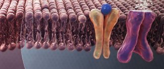
Lymphadenopathy develops due to the accumulation of a certain type of cell in the lymphadenoid tissue. The reaction is often provoked by increased blood flow, an increase in the number of lymphocytes and macrophages in response to the appearance of foreign genes in the body. In just one week, the node can increase five to fifteen times. A pathological condition is spoken of if, along with an excess of size, there is a change in the density, mobility and surface of the structure. When palpated, it can be quite painful or painless.
Reasons for contacting our center for diagnosing lymphadenopathy in Moscow: independent identification of large nodes that do not hurt, a feeling of intense pain during palpation, other symptoms in the form of a rash, increased body temperature, weight loss, and rapid fatigue. Particularly dangerous can be nodes that do not go away for more than two months, located in different areas, with a diameter of more than two centimeters, the enlargement of which occurred for no apparent reason.
Survival of patients with retroperitoneal lymphadenopathy after cytoreductive surgery and chemotherapy
The effect of treatment should always exceed the possible risks associated with treatment. This thesis is especially acute in the treatment of cancer patients.
To address this in patients with carcinomatosis secondary to colorectal cancer, LR van der Werf and colleagues conducted a multicenter retrospective study of the treatment outcomes of these patients with CT-visualized retroperitoneal lymphadenopathy.
One of the main treatment methods for patients with carcinomatosis is cytoreductive surgery followed by the introduction of chemotherapy drugs heated to 41°C into the abdominal cavity, the so-called hyperthermic intraperitoneal chemotherapy (HIPEC).
If untreated, the median survival for these patients is about 6 months. And when performing cytoreductive surgery and hyperthermic intraperitoneal chemotherapy (CRS-HIPEC), some researchers indicate a median survival of up to 48 months.
However, these manipulations are associated with the risks of developing severe complications, such as the formation of intestinal fistulas and anastomotic failure. This is especially true for patients with extra-regional metastases. This also includes patients with visualized retroperitoneal lymphadenopathy, so these patients may be denied CRS-HIPEC due to unsatisfactory treatment results. Most often, CT or PET scanning is used to diagnose retroperitoneal lymphadenopathy, since access for biopsy is difficult. But enlargement of the retroperitoneal lymph nodes is not always an accurate criterion for their metastasis. This may be an inflammatory response to a tumor or infection. Therefore, the appropriateness of excluding these patients for CRS-HIPEC is debatable.
According to the study, 25 of 401 patients (6.1%) had lymphadenopathy of the retroperitoneal lymph nodes on preoperative CT imaging. In the group with lymphadenopathy, one-, three- and five-year survival rates were 80%, 59%, 38%, and without - 90%, 50%, 36%. The median overall survival was 47 months in the group without lymph node involvement versus 35 months with lymph node involvement (p=0.70), and the median disease-free survival was 15 versus 14 months (p=0.81), with no statistically significant difference between the groups . In multivariate analysis, retroperitoneal lymphadenopathy had no significant effect on survival.
Lymphadenopathy: classification and causes
The classification of lymphadenopathy is based on a number of parameters, starting with the location of the affected structures and ending with their pain and size:
| Localization of the affected node/nodes | Etiological factors |
| Under the lower jaw | Diseases of the eyes, ear, throat, nose, damage to the skin in the head or neck area, dental problems. |
| Neck |
|
| Supraclavicular region | Malignant neoplasms:
|
| Armpits |
|
| Groin |
|
Causes of enlarged cervical lymph nodes
This group of lymph nodes drains the area of the head, neck, upper chest, and proximal parts of the upper extremities. Accordingly, enlarged cervical nodes most often indicate the presence of thyroid diseases, inflammatory processes in the oropharynx, bacterial and viral infections. The symptom is pathognomonic for rubella, develops with measles, and can be detected with more rare infectious pathologies - psittacosis, Ebola and Marburg fever, mycoplasma infections.
Thyroid cancer
Thyroid neoplasia accounts for about 1.5% of all malignant neoplasms and in most cases is asymptomatic. The most typical variant is papillary cancer. Follicular tumors are quite common. With these space-occupying formations, there is always an enlargement of the cervical lymph nodes, which indicates increased proliferation of malignant thyrocytes and metastasis of tumor cells. Typically, the lymph node remains soft and mobile for a long time, since the process does not affect the capsule and surrounding tissues.
Patients, as a rule, consult a doctor about an accidentally discovered nodule in the thyroid gland when it reaches 1 cm or more. As the tumor grows, other manifestations occur: cough, hoarseness, associated with compression of adjacent anatomical structures. Large tumors can compress the airways, causing shortness of breath and suffocation. When cancer spreads beyond the organ capsule, an expansion of the subcutaneous venous network and deformation of the contours of the neck occurs. In elderly patients, cachexia increases.
Cervical lymphadenopathy is one of the signs of thyroid lymphoma - an aggressive neoplasia characterized by intensive growth and involvement of neighboring organs in the pathological process. The disease often develops against the background of autoimmune thyroiditis. The tumor grows rapidly, occupying an entire lobe of the organ. Patients often themselves discover a node of woody density, which is combined with enlargement and hardening of the cervical lymph nodes on the affected side. Compression of surrounding tissues provokes dysphagia, paresis of the vocal cords, and displaces the esophagus and trachea.
Organic diseases of the thyroid gland
In other lesions of the endocrine gland, enlargement of the lymph nodes is caused by an increase in blood flow by 10-15 times, increased production and differentiation of normal lymphocytes in response to stimulation by foreign antigens. The size of the lymph nodes is more than 1 cm, they are elastic, not fused with the surrounding tissues, and are sometimes sensitive to palpation. The symptom is characteristic of acute inflammatory processes, but also occurs in benign neoplasms and chronic autoimmune thyroiditis. Cervical lymphadenopathy is caused by:
- Acute thyroiditis
. The disease begins suddenly with a sharp pain in the thyroid gland, which radiates to the lower jaw and ear. Lymph nodes enlarge on both sides, become very painful, and redness of the skin is noted. Purulent thyroiditis occurs with an increase in temperature to febrile levels and severe symptoms of intoxication. There may be complaints of a feeling of pressure and fullness in the neck, and increased symptoms when coughing. - Nodules and cysts.
According to statistics, various benign formations of the thyroid gland are detected in 10% of the population, but more often they are asymptomatic. Enlargement of lymph nodes in the cervical region occurs with inflammation or suppuration of cysts, hormonally active neoplasia. Lymph nodes are elastic, practically painless, the skin over them is not changed. A detailed clinical picture of thyroid damage is observed with overproduction of hormones - thyrotoxicosis.
Rubella and measles
Cervical lymphadenopathy involving the posterior cervical and occipital nodes is an important symptom of rubella. Lymph nodes are moderately enlarged in size, painless, and not fused to the surrounding skin. For children, the appearance of “bullet” lymph nodes is typical - multiple small formations in the neck. Simultaneously with lymphadenopathy, a rash occurs - pinpoint or papular rashes are localized on the extensor surfaces of the limbs, in the torso and head. The rash disappears after a few days, leaving no peeling or pigmentation.
Lymphadenitis with enlargement of the cervical lymph nodes develops in the catarrhal stage of measles. Lymphadenopathy is combined with rhinitis, conjunctivitis, hyperemia of the pharynx and puffiness of the face. In adults, the manifestations of the catarrhal period are less pronounced. A pathognomonic sign of measles is Belsky-Filatov-Koplik spots on the mucous membrane of the cheeks. After 4-5 days, the second wave of fever begins, which coincides with the appearance of a maculopapular rash. On the 1st day, the rash is located on the face and neck, by the end of the first day it spreads to the torso, and on the third day the rash spreads to the limbs.
Other infectious diseases
Cervical lymphadenopathy is detected in various infections occurring in the oral cavity and ENT organs. Symptoms may occur with systemic bacterial and protozoal lesions - anginal-bubonic form of tularemia, sleeping sickness, diphtheria. Enlargement of lymph nodes is associated with the primary penetration and proliferation of pathogenic microorganisms, intensive proliferation and accumulation of specific clones of lymphocytes in the follicular and paracortical zones. With damage to the cervical lymphoid formations, the following occurs:
- Congenital listeriosis
. Infectious pathology develops during transplacental or intrapartum infection of the child and manifests itself in the first days after birth. Damage to the cervical nodes is combined with febrile body temperature, roseola or hemorrhagic rash, granulomas on the oral mucosa. The late form is accompanied by muscle tremors, convulsions, enlargement of the liver and spleen. - Syphilis
. After entering the body, Treponema pallidum multiplies in regional lymph nodes, causing them to enlarge. Cervical lymphadenopathy is often observed when the pathogen penetrates through the mucous membrane of the oral cavity or lips, where the primary affect is localized - chancroid. A month later, the chancre disappears on its own, then a polymorphic rash appears, which indicates the generalization of the infection and the development of secondary syphilis. - Brucellosis
. In the prodromal period, patients complain of myalgia, arthralgia, and headaches. Then a fever occurs, lasting from several days to 3 weeks and alternating with heavy sweats. At high temperatures, there is facial hyperemia, enlargement of the cervical and axillary lymph nodes, which can be painful on palpation. In the acute form, small fibrous formations appear along the tendons. - Inguinal lymphogranulomatosis
. Enlargement of the lymph nodes of the neck and submandibular region is observed in the secondary period when the primary affect (ulcer) is localized in the mucous membrane of the mouth and pharynx. As the disease progresses, lymphoid formations turn into large-lumpy tumors and lose mobility. Subsequently, the nodes suppurate, fever and intoxication develop, and fistulas form. - Tonsillitis, pharyngitis
. The reaction of the cervical lymph nodes is detected in tonsillitis, which is due to increased antigenic stimulation of lymphoid formations. The symptom is accompanied by sore throat, redness of the throat and tonsils, and fever. You may notice yellowish dots or widespread plaque on the surface of the tonsils. Enlarged lymph nodes are also found in herpetic pharyngitis, which is characterized by a vesicular rash on the pharyngeal mucosa.
Severe cases of tonsillitis can be complicated by a retropharyngeal abscess, a purulent inflammation of the pharyngeal tissue. In addition to lymphadenopathy of the upper cervical and occipital lymph nodes, the patient is bothered by sharp pain in the throat, difficulty swallowing, and if the abscess is large, breathing disorders are possible. The general condition is disturbed, body temperature rises to 39-40° C. Enlarged lymph nodes, combined with damage to the jugular vein and septicemia, are pathognomonic for Lemierre's syndrome. In children, cervical lymphadenopathy often indicates adenoiditis.
Head and neck tumors
Lymph from the face and neck goes directly to the cervical lymph nodes, therefore, for various malignant tumors of this area, they are a typical site of metastasis. Lymph nodes are usually woody in density, tightly connected to the skin and surrounding tissue, and are not painful. Lymphadenopathy in certain types of tumors serves as the initial sign of the disease, when the primary tumor does not yet cause clinical symptoms. Enlarged lymph nodes are observed with such malignant neoplasias as:
- Neoplasms of the jaws.
The tumor may be characterized by exophytic growth with the formation of a protruding node with ulcerations. It may occur as a long-term non-healing ulcer with purulent discharge. Bone damage (osteosarcoma) is indicated by shooting pains, loosening and loss of teeth. The lymph nodes are enlarged on both sides, have a rocky density, and are fused to the skin and subcutaneous tissue. - Tongue cancer
. Although with neoplasia of this localization there is often an enlargement of the submandibular lymph nodes, sometimes metastasis also occurs in the cervical group. Neoplasia of the tongue externally looks like a diffuse thickening of the organ with the formation of ulcers or local tissue growth. Pain syndrome appears early, eating disorders are pronounced, and weight loss progresses. - Neoplasms of ENT organs.
Cervical lymphadenopathy is a symptom of epithelial tumors of the nasopharynx, which also manifest as nosebleeds and difficulty breathing. The symptom is determined by esthesioneuroblastoma and is combined with anosmia, nasal congestion, and mucous discharge. Sometimes unilateral enlargement and hardening of the lymph nodes indicates ear neoplasia (basal cell carcinoma, epithelioma, sarcoma). - Eye tumors.
Enlarged nodes are observed at an advanced stage of conjunctival neoplasms, when malignant cells grow into the surrounding tissues and spread through the lymphogenous and hematogenous routes. Enlarged lymph nodes of the submandibular region and neck occur in ocular melanoma, an aggressive neoplasm of pigment cells that rapidly progresses with the development of distant metastases. - Timoma
. An increase in cervical lymphoid structures is detected in benign and malignant tumors of the thymus. Invasive growth of the tumor causes compression syndrome with intense chest pain, dry cough, and difficulty breathing. When peripheral nerves are compressed, Horner's syndrome, hoarseness, and dysphagia occur. About 30% of cases of the disease are accompanied by myasthenia gravis.
Systemic lesions of lymphoid tissue
Enlarged lymph nodes in the neck can be the first sign of lymphogranulomatosis - malignant hyperplasia of lymphoid tissue with the formation of specific granulomas. The disease is characterized by the appearance of dense, painless nodes, which are arranged in the form of a chain. In the local form of the lesion, one group of lymph nodes enlarges; in the generalized form, total lymphadenopathy develops with damage to the internal organs. Cervical lymph nodes are affected in chronic lymphocytic leukemia, an autoimmune lymphoproliferative syndrome.
Lymphadenopathy: diagnosis
Before starting treatment of lymphadenopathy in adults, CELT hematologists conduct comprehensive studies to accurately diagnose and determine the cause of the development of the pathological condition. The following node parameters must be taken into account:
- Dimensions - determine the degree of the disease (first - 5 mm-14 mm, second - 15 mm-24 mm, third - 25 mm or more);
- Painful symptoms - most often it occurs during inflammatory processes, in particular purulent lymphadenitis;
- Quantity – one, two or several nodes located in one area can be affected. Multiple lesions are a sign of an active pathological process;
- Density - usually inflamed nodes are soft, their fluctuation is a sign of a purulent process, tight plasticity - lymphoma, hardness - metastases;
- Connectivity - several nodes combined into one whole can be a sign of tuberculosis, Besnier-Böck-Schaumann disease, or damage to lymphatic tissue.
In addition to the examination, the patient is prescribed diagnostic tests:
- Detailed blood test;
- Test for hepatitis and HIV;
- Ultrasound scanning of internal organs and affected nodes;
- Histology of biopsy samples from the affected node;
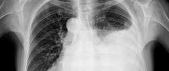
- X-ray examination;
- Magnetic resonance or computed tomography.
Causes
There are many reasons for increasing size, since any pathological process in the human body leads to a reaction of the lymphatic system.
- Infections. One of the most common causes of lymphadenopathy. This includes all infections that can be in the human body: bacterial, viral, parasitic and fungal.
- oncological diseases. The second most common reason. It may be a reaction to the primary cancer or metastases. Lymphadenopathy can develop both in a malignant and benign process.
- immune diseases. This includes all systemic diseases: rheumatoid arthritis, dermatomyositis, systemic lupus erythematosus and others.
Regardless of the cause of the development of lymphadenopathy, consultation and examination by a doctor with a thorough diagnosis is necessary.
Lymphadenopathy: treatment
The development of treatment tactics is carried out by CELT specialists based on the diagnostic results obtained and the patient’s individual indicators. First of all, efforts are directed toward eliminating the pathological condition that has become the initiating factor for the development of lymphadenopathy. If purulent lesions of the lymphatic structures or bacterial infections are detected, the patient is prescribed antibacterial therapy. If indicated, surgical removal of the node is possible.
If the etiological factors involve damage by viral agents, the patient is prescribed antiviral and immunomodulatory pharmacological drugs. If he suffers from pain - painkillers. Most often, lymphadenopathy goes away within one to one and a half months after the cause that caused it is eliminated. Otherwise, the hematologist prescribes a biopsy and develops a new treatment strategy.
The hematology department of our clinic receives candidates, doctors and professors of medical sciences. Their practical and scientific work experience ranges from twenty-five years. You can make an appointment with them online or by contacting our information line operators. If necessary, you can make an appointment with specialists at any of our departments and undergo the necessary procedures. In particular, septoplasty is available in the otolaryngology department, which can improve the patient’s quality of life.
At CELT you can consult a hematologist.
- Initial consultation – 3,500
- Repeated consultation – 2,300
Make an appointment
By making an appointment with a hematologist, you can get a comprehensive consultation. The doctor is competent to treat various blood diseases, most of which can be identified in the early stages and prescribe timely treatment to cope with the disease quickly and easily.
Survey
Enlargement of the cervical lymph nodes, which is not accompanied by other symptoms, is an indication for consultation with a hematologist. When lymphadenopathy is combined with signs of damage to the respiratory and digestive systems, consultation with other specialists may be required. Diagnosis involves assessing the condition of pathologically changed lymph nodes and identifying the cause of the condition. The most valuable are:
- Ultrasonography
. Ultrasound of the lymph node is performed to study the morphological structure of the affected tissue; the method allows you to detect hyperechoic and hypoechoic zones. Additionally, ultrasound of the thyroid gland and sonography of the thymus are recommended to determine the root cause of lymphadenopathy. - Blood tests
. Infectious and hematological diseases, in which enlargement of the cervical lymph nodes is possible, are characterized by changes in blood counts. Patients are prescribed a standard general analysis, biochemical study, and determination of the level of acute phase indicators. - Lymph node biopsy.
Collection of cytological material from the affected organ followed by histological examination is recommended to verify the diagnosis. The method is primarily used for differential diagnosis of malignant tumors of lymphoid tissue and metastases with other diseases. - Lymphography
. A radionuclide study of the lymphatic system using the introduction of a special radiopharmaceutical is prescribed to study the pathways of lymphatic drainage and exclude oncopathology. The method is widely used for the neck area, since standard radiography is difficult.
A further list of studies is formed taking into account the patient’s complaints. Often an examination of the upper respiratory tract is required - pharyngoscopy, rhinoscopy, laryngoscopy. To confirm the infectious etiology of the process, specific serological reactions are performed (RIF, ELISA, PCR). If a connection between lesions of the cervical lymphatic structures and pathology of the thyroid gland is suspected, a blood test for triiodothyronine and thyroxine and scintigraphy with radioactive iodine are performed.