The name “Hemothorax” refers to the accumulation of blood in the pleural cavity - the result of internal bleeding. Leads to compression of one of the lungs and displacement of the mediastinal organs to the opposite side.
According to statistics, hemothorax due to chest trauma develops in every fourth patient. It can develop simultaneously with pneumothorax, in which case it is called hemopneumothorax. In any case, the accumulation of blood in the pleural cavity is an acute condition that is dangerous to the health and life of the patient and requires emergency medical care.
What is the danger of not treating hemothorax in a timely manner? Compression of the lung can lead to the development of acute respiratory failure in the patient. And if the internal bleeding is severe enough, it can cause hemorrhagic shock. And within two to three hours from the start of bleeding into the pleural cavity, inflammation develops in it - the so-called hemopleuritis.
1.General information
Hemothorax in medicine is an emergency condition caused by massive hemorrhage into the pleural cavity (the pleura is a closed two-lobed serous membrane of the lungs; the pleural cavity is a narrow space between the internal, visceral, covering the lung, and the external, parietal lobes of the pleura, adjacent to the chest).
Hemothorax is considered the second (after pneumothorax) complication of thoracic injury. Estimates of the frequency of occurrence vary; According to current data, hemothorax is accompanied by 25% to 37% of isolated blunt thoracotraumas, and about 2/3 of all cases of registered polytrauma involving the chest.
A must read! Help with treatment and hospitalization!
Symptoms of post-traumatic hemothorax
The manifestations of this condition can be more or less intense depending on how severe the internal bleeding is and how severe the compression of the lung is. If the hemothorax is minor, then there may be no characteristic symptoms; that something is wrong with the patient is indicated only by pain in the chest, shortness of breath and cough.
If a sufficiently large volume of blood has accumulated in the pleural cavity, then the manifestations are more pronounced. The patient suffers from severe chest pain, which can radiate to the back and upper limbs, worsening when coughing and during physical activity. Characterized by severe shortness of breath and general weakness, a strong decrease in blood pressure. The patient is forced to maintain a sitting position: this makes it a little easier for him.
Severe forms of hemothorax - for example, after a serious chest injury - are characterized by symptoms of acute internal bleeding. These include dizziness, lightheadedness, severe hypotension, increased heart rate, and often loss of consciousness. Moreover, if internal bleeding is accompanied by rib fractures, emphysema and severe hematomas appear under the patient’s skin, and the chest may be deformed. A violation of the integrity of the lung may be indicated by the development of hemoptysis in the patient.
2. Reasons
The most common cause is mechanical trauma to the chest (in the broadest sense and spectrum) with disruption of the integrity of large blood vessels.
Two other statistically significant etiopathogenetic factors are background primary diseases (mainly oncological, cardiovascular and infectious, less often hematological) and iatrogenic complications during thoracic surgery.
Visit our Thoracic Surgery page
Detection and treatment of hemothorax at ON CLINIC Ryazan
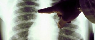
To clarify the diagnosis previously made based on symptoms, instrumental studies such as ultrasound of the chest, radiography, and thoracoscopy can be used. The presence of blood in a diagnostic puncture of the pleural cavity is unambiguous evidence of its accumulation inside.
Small hemothorax in most cases can be successfully treated conservatively. Massive antibiotic therapy, as well as symptomatic treatment with the mandatory use of blood transfusion and immunomodulatory drugs help eliminate all manifestations of the disease and prevent the development of complications.
Surgical treatment of hemothorax involves ensuring the outflow of blood from the pleural cavity, followed by the introduction of an antiseptic solution and antibiotics inside to prevent inflammation, as well as drugs to dissolve blood clots.
Indications for emergency thoracotomy for hemothorax are damage to the organs of the chest cavity and/or large blood vessels. This is necessary to restore the integrity of the vessel and suturing damaged organs. At the same time, the pleural cavity is sanitized and clots of coagulated blood are removed. If a patient has developed purulent inflammation of the pleural cavity, its treatment should be identical to the treatment of purulent pleurisy.
3. Symptoms and diagnosis
There are many classifications of hemothorax, based on various criteria and allowing to take into account the variety of possible manifestations. The severity of symptoms depends on the volume of hemorrhage, the nature of the causes and the presence of associated injuries.
In general, there is pain that increases with coughing, shortness of breath, hyperhidrosis, dizziness and tinnitus, cyanotic pallor, tachycardia, decreased blood pressure, and, less commonly, hemoptysis. In general, the symptoms are not very specific, often moderate or mild, and sometimes practically absent.
However, in severe cases, a drop in blood pressure can result in hypotonic collapse or hypovolemic shock, and a lack of cellular oxygenation can result in fatal systemic hypoxia. Acute cardiac and respiratory failure, a sharp reduction in circulating blood volume with massive blood loss, the likelihood of pulmonary hemorrhage - together, all this makes hemothorax a particularly dangerous condition with unpredictable dynamics, which requires assistance according to an emergency protocol.
The diagnosis is established on the basis of available anamnestic information, clinical manifestations, results of physical, laboratory and instrumental (if the patient’s condition allows and there is at least a minimum amount of time) examination. The standard of express diagnostics in this case includes plain radiography, CT, ultrasound, pleural puncture, diagnostic thoracoscopy and clinical blood test in cito mode (Latin for “urgent”).
About our clinic Chistye Prudy metro station Medintercom page!
Hemothorax
Hemothorax is one of the diseases caused by complications of open or closed chest injury. This condition is extremely dangerous and requires immediate medical attention.
What is Hemothorax
Hemothorax is a pathology expressed by the accumulation of blood in the pleural cavity. As a rule, the cause of hemothorax is rupture of blood vessels in the lung or chest tissue. In this case, the volume of hemorrhage can reach two or more liters. With extensive disease, there is a violation of the integrity of the intercostal arteries, sometimes the aorta or other large vessels of the chest.
Important! Extensive hemothorax is accompanied by large blood loss, gradually increasing compression of the lung and the development of respiratory failure.
Kinds
Depending on the causes of hemothorax, it is classified as follows:
- Traumatic - caused by internal damage to the sternum due to a penetrating wound, rib fracture or severe bruise. This category of injury is characterized by damage to the blood vessels in the space between two ribs, resulting in blood leaking into the pleura.
- Pathological - occurs with aortic aneurysm, tuberculosis, pulmonary abscess, pleural or lung cancer.
- Iatrogenic – occurs against the background of complications caused by the consequences of surgical intervention in the pleura.
Depending on the duration of bleeding, hemothorax can be:
- stable;
- growing.
Depending on the volume of blood loss, several types of this pathology are distinguished:
- small – up to 500 ml;
- medium – up to 1 liter (the blood level reaches the lower edge of the 4th rib);
- subtotal – up to 2 l (blood accumulation is fixed at the level of the second rib);
- total – over 2 liters (blood completely fills the lung).
Causes
In most cases, the occurrence of hemothorax is caused by an injury: the accumulation of blood in the pleural cavity in 60% of cases is caused by penetrating wounds of the chest, in 8% by non-penetrating injuries.
The main causes of hemothorax:
- tissue damage due to rib fractures;
- gunshot and knife wounds;
- blunt wounds resulting from bruises and leading to rupture of blood vessels;
- rupture of aortic aneurysm;
- pulmonary tuberculosis;
- malignant processes occurring in the lungs;
- thoracentesis;
- lung abscess;
- complications after surgery;
- dysfunction of the coagulation system;
- drainage of the pleural cavity;
- incorrectly performed central venous catheterization.
Important! Blood entering the pleural cavity compresses the lung, which leads to respiratory dysfunction.
Symptoms and signs
Small hemothorax is accompanied by cough, respiratory discomfort and slight shortness of breath. These symptoms do not affect the patient's activity in any way.
Average hemothorax is characterized by a more pronounced clinical picture:
- condition of moderate severity;
- intense cough;
- chest congestion;
- severe shortness of breath, increasing with physical exertion.
The symptoms of subtotal and total hemothorax are similar to each other, but at the same time they differ in severity:
- cyanotic staining of mucous membranes and skin;
- hemodynamic disorders and respiratory failure, causing an extremely serious condition;
- shortness of breath caused by even slight physical exertion;
- severe hypotension;
- dizziness;
- feelings of weakness;
- frequent thready pulse;
- coughing;
- chest pain;
- the occurrence of suffocation in a lying position.
Which doctor treats
A pulmonologist treats hemothorax.
Diagnostic methods
Basic diagnostic tests:
- examination of the patient for the presence of injury, wound;
- X-ray;
- MRI or (if necessary);
- puncture of the pleural cavity to detect suppuration (Petrov’s test);
- Rouvilois-Grégoire test.
By registering at the Health Clinic on Kurskaya, the patient will undergo the necessary diagnostics: MRI, CT, ultrasound, after which he will be immediately referred to a doctor to develop an effective treatment program.
Treatment methods
Hemothorax therapy includes several measures:
- treatment of chest wounds;
- pain relief;
- suturing;
- removal of blood from the pleural cavity by drainage;
- replenishment of lost blood volume;
- antibacterial and antishock therapy.
During the treatment process, the patient is prescribed analgesics, hemostatic and anti-inflammatory drugs, antiseptics, antibiotics and proteolytic enzymes.
Important! Depending on the severity of the pathology, surgical interventions are performed.
results
The prognosis of the disease directly depends on the nature of the injury and how timely treatment was started. Timely prescribed therapy allows for complete recovery in most patients. Lack of medical care or its untimely provision causes serious complications. In advanced cases, death occurs.
Rehabilitation and lifestyle restoration
During the rehabilitation period, the patient needs the following:
- regular examination by a doctor;
- proper nutrition;
- maintaining a healthy lifestyle;
- systematic swimming and breathing exercises;
- carrying out activities aimed at preventing cancer, aneurysm and tuberculosis.
Lifestyle with Hemothorax
People with hemothorax should avoid situations that could cause chest injury. The appearance of the first signs of bleeding in the pleural cavity requires immediate consultation with a doctor.
| Name of service | Price in rubles | Price until 19.12. |
| CT scan of the chest in the morning and evening hours. Promotion | 4 990 | 2 900 |
| CT scan of the chest | 4 990 | 2 790 |
| CT scan of the thoracic spine | 4 990 | 2 890 |
| X-ray of the sternum | 2250 | |
| X-ray of the thoracic spine with functional tests (4 images) | 4190 | |
| X-ray of the thoracic spine (2 projections) | 2560 | |
| X-ray of the chest organs (1 projection) | 1300 | |
| Plain radiography of the lungs 1 projection | 1300 | |
| MRI of the thoracic spine | 4 650 | 3 000 |
If you do not find a service in the price list, please call us and we will provide you with the necessary information.
Our doctors will help you:
General practitioner, immunologist
Zhemchugov Vladislav Evgenievich
4.Treatment
The patient is unconditionally hospitalized in all cases of suspected hemothorax. First-line measures include stopping bleeding, draining the pleural cavity, preventing thrombus formation and lysis of already formed blood clots, blood replacement transfusions according to indications, eradication of infectious pathogens, restoration of respiratory function, cardiac activity and hemodynamics in full.
Sometimes, with the so-called small hemothorax, these problems are solved conservatively; sometimes it is enough to use a high-tech minimally invasive intervention, but in some cases the only choice is thoracic surgery. The nature and scope of the operation is determined (often already on the table) by the causes of hemothorax, the current clinical situation, the patient’s condition and other individual characteristics of the case.
Causes of occurrence and mechanisms of development
Depending on the cause of hemothorax, there are:
- traumatic – due to a chest injury;
- pathological - due to a pathological process that has developed in the wall or organs of the chest;
- iatrogenic – as a consequence of medical interventions;
- spontaneous - in which blood flows spontaneously into the pleural cavity, the reasons for this phenomenon have not been established.
Iatrogenic hemothorax is actually a type of traumatic hemothorax. Most often it occurs:
- after surgical interventions - due to too extensive forced tissue trauma, or if the bleeding was not stopped properly;
- during pleural puncture or thoracentesis, if they were performed with technical errors, or there were factors complicating the procedure;
- at the time of insertion of the catheter into the central venous vessel.
The following forms of hemothorax are distinguished separately:
- clotted - observed after surgical interventions, when the patient is given coagulant therapy according to indications (it is aimed at increasing blood clotting - in particular, to prevent bleeding). Due to the use of coagulants, blood discharge that enters the pleural cavity coagulates faster than with normal hemothorax ;
- pneumohemothorax - blood and air accumulate in the pleural cavity at the same time. It is observed in cases of traumatic rupture of the lung, melting of the focus of tuberculosis and injury to the chest with a sharp massive object.
Based on the attachment of an infectious agent, the following forms of hemothorax are distinguished:
- uninfected;
- infected. It is often observed with coagulated hemothorax, when the infection quickly “settles” on the intrapleural blood clot, and this, in turn, provokes a subsequent purulent process - pyothorax (pus in the pleural cavity) or pleural empyema (purulent diffuse lesion of the pleural sheets).
The list of the most common causes of hemothorax looks like this:
chest wounds - most often gunshot, knife, compression (if the chest was pressed by a heavy massive object);- rib fractures (as a result of an accident or too intense pulmonary-cardiac resuscitation);
- aortic aneurysm;
- pulmonary tuberculosis;
- malignant neoplasms of the chest wall or chest organs (lungs, pleura or mediastinal organs), especially at the stage of decay;
- lung abscess;
- deterioration of blood clotting properties (occurs with coagulopathy, hemorrhagic diathesis, and so on).
The immediate cause of hemothorax is a violation of the integrity of the vascular wall:
- chest;
- lung
Less commonly, hemorrhage occurs due to trauma to the vessels of the mediastinal organs - the thymus gland (or the fatty tissue that replaces it), that part of the aorta that is located outside the heart sac, trachea, esophagus, lymphatic ducts, blood vessels and nerve structures. They are partially covered by the lungs, which, when exposed to a traumatic factor, mainly take the blow.
Hemothorax is often unilateral. Bilateral damage occurs due to a pronounced traumatic factor:
- in production (when falling from a height);
- in case of accidents (road accidents);
- during natural disasters (due to house collapses);
- during hostilities;
- when playing sports (especially using strength methods).
Bilateral hemothorax in 90-95% of cases means severe. This is caused by damage:
- intercostal arteries;
- aorta;
- vena cava.
In these cases, the amount of blood poured into the pleural cavity can reach two liters or more. At first, blood fills the diaphragmatic pockets, but since the space of the pleural cavity is quite narrow, it fills quickly, the blood begins to compress one or both lungs, which is why they cannot expand normally.