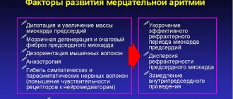
Heart block is an abnormal heart rhythm in which the heart beats too slowly. Why is this happening and what to do? Let's find out together with the experts. Each heartbeat occurs in the heart's upper right chamber (atrium) in the sinus node, a bundle of specialized cells that act as the heart's natural pacemaker. When it beats, an electrical signal from the upper chambers goes to the lower chambers, which causes the heart to contract and pump blood. Heart block disrupts this normal rhythm.
Degrees of heart block in adults
Heart block can be first, second, or third degree, depending on the electrical signal abnormality.
First degree heart block. In this case, the electrical impulse still reaches the ventricles, but travels through the AV node more slowly than usual.
Second degree heart block. It is divided into two categories:
- Type I, also called Mobitz block I or Wenckebach AV block, is a less severe form of second-degree heart block in which the electrical signal on its way to the ventricles becomes slower and slower;
- type II, also known as Mobitz block II - in this case, some electrical signals do not reach the ventricles at all, and the heartbeat may be slower than usual.
Third degree heart block. In this embodiment, the electrical signal from the atria to the ventricles is completely blocked. To compensate, part of the ventricles acts as a pacemaker, creating electrical signals. These signals cause the heart to pump blood, but much more slowly than normal.
Classification
There are several types of heart block. They are divided based on the location of the area where the conduction function of the heart is difficult or completely blocked. Due to the development of the disease in different parts of the heart, each blockade has its own characteristic features.
Photo: psodaz / freepik.com
Sinoatrial blockade
Sinoatrial block or sick sinus syndrome (SSNS) develops as a result of complete or partial dysfunction of the sinus node. This can occur both as a result of physiological disorders and due to other factors not related to the state of the body (extracardiac factors).
The main cause of SSSS can be considered fibrosis (development of connective tissue and its scarring) of the sinus node tissue. This pathology can be caused by inflammatory processes, increased tone of the vagal nerve, or taking medications.
SSSU can manifest itself:
- shortness of breath;
- general weakness of the body;
- pain in the heart;
- dizziness and loss of consciousness;
- low blood pressure;
- arrhythmia.
Interatrial block
Interatrial block develops when the propagation of electrical impulses is disrupted, both within and between the atria. The propagation of excitation occurs along special conduction pathways, which are circular and longitudinal muscle bundles, usually not described as part of the specialized conduction system of the heart.
One of the mechanisms of changes in the electrophysiological properties of the atrial myocardium, leading to a slowdown in conduction, is the development of fibrosis. There is a violation of the integrity of myocardial cells with the deposition of collagen in the intercellular space, which prevents the movement of impulses throughout the heart.
Accompanied by general weakness, pain in the chest and abnormal heart rate.
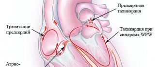
Atrioventricular block
Atrioventricular (AV) block is a partial or complete interruption of the transmission of impulses from the atria to the ventricles, leading to disturbances in heart rate and blood flow rate.
There are 3 types of atrioventricular block:
- 1st degree - due to a slow transition of impulses from the atria to the ventricles;
- 2nd degree - characterized by blocking of some impulses;
- 3rd degree - (complete atrioventricular block), associated with blocking of all impulses coming from the atria to the ventricles.
It occurs due to damage to the most sensitive area of the conduction system of the heart - the atrioventricular junction.
Intraventricular block
Intraventricular block occurs when the His bundle, the conducting region of the heart located in the region of the ventricles, is disrupted. The bundle of His consists of 3 branches: the left and right anterior branches and the posterior branch. The posterior branch is also divided into right and left.
Figure 1. Differences in ECG readings with normal rhythm and with blockades.
Intraventricular block is classified based on damage to 1 of 3 branches:
- Blocking of the anterior right branch - the innervation of the right ventricle is disrupted;
- Blocking of the anterior left branch - in case of disruption of the innervation of the left ventricle;
- Blocking of the posterior branch - the work of the His bundle is disrupted to the greatest extent, since along this branch impulses go from the atria to the ventricles, and are subsequently divided into right and left;
- Three-bundle block is associated with disruption of the conduction of the entire His bundle.
Causes of heart block in adults
The most common cause of heart block is a heart attack.
Other causes include heart disease, problems with the structure of the heart (due to defects) and rheumatism. The blockage can also be caused by damage during open heart surgery, as a side effect of certain medications or exposure to toxins. Heart block can be congenital, but it is a rare pathology. The risk of heart block increases with age and the presence of heart disease.
First-degree heart block is common in well-trained athletes, teenagers, young adults, and people with a highly active vagus nerve.
Treatment
Blockades of individual branches of the cardiac conduction system sometimes do not require special treatment, but may indicate the presence of some kind of heart disease that requires therapy. Some blockages are eliminated by prescribing medications. On the other hand, there are medications that themselves contribute to the occurrence of blockades. Therefore, before “treating” the blockade, it is necessary to clarify whether the patient is taking any medication, or a combination of drugs that affects intracardiac conduction, their dosage and time of administration.
Blockades of the 3rd degree, or complete blockades, as a rule, significantly worsen the patient’s well-being and also affect the prognosis of the disease. In these cases, implantation of an artificial pacemaker (pacemaker) is absolutely necessary.
In the multidisciplinary CELT clinic, a complete examination of the patient is possible to identify and timely treat blockades and, if necessary, implantation of an artificial pacemaker (PAC).
Make an appointment through the application or by calling +7 +7 We work every day:
- Monday—Friday: 8.00—20.00
- Saturday: 8.00–18.00
- Sunday is a day off
The nearest metro and MCC stations to the clinic:
- Highway of Enthusiasts or Perovo
- Partisan
- Enthusiast Highway
Driving directions
Diagnostics
First, the cardiologist will review the patient's medical history and ask questions about your general health, diet and activity level, and family medical history.
In addition, he will ask about the medications the patient is taking, as well as about bad habits. The doctor will then listen to your heart and check your pulse for signs of heart failure, such as fluid retention in your legs and feet.
In addition, to diagnose heart block, use:
- ECG – this will show the heart rate and rhythm, as well as the timing of electrical signals as they pass through the heart;
- A Holter monitor is a portable ECG that is attached to the patient’s body and records heart activity from 24 to 48 hours;
- An event monitor is another type of portable ECG that is worn over an extended period, where the device records heart activity at specific times rather than continuously;
- Electrophysiology test - This test uses thin flexible wires (catheters) that are placed on the surface of the heart to record its electrical activity.
Advantages
Excellent technical base
Powerful equipment - expert class ultrasound machines with 3D scanning capabilities, monitors/defibrillators, Holter monitors, as well as our own laboratory make it possible to accurately determine the causes of pathology and monitor the progress of treatment.
Highly qualified personnel
A complete examination of patients and treatment is carried out by experienced cardiologists, arrhythmologists, therapists, and functional diagnostics doctors. Candidates of Medical Sciences, doctors of the highest category are regular participants in European and domestic congresses of cardiologists, as well as participants in international clinical studies, and have numerous publications in scientific journals.
Modern diagnostic methods
To diagnose the disease, extensive laboratory testing, ECG at rest, with stress and 24-hour Holter ECG monitoring, ultrasound, Doppler and Duplex examination of the carotid arteries are performed. If necessary, it is possible to conduct an ECG at home using a portable device.
Popular questions and answers
Cardiologist Stanislav Snikhovsky answered our questions about heart block.
What complications can occur with heart block?
Clinically, all heart blocks are manifested by slowing of the pulse.
When the pulse rate decreases, fainting conditions associated with a lack of blood circulation to the brain may develop. Patients may complain of irregular heartbeat, headache and shortness of breath. With complete heart block (pulse about 40 beats per minute or less), Morgagni-Edams-Stokes syndrome develops, which is manifested by convulsions and loss of consciousness. Complete heart block causes rapid development of heart failure and can be fatal.
When to call a doctor at home if you have a heart block?
If there is sudden general weakness or fainting, a rare (less than 50 beats per minute) or very weak pulse, a feeling of irregular heartbeat, sudden shortness of breath and a feeling of lack of air, you should immediately call an ambulance.
Degrees and their symptoms
Heart blocks are classified not only by the place of its development, but also by the degree of severity, which reflects the danger of the disease to human health and approaches to treating a particular stage of heart block. There are three main stages of heart block
First degree heart block
The first degree is the mildest form of manifestation. There is a delay of a fraction of a second in the time required for electrical impulses to travel through the atrioventricular junction. Complete blocking does not occur and the impulses reach the desired point.
First-degree heart block usually does not cause any noticeable symptoms and is diagnosed by examining other medical conditions in the person. Treatment may be limited to eliminating risk factors that affect heart health.
Second degree heart block
As the second degree develops, there is a series of increasing delays in the time it takes for the atrioventricular junction to conduct an impulse into the ventricles until the heart beat is missed. Partial blocking of impulses is possible.
The second degree is divided into 2 types:
- The first type of Mobitz. This is a less serious type of second-degree heart block. Characterized by slow conduction of impulses. May cause symptoms of mild dizziness and weakness. Usually does not require treatment.
- Mobitz's second type. Electrical signals reach the ventricles slowly or may be completely blocked. This type of heart block can often progress to third degree heart block. May cause symptoms of dizziness and fainting, accompanied by bradycardia. Requires medical consultation and full treatment.
Third degree heart block
Third degree heart block is the most dangerous degree for a person’s health and life and requires immediate medical attention. It is characterized by complete blocking of the transmission of electrical impulses from the atria to the ventricles due to the lack of communication between the atrioventricular junction and the ventricles.
In this case, the ventricles can partially take over the function of a pacemaker, but they are not able to ensure full functioning of the heart.
Third degree blocks can be congenital or acquired as a result of other heart diseases or injuries.
Symptoms include:
- shortness of breath;
- weakness;
- fainting conditions;
- chest pain;
- pale skin, spots;
- arrhythmia.
Many cases of third-degree congenital heart block are diagnosed during pregnancy because ultrasound scans can often determine whether the baby has bradycardia.
Important! If the diagnosis is missed during pregnancy, symptoms of third-degree congenital heart block usually do not appear until the baby is older and his heart is more developed.
Mechanism of occurrence
Normally, electrical impulses pass through the myocardium at a certain speed and in a strictly established sequence. The signal path begins in the right atrial appendage - in the sinus node. From here, the excitation gradually spreads through the tissues of the atria and slows down briefly in the atrioventricular node. Next, the impulse spreads along the branches of the His bundle, which cover the right and left ventricles. The conducting system ends with small Purkinje fibers.
The problem occurs when the conduction of the impulse slows down or is completely blocked at a certain point. The cause may be functional and organic changes. In the first case, the impulse reaches cells that are in the refractory (inactive) phase - and its further passage is disrupted. The next signal can travel through the tissues without obstruction. With organic changes (for example, in the case of scar formation after a heart attack), the impulse will “stumble” over an obstacle, and the failure will become persistent.
If we talk about the pathophysiology of disorders, we should note the work of Na+ channels in cardiomyocytes. As long as these pathways are open, the impulse can penetrate the cells without obstacles. But, if the channels are inactivated, the signal transmission slows down or stops. This happens, for example, in the zone of myocardial ischemia - where the blood supply to the tissues stops.
Signs of heart block are nonspecific and are not always noticeable without special examination. The problem can be identified using an ECG. The film shows how the impulse travels through the heart muscle, whether there are obstacles to tissue excitation, and in which zone they are localized. Electrocardiography is the main method for making a diagnosis and prescribing treatment.
What is a blockade?
The work of the heart is based on its ability to conduct electrical impulses. They cause muscle contraction, resulting in blood movement. From the left ventricle it enters the systemic circulation through the aorta, then flows into the right atrium, from which it moves into the right ventricle. From here the pulmonary circulation begins, as a result of which fluid enters the left atrium. Thanks to the operation of the valve apparatus, it enters the left ventricle, and the process repeats.
Automaticity of the heart is possible thanks to a special mechanism of contraction of its structures. First, the atria contract, then the ventricles (these two phases represent systole), and the simultaneous relaxation of all muscles is called diastole. Electrical impulses that cause muscle tissue contraction are generated in the following structures:
- sinoatrial node;
- atrioventricular node;
- bundle of His, from which small branches arise - Purkinje fibers.
The most informative way to diagnose cardiac conduction is electrocardiography. The device has a complex structure, thanks to which it can capture electrical impulses arising in the heart, amplify them and output them in the form of a record on paper. The periods of occurrence of impulses look like teeth. A cardiologist deciphers the blockade.
Causes of conduction disorders
A healthy heart works without stopping, and electrical impulses are constantly generated in it. Minor conduction disturbances that do not manifest themselves clinically may be a normal variant in individuals who experience severe physical exertion, as well as in congenital anomalies of the valve apparatus. More serious pathologies are associated with organic damage to myocardial tissue.
Diseases that can provoke blockades include:
- myocarditis - inflammation of the muscular lining of the heart caused by bacterial or viral infection, as well as autoimmune processes;
- cardiomyopathy - pathologies associated with thickening or thinning of the walls of the heart (can occur with ischemia, chronic hypertension, endocrine or metabolic disorders);
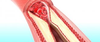
- cardiosclerosis - replacement of normal myocardial tissue with scar, which occurs after a stroke or heart attack;
- myocardial infarction;
- heart defects (congenital or acquired);
- acute intoxication.
The figure shows a simplified block diagram of the electrocardiograph
REFERENCE! An ECG study allows you to assess the degree of cardiac conduction, but does not indicate the cause of the disorders. If chronic diseases of the cardiovascular system are suspected, an additional set of tests is prescribed.
Doctor's advice: how to properly see a doctor if there is a blockage
After the diagnosis is established, the patient remains in outpatient treatment or is hospitalized in a hospital. Tactics are determined by the severity of the blockade. After achieving remission, the patient should not be left without specialist supervision. We recommend:
- If your condition is stable and there are no complaints, visit a cardiologist and do an ECG every 6 months.
- If the condition worsens, new complaints appear or existing disorders progress, make an appointment with a doctor as soon as possible.
If the doctor prescribes drug therapy, you should adhere to it and not violate the drug dosage schedule. Stopping the medication on your own is unacceptable - this leads to the development of complications.
If the patient has had a pacemaker installed, the monitoring tactics change. You should visit your doctor 3, 6 and 12 months after surgery to make sure that the device is working smoothly. The further observation schedule will depend on the patient’s condition.
Symptoms, causes, diagnosis, treatment features
The heart is the main supplier of oxygen to the human blood. When the blockade occurs, the conduction system does not work properly, causing the heart to contract in a slow manner - with a frequency of 50 beats per minute or less.
Heart block, the symptoms of which are determined by the degree of the disease, is caused by a violation of intrauterine development, a hereditary predisposition. The cause may be an overdose of drugs or any damage to the heart (cardiosclerosis, myocarditis, myocardial infarction, coronary heart disease), as a result of which the myocardial muscle tissue is replaced by scar (connective) tissue. Severe physical activity, for example, in professional athletes and high blood pressure, can also cause heart rhythm disturbances.
Clinical manifestations of the disease vary. For example, blockade of the right bundle branch is asymptomatic and is most often detected by ECG, and complete blockage of the left bundle branch is accompanied by suffocation, blue discoloration of the fingers, lips, face, and cough with foamy sputum.
Prevention
- Elimination of cardiac pathology.
- Exclusion of extracardiac factors of arrhythmia or blockade (intoxication, autonomic dysfunction, stress, etc.).
- Limit intake of caffeine-containing products and alcohol.
We will help make a diagnosis at an early stage of the development of blockade, tachycardia or other disorders and provide timely treatment for cardiac arrhythmia. Further observation will allow maintaining a good quality of life. For questions regarding the treatment of cardiac arrhythmia in Moscow, please contact us by dialing the number +7.
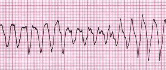
ECG with complete blockade
ECG signs are the most accurate objective confirmation in diagnosing blockade. Typical:
- unchanged atrial waves at regular intervals, emphasizing the preserved atrial rhythm;
- the distances between the ventricular complexes are also equal, but they have an independent rare rhythm;
- the P wave (atrial) can be located anywhere and is in no way connected with R.
When the His bundle is destroyed, impulses can arise in one of the legs and be transmitted first to one ventricle, then to the other. The ECG picture will be similar to ventricular extrasystoles.
If the pathological process has not yet ended with scarring, then the ECG can be used to observe the transition from an incomplete blockade to the formation of a complete one.