Detailed description of the study
Dehydrotestosterone is the active form of testosterone, a male sex hormone related to androgens, which is formed from cholesterol by Leydig cells in the testicles in men, and in small quantities in the ovaries in women.
In peripheral tissues, 60-70% of dehydrotestosterone is formed from testosterone under the action of the enzyme 5-alpha reductase in men, the rest is synthesized directly in the testicles. This hormone performs a number of functions, including:
- participation in sperm maturation;
- regulation of metabolic processes in nervous, muscle, fat and bone tissue;
- stimulation of muscle tissue growth;
- stimulation of the formation of erythrocytes - red blood cells;
- Hair Growth;
- regulation of normal prostate function.
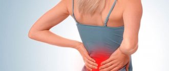
There is androgenetic alopecia, the most common form of alopecia (hair loss), associated with a hereditary predisposition, which affects up to 80% of men and 50% of women during their lifetime. It is characterized by a gradual decrease in the diameter, length and pigmentation of hair. Thinning hair is the result of excess exposure of hair follicles to dehydrotestosterone.
Specialists prescribe the determination of this hormone in the blood before prescribing testosterone therapy for the treatment of male hypogonadism (insufficient amounts of male sex hormones and pathological changes in the genital organs due to this), the study of androgen deficiency in severe diseases, aging and underdevelopment of the penis (microphallus) in infancy.
Dehydrotestosterone has been proposed as an androgen replacement therapy with possible advantages over testosterone in patients with gynecomastia (breast enlargement) and microphallos. Dehydrotestosterone is also determined when prescribing treatment for prostatic hyperplasia with a 5-alpha reductase inhibitor, since the content of this hormone helps to assess the effectiveness of therapy.
However, not only men need to determine this hormone. Hyperandrogenism (excess of androgens, including dehydrotestosterone), one of the common endocrine pathologies in women of reproductive age. In their body, there is excess production of androgens by the adrenal glands and ovaries. Hyperandrogenism is associated with polycystic ovary syndrome (PCOS), the risk of which in women is 7-12%.
Hyperandrogenism is associated with the development of the following diseases and conditions: infertility, endometrial cancer, menstrual irregularities, lipid and carbohydrate metabolism in the body, cardiovascular diseases.
The most common clinical manifestations of hyperandrogenism include hirsutism (excessive male pattern hair growth in women and children), virilization (appearance of masculine features in women), acne vulgaris, and androgenetic alopecia (baldness). Almost 80-90% of women with hirsutism are ultimately diagnosed with hyperandrogenism.
Hirsutism can sometimes be associated with excessive sensitivity of the androgen receptor, but the level of male hormones will be normal.
A test for dihydrotestosterone helps identify the causes of hirsutism and is also important in the diagnosis of hypogonadism, prostate diseases, androgenic alopecia, and gynecomastia.
What is dihydrotestosterone?
Dihydrotestosterone is a hormone that stimulates the development of male qualities (androgen). It is produced by converting the better known androgen, testosterone. Almost 10% of the testosterone produced by adults every day is converted into dihydrotestosterone by the testicles and prostate gland (in men), ovaries (in women), skin and other parts of the body.
However, before puberty the rate is much lower, and it is thought that increased production of dihydrotestosterone may be responsible for the onset of puberty in boys, causing the development of the reproductive organs (penis, testicles and scrotum) and the growth of pubic and body hair.
This hormone also causes prostate growth and is thought to, when combined with testosterone, cause the expression of male sexual behavior. Dihydrotestosterone is many times stronger than testosterone, and many of the effects testosterone has on the body only occur after it is converted to dihydrotestosterone.
Less is known about the importance of dihydrotestosterone for women, but it is known to cause the growth of pubic hair seen in girls after puberty and can help determine the age of puberty in girls.
References
- Marchetti PM, Barth JH. Clinical biochemistry of dihydrotestosterone. Ann Clin Biochem. 2013 Mar;50(Pt 2):95-107. doi: 10.1258/acb.2012.012159. Epub 2013 Feb 21. PMID: 23431485.
- Gorityala S, Yang S, Montano MM, Xu Y. Simultaneous determination of dihydrotestosterone and its metabolites in mouse sera by LC-MS/MS with chemical derivatization. J Chromatogr B Analyt Technol Biomed Life Sci. 2021 Jul 15;1090:22-35. doi: 10.1016/j.jchromb.2018.05.008. Epub 2021 May 16. PMID: 29778874; PMCID: PMC6326363.
- 3. Kim MK, Zhao C, Kim SD, Kim DG, Park JK. Relationship of sex hormones and nocturia in lower urinary tract symptoms induced by benign prostatic hyperplasia. // Aging Male. 2012. Vol. 15, N 2. P. 90-95.
- 4.Li J, Ding Z, Wang Z, Lu JF, Maity SN, Navone NM, Logothetis CJ, Mills GB, Kim J. Androgen regulation of 5α-reductase isoenzymes in prostate cancer: implications for prostate cancer prevention. // PLoS One. 2011. Vol. 6, N 12. P. 28840.
What happens if I have too little dihydrotestosterone?
Dihydrotestosterone is thought to have less of an effect on women, and as a result they are thought to be relatively unaffected by too little dihydrotestosterone. However, it is possible that the onset of puberty may be delayed in girls with low dihydrotestosterone levels, and the amount of pubic and body hair may also be reduced in adult women.
In contrast, low dihydrotestosterone levels in men can have dramatic consequences. If, for example, there is too little dihydrotestosterone while male fetuses are still in the womb, they may not be “masculinized” and their genitalia may appear similar to those of girls of the same age.
Later, boys with low DHT may experience some of the changes typically seen during puberty (such as muscle growth and sperm production), but they will not develop normal body hair growth or genital development.
ONLINE REGISTRATION at the DIANA clinic
You can sign up by calling the toll-free phone number 8-800-707-15-60 or filling out the contact form. In this case, we will contact you ourselves.
Hyperandrogenism. Treatment
A woman is primarily concerned about how she looks. In order to relieve the patient of the symptoms of hyperandrogenism, it is necessary to prescribe treatment for the underlying disease that caused the corresponding changes. If she has been diagnosed with polycystic ovary syndrome, then she is prescribed, first of all, medroxyprogesterone, which is especially indicated for hirsutism. Hormonal combined contraceptives reduce hair growth and cure acne in most patients.
Under the influence of these drugs, periods are evened out and blood loss during menstruation is reduced. Hyperandrogenism is also treated with the following drugs: corticosteroids, ketokenazole, spironalactone. For hyperandrogenism with PCOS, surgical treatment is used. Most often, cysts are cauterized using an electrocoagulator.
Treatment of hyperandrogenism with pharmacological drugs for Itsenko-Cushing syndrome is considered a preparatory stage before surgery. The adenoma is surgically removed. Radiation therapy is indicated for pituitary tumors and is not very effective. In the case of congenital adrenal hyperplasia, the only drug used to treat hyperandrogenism is dexamethasone.
If hyperandrogenism in women is caused by hormone-synthesizing tumors, then treatment is usually carried out in oncology centers. The tumor is removed surgically. Surgical treatment is combined with radiation and chemotherapy. Hormonal medications must be prescribed. In case of ovarian hyperplasia, wedge resection should be performed. In severe cases, both ovaries are removed.
The role of androgens in women: what do we know?
Until recently, androgens in women were considered only as a cause of various metabolic and functional disorders, but their role in the female body is still not fully understood. Using polycystic ovary syndrome (PCOS) as an example, it is well known that elevated androgen levels often correlate with anovulation, infertility, as well as disorders of fat and carbohydrate metabolism [1]. At the same time, antiandrogen therapy did not solve these problems [2–4]. Androgens are perceived by most clinicians as “male” sex hormones, but is this true? In the last decade, androgen deficiency conditions in women, which can lead to a deterioration in the quality of life and sexual disorders, have been actively studied [5–7]. The influence of androgens on libido and a sense of well-being in women has now been proven [7–10], but their role in the genesis of metabolic disorders is still not fully known. The issues of the influence of androgens on bone, muscle tissue and hematopoiesis in the female body also remain unresolved.
Production and transport of androgens in the female body
The pituitary gland regulates androgen secretion in women through the production of luteinizing hormone (LH) and adrenocorticotropic hormone (ACTH). The main androgens in serum in women with a normal menstrual cycle are testosterone and dihydrotestosterone. Dehydroepiandrosterone sulfate (DHEA-S), dehydroepiandrosterone (DHEA) and androstenedione are considered prohormones because only conversion to testosterone reveals their full androgenic properties. DHEA is produced mainly in the zona reticularis of the adrenal glands, as well as in the theca cells of the ovaries [11]. Testosterone is synthesized as follows: 25% is synthesized in the ovaries, 25% in the adrenal glands, the remaining 50% is produced as a result of peripheral conversion mainly in adipose tissue from androgen precursors, which are produced by both glands [12]. Healthy women of reproductive age produce 300 mcg of testosterone daily, which is approximately 5% of the daily production in men [13]. Unlike the fairly dramatic decline in estrogen production associated with menopause, levels of androgen precursors and testosterone decline gradually with age. A decrease in DHEA-S levels occurs as a result of decreased adrenal function. Concentrations of DHEA-S, which is not bound to any protein and does not change during the menstrual cycle, are approximately 50% in women aged 40–50 years compared to the concentrations observed in 20-year-old women [14–16]. Similar dynamics were also noted in the secretion of testosterone [17].
It is known that androgens are precursors of estrogens, which are formed from testosterone by aromatization in granulosa and theca cells of the ovaries, as well as in peripheral tissues.
In plasma, testosterone is predominantly in a bound state, with 66% bound to sex hormone binding globulin (SHBG), 33% bound to albumin, and only 1% in an unbound state [17]. Some diseases (thyrotoxicosis, liver cirrhosis), as well as taking estrogens as part of combined oral contraception (COCs) and hormone replacement therapy (HRT) can lead to a significant increase in SHBG and a decrease in the free fraction of testosterone [18]. Consequently, pathology of the pituitary gland, ovaries, adrenal glands, as well as diseases accompanied by a deficiency of adipose tissue or an increase in SHBG, can lead to the development of androgen deficiency conditions in women.
The final metabolites of testosterone are 5-alpha-dehydrotestosterone and estradiol, the amount of which is several times less than testosterone, from which we can conclude that the concentration of androgens in women is several times higher than the concentration of estrogens. Thus, studying the role of androgens, as well as replacement therapy for androgen deficiency conditions in women, including those receiving HRT with estrogens and progestins with insufficient effect, has a convincing biological basis.
The effect of androgens on fat and carbohydrate metabolism
One of the discussed side effects of testosterone is the negative effect on lipid metabolism, which consists of a decrease in high-density lipoproteins (HDL). Many studies have noted that higher levels of total testosterone and free androgen index were directly proportional to total cholesterol, low-density lipoprotein (LDL) and triglycerides, on the one hand, and lower levels of HDL, on the other [19–21]. This relationship was most clearly observed in women with PCOS [22]. Studies with oral methyltestosterone also showed a significant reduction in HDL cholesterol with normal or reduced LDL levels [23]. For many years, this fact was the main argument of opponents of the use of androgens in women.
At the same time, when using parenteral forms of testosterone (implants, intramuscular injections and transdermal drugs), no decrease in HDL was observed [24], and in women receiving estrogen replacement therapy, when adding testosterone undecanoate daily and even when supraphysiological concentrations of testosterone were achieved, significant reduction in total cholesterol and low-density lipoproteins [25].
Bell R. et al. examined 587 women aged 18 to 75 years who did not present any complaints. There was no statistically significant relationship between the concentrations of endogenous testosterone, its adrenal precursors and HDL levels, while SHBG levels were inversely related to LDL and triglyceride levels [26].
A population-based study in Sweden found that women with low androgen levels had higher cardiovascular morbidity, including those on HRT, even if they controlled lipid levels. However, logistic regression analysis showed that the concentration of total testosterone was directly proportional to HDL and LDL in all women, while the level of androstenedione was positively associated with HDL and negatively with triglycerides [27].
Interestingly, levels of DHEA-S, total and free testosterone, and the free androgen index are inversely correlated not only with body mass index, but also with waist-to-hip ratio in both men and women [28, 29], however in the female population this pattern was less pronounced [28].
For many years, an association between hyperandrogenism and insulin resistance has been found in women with PCOS [1], but research data have shown that therapy with flutamide and gonadotropin-releasing hormone agonists did not improve insulin sensitivity in these patients [5–7]. Conflicting data that were obtained in women without PCOS in some studies did not confirm the relationship of testosterone with insulin resistance [30, 31]. Removal of an androgen-producing tumor in a patient with severe hyperandrogenism after 9 months led to a marked deterioration in peripheral insulin sensitivity [32].
Androgens and cardiovascular morbidity in women
Most often, researchers associate the effect of androgens on cardiovascular risk with the clinical model of hyperandrogenism in PCOS. In women with PCOS, there was an increase in the level of endothelin-1, a marker of vasopathy, free testosterone, and insulin. Administration of metformin, which increases the sensitivity of peripheral tissues to insulin, for 6 months contributed to a significant decrease in endothelin-1 levels, a decrease in hyperandrogenism and hyperinsulinemia, and an improvement in glucose utilization [33]. A meta-analysis of randomized clinical trials also showed that metformin therapy in patients with PCOS resulted in a decrease in androgen levels [34], suggesting a primary role of hyperinsulinemia in increasing androgen secretion in women.
The thickness of the intima-media of the carotid arteries, determined using ultrasonography, is one of the most popular markers used by researchers to determine the severity of atherosclerosis [35]. A large number of publications focused on measuring intima-media thickness and determining androgen levels once again confirms this. Bernini et al. 44 patients with physiological menopause were examined. The levels of total and free testosterone, androstenedione were studied, and the thickness of the intima-media of the carotid arteries was measured. An inverse correlation was noted between androgen levels and intima-media thickness, a sign that most reflects atherosclerotic changes in blood vessels: in women with the smallest intima-media thickness, androgen levels were in the upper third of the normal range, and in women with the greatest, in the lower quarter. Based on the study, the authors concluded that androgens may have a beneficial effect on the carotid artery wall in postmenopausal women [36]. Other authors came to a similar conclusion in their studies [37–39].
Hak et al. studied the relationship between the levels of total and bioavailable testosterone and the intima-media thickness of the abdominal aorta in men and women. While in men there was a clear inverse correlation between the levels of total and free testosterone, in women the levels of these androgens were positively correlated with aortic atherosclerosis, but this correlation became statistically insignificant after taking into account other cardiovascular risk factors [40].
An important factor in the development of serious cardiovascular complications is vasospasm. Worboys S. et al. investigated the effects of parenteral testosterone therapy in women receiving HRT with estrogens and progestins. We examined 33 postmenopausal women receiving HRT with testosterone implants (50 mg) for more than 6 months. The control group consisted of 15 women who did not receive any therapy. Ultrasound was used to study brachial artery diameter, reactive hyperemia (endothelium-dependent vasodilation) and the effect of nitroglycerin (endothelium-independent vasodilation). In the study group, there was an increase in testosterone levels, which was associated with a 42% increase in endothelium-dependent vasodilation. No changes were noted in the control group. Similar data were obtained regarding endothelium-independent vasodilation. The authors concluded that parenteral testosterone therapy in postmenopausal women receiving long-term HRT improves both endothelium-dependent and endothelium-independent vasodilation of the brachial artery [42].
The effect of androgens on the musculoskeletal system in women
A number of studies have shown a positive effect of endogenous androgens on bone mineral density (BMD) in postmenopausal women. E. C. Tok et al. examined 178 postmenopausal women who had never received HRT [43]. Androgen levels (DHEAS, androstenedione and free testosterone) and their correlation with BMD measured by dual-energy x-ray absorptiometry were examined. It was noted that DHEAS and free testosterone levels were positively associated with lumbar spine and femoral neck BMD. At the same time, data analysis using linear regression showed a different effect of androgens on bone tissue. Thus, free testosterone was independently associated with lumbar spine mineral density (trabecular bone tissue), while DHEAS was independently associated with femoral neck mineral density (cortical bone tissue). According to the authors, different androgens have different effects on different types of bone tissue. S. R. Davis et al. in their study showed that among two groups of postmenopausal women receiving HRT with estrogens and estrogens in combination with testosterone, BMD was significantly higher in group 2 [44].
Women with androgen deficiency associated with HIV infection are more likely than the general population to develop osteoporosis and increase the risk of fractures. In a study by S. Dolan et al. it was noted that the risk of osteopenia and osteoporosis in such patients was associated with low levels of free testosterone [45].
The influence of androgens on hematopoiesis
The effects of testosterone on erythropoietin were noted back in the 60s of the 20th century [46]. L. Ferrucci at al. in a study of 905 patients over 65 years of age (exclusion criteria were cancer, chronic renal failure, and taking medications that affect hemoglobin concentration), it was found that the hemoglobin level correlated with the level of free testosterone in both men and women; in addition, it was noted that that with low testosterone levels, the three-year risk of developing anemia was higher than with normal levels (4.1 times in women and 7.8 times in men) [47]. Another study in women with anemia associated with HIV infection showed a similar pattern [48]. In women with PCOS receiving antiandrogen therapy, a clear positive association was also found between free testosterone concentrations and hemoglobin and hematocrit levels [49].
Reasons for the development of androgen deficiency conditions in women
Androgen deficiency in women is characterized by decreased libido, decreased feelings of well-being, depression, decreased muscle mass, and prolonged unreasonable fatigue in combination with low levels of total and free testosterone with normal estrogen levels [50]. Among the causes of androgen deficiency are ovarian, endocrine, chronic diseases and medications [18, 50] (.).
The laboratory criterion for androgen deficiency in women is the concentration of total testosterone in the lower quartile or below the lower limit of the normal range [50].
Effects of androgen replacement therapy
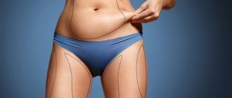
Testosterone therapy in women was first used in 1936 to relieve vasomotor symptoms [51]. Currently, testosterone is used as off-label therapy in many countries for various diseases and conditions in women. A new era began in 2006, when the use of a patch containing 300 μg of testosterone was officially approved by the European Medicines Agency for the treatment of sexual dysfunction in women after oophorectomy [52]. Testosterone can be used either as an addition to traditional HRT [27, 53] or as monotherapy [54]. Randomized, placebo-controlled trials showed that transdermal testosterone monotherapy at a physiological dose of 300 mcg twice a week for 18 months in women with androgen deficiency caused by both hypopituitarism and HIV infection led to significant increases in BMD, muscle mass and strength, and also improved indexes of depression and sexual function in such patients. At the same time, fat mass indicators did not change, and side effects were minimal [55–57]. It was also noted that transdermal testosterone therapy in women with androgen deficiency caused by HIV-associated weight loss syndrome did not impair insulin sensitivity, total adipose tissue mass, regional distribution of subcutaneous fat, and did not affect markers of inflammation and thrombolysis [58]. ]. In addition, testosterone gel applied to the anterior abdominal wall led to a decrease in abdominal subcutaneous fat and a decrease in total body weight in postmenopausal women [59]. Topical application of androgen cream was effective against atrophic vaginitis and dyspareunia in postmenopausal patients [60, 61].
Combining testosterone with traditional HRT
One of the most commonly used estrogen-androgen drugs in women in the United States is Estratest, which contains conjugated equine estrogens and methyltestosterone. As shown by the WHI data, conjugated estrogens are not the drug of choice for HRT due to the relative increase in the risk of breast cancer and cardiovascular complications in older women. Therefore, the optimal drug for estrogen-progestin replacement therapy should meet safety criteria for the mammary glands and endometrium, not have a negative effect on lipid and carbohydrate metabolism, not increase the risk of cardiovascular complications and have a positive effect on bone metabolism.
Of the drugs containing native sex hormones, the drug of choice is Femoston, used for hormone replacement therapy in peri- and postmenopause and the only one on the modern market available in three dosages: 1/5, 1/10 and 2/10. Femoston is a combination drug that contains 17-beta-estradiol - a natural estrogen - and dydrogesterone - a pure analogue of natural progesterone that does not lose its activity when administered orally.
The use of dydrogesterone in combination with 17-beta-estradiol enhances the protective effect of estrogens on bone tissue. If estrogens act to reduce bone resorption, in vitro studies suggest that dydrogesterone may promote bone formation [62]. In addition, dydrogesterone does not have hormonal side effects and does not have a negative effect on the blood coagulation system, carbohydrate and lipid metabolism [63]. The results of clinical studies of Femoston have shown its high effectiveness for the treatment of menopausal disorders in perimenopausal women, safety and good tolerability, acceptability and ease of use. The drug helps reduce the atherogenic potential of the blood, and therefore can have a real preventive effect on the incidence of cardiovascular diseases. The combination of 17-beta-estradiol with dydrogesterone has a better effect on the lipid profile than some other HRT regimens. In a double-blind study, a comparative study of the effect of two options for HRT was carried out: Femoston 1/5 and conjugated equine estrogens orally (0.625 mg) + norgestrel (0.15 mg). Both options had an equally positive effect on LDL levels (decrease by 7% over 6 months), but in terms of their effect on HDL levels, Femoston 1/5 was significantly more effective (increase by 8.6% and decrease by 3.5%, respectively; p < 0.001 ) [64]. All this significantly reduces the risk of cardiovascular diseases in postmenopausal women. Thus, the combined use of Femoston with androgens may have potential benefits in women with psycho-emotional and sexual disorders caused by androgen deficiency and not relieved by traditional HRT.
Conclusion
Currently, in world practice there is extensive experience in the use of estrogen and estrogen-progestogen replacement therapy, however, more and more data have accumulated that in a number of patients, without correction of age-related androgen deficiency, it is not possible to improve the quality of life. Androgen replacement therapy, due to the lack of knowledge about the role of androgens, as well as due to the large number of prejudices about androgens as “male” sex hormones, is still not widely used. Currently, there are no algorithms for the use of androgens in women, the dosages at which the greatest effectiveness would be achieved with the least side effects are unknown, and the safety issues of long-term use of androgens have not been sufficiently studied.
Literature
- Azziz R., Nestler JE, Dewailly D. Androgen excess disorders in women. Second Edition. Humana Press, 2007.
- Diamanti-Kandarakis E., Mitrakou A., Hennes MM, Platanissiotis D., Kaklas N., Spina G., Georgiadou E., Hoffmann RG, Kissebah AH, Raptis S. Insulin sensitivity and antiandrogenic therapy in women with polycystic ovary syndrome / / Metabolism, 1995, vol. 44, p. 525–531.
- Dunaif A., Green G., Futtermeit W., Dobrjansky A. Suppression of hyperandrogenism does not improve of peripheral or hepatic insulin resistance in the polycystic ovary syndrome // J Clin Endocrinol Metab, 1990, vol. 70, p. 699–704.
- Lasco A., Cucinotta D., Gigante A., Denucco G., Pedulla M., trifiletti A., Fristina N. No changes in peripheral insulin resistance in polycystic ovary syndrome after long-term reduction of endogenous androgens with leuprolide // Eur J Endocrinol, 1995, vol. 133, p. 718–722.
- Sherwin BB Use of combined estrogen-androgen preparations in the postmenopause: evidence from clinical studies // Int J Fertil Womens Med, 1998, vol. 43, p. 98–103.
- Guay AT Decreased testosterone in regularly menstruating women with decreased libido: a clinical observation // J Sex Marital Ther, 2001, vol. 27, p. 513–519.
- Braunstein GD, Sundwall DA, Kate M., Shifren JL, Buster JE, Simon J. A, Bachman G., Aguirre OA, Lucas JD, Rodenberg C., Buch A., Watts NB Safety and efficacy of a testosterone patch for the treatment of hypoactive sexual desire disorder in surgically menopausal women: randomized placebo-controlled trial // Arch Intern Med, 2005, vol. 165, p. 1582–1589.
- Buster JE, Kingsberg SA, Aguirre O., Brown C., Breaux JG, Buch A., Rodenberg CA, Wekselman K., Casson P. Testosterone patch for low sexual desire in surgically menopausal women: A randomized trial // Obstet Gynecol, 2005, vol. 105, p. 944–952.
- Davis SR, Bouchard C., Kroll R., Moufarege A., Von Schoultz B. The effect of a testosterone transdermal system on hypoactive sexual desire disorder in postmenopausal women not receiving systemic estrogen therapy, the aphrodite study. 82nd Annual Meeting of the Endocrine Society; Boston USA, 2006.
- Nathorst-Boos J., Floter A., Jarcander-Rollf M. Treatment with percutaneous testosterone gel in postmenopausal women with decreased libido-effects on sexuality and psychological well-being // Maturitas, 2006, vol. 53, p. 11–18.
- Burger HG Androgen production in women // Fertil Steril, 2002, vol. 77 (Suppl 4), p. 3–5.
- Balthazart J. Steroid control and sexual differentiation of brain aromatase // J Steroid Biochem Mol Biol, 1997, vol. 61, p. 323–339.
- Southren AL, Gordon GG, Tochimoto S. Further study of factors affecting the metabolic clearance rate of testosterone in man // J Clin Endocrinol Metab, 1968, vol. 28, p. 1105–1112.
- Guay A., Munarriz R., Jacobson J., Talakoub L., Traish A., Quirk F., Goldstein I., Spark R. Serum androgen levels in healthy premenopausal women with and without sexual dysfunction: Part A. Serum androgen levels in women aged 20–49 years with no complaints of sexual dysfunction // J Impot Res, 2004, vol. 16, p. 112–120.
- Labrie F., Belanger A., Cusan L., Gomez JL, Candas B. Marked decline in serum concentrations of adrenal C19 sex steroid precursors and conjugated androgen metabolites during aging // J Clin Endocrinol Metab, 1997, vol. 82, p. 2396–2402.
- Orentreich N., Brind JL, Riser RL, Vogelraan JH Age changes and sex differences in serum dehydroepiandrosterone sulfate concentrations through out adulthood // J Clin Endocrinol Metab, 1984, vol. 59, p. 551–555.
- Pancer C., Guay A. Testosterone replacement therapy in naturally and surgically menopausal women // J Sex Med, 2009, vol. 6, p. 8–18.
- Riverra-Woll LM, Papalia M., Davis SR, Burger HG Androgen insufficiency in women: diagnostic and therapeutic implications // Human Reproduction Update, 2004, vol. 10, no. 5, p. 421–432.
- Mudali S., Dobs AS, Ding J., Cauley JA, Szklo M., Golden SH Endogenous postmenopausal hormones and serum lipids: the Atherosclerosis Risk in Communities Study // J Clin Endocrinol Metab, 2005, vol. 90, p. 1202–1209.
- Lambrinoudaki I., Chrisotdoulakos G., Rizos D., Economou E., Argeitis J., Vlachou S., Creatsa M., Kouskouni E., Botsis D. Endogenous sex hormones and risk factors for atherosclerosis in healthy Greek postmenopausal women // Eur J Endocrinol, 2006, vol. 154, p. 907–916.
- Debing E., Peeters E., Duquet W., Poppe K. Velkieners B., Brande P. Van de. Endogenous sex hormone levels in postmenopausal women undergoing carotid artery endarterectomy // Eur J Endocrinol, 2007, vol. 156, p. 687–693.
- Vrionidou A., Papatheodorou A., Tavridou A., Terzi Th., Loi V., Vatalas I.-A., Batakis N., Phenekos C., Dyonissou-Asteriou A. Association of hyperandrogenemic and metabolic phenotype with carotid intima- media thickness in young women with polycystic ovary syndrome // Obst gyn Surv, 2006, vol. 61, No. 2, r. 104–106.
- Hickok L.R., Toomey C., Speroff L. A comparison of esterified estrogens with and without methyltestosterone: effects on endometrial histology and serum lipoproteins in postmenopausal women // Obstet Gynecol, 1993, vol. 82, p. 919–924.
- Shifren JL, Davis S., Moreau M., Waldbaum A., Bouchard C., DeRogatis L., Derzhko C., Baernson P., Kakos N., O'Neill S., Levine S., Wekselman K., Buch A., Rodenberg C., Kroll L. Testosterone patch for the treatment of hypoactive sexual desire disorder in naturally menopausal women: results from the INTIMATE NM 1 study // Menopause, 2006, vol. 13, p. 770–779.
- Floter A., Nathorst-Boos J., Carlstrom K., von Schoulz B. Serum lipids in oophorectomized women during estrogen and testosterone replacement therapy // Matutritas, 2004, vol. 47, no. 2, p. 123–129.
- Bell RG, Davison SL, Papalia M.-A., McKenzie D., Davis S. Endogenous androgen levels and cardiovascular risk profile in women across the adult life span // Menopause, 2007, vol. 14, no. 4, p. 630–638.
- Khatibi A., Agardh C.-D., Shakir YA, Nerbrand C., Nyberg P., Lidfeldt J., Samsioe G. Could androgen protect middle aged women from cardiovascular events. A population-based study of Swedish women. The Women's Healh in the Lund Area (WHILA) study // Climacteric, 2007, vol. 10, no. 5, p. 386–392.
- Manolakou P., Angelopoulou R., Bakoyiannis C., Bastounis E. The effects of endogenous and exogenous androgens on cardiovascular disease risk factors and progression // Reprod Biol Endocr, 2009, vol. 7, p. 44.
- Bernini GP, Moretti A., Sgro M., Argenio GF, Barlascini CO, Cristofani R., Salvetti A. Influence of endogenous androgens on carotid wall in postmenopausal women // Menopause, 2001, vol. 8, p. 43–50.
- Evans DJ, Hoffman RG, Kalkhoff RK, Kissebach AH Relationship of androgenic activity to body fat topography, fat cell morphology, and metabolic aberrations in premenopausal women // J Clin Endocr Metab, 1983, vol. 57, p. 304–310.
- Peiris AN, Mueller RA, Struve MF, Smith GA, Kissebah AH Relationship of androgenic activity to splanchnic insulin metabolism and peripheral glucose utilization in premenopausal women // J Clin Endocr Metab, 1987, vol. 64, 162–169.
- Volpi E., Lieberman SA, Ferrer DM, Gilkison Ch. R., Rassmussen BB, Nagamani M., Urban RG The relationship between testosterone body composition, and insulin resistance. A lesson from a case of extreme hyperandrogenism // Diabetes Care, 2005, vol. 28, no. 2, p. 429–432.
- Diamanti-Kandarakis E., Spina G., Kouli Ch., Migdalis I. Increased endothelin levels in women with polycystic ovary syndrome and the beneficial effect of metformin therapy // Journ Clin Endocr Met, 2001, vol. 86., No. 10, p. 4666–4673.
- Barba M., Schunemann H., Sperati F., Akl E., Mussico F., Guyatt G., Muti P. The effects of metformin on endogenous androgens and SHBG in women: a systematic review and meta-analysis // Clin Endocr , 2009, vol. 70, no. 5, p. 661–670.
- Manolakou P., Angelopoulou R., Bakoyiannis Ch., Bastounis E. The effects of endogenous and exogenous androgens on cardiovascular disease risk factors and progression // Reprod Biol Endocrinol, 2009, vol. 7, p. 44–52.
- Bernini GP, Sgro M., Moretti A., Argenio GF, Barlascini CO, Cristofani R., Salvetti A. Endogenous androgens and carotid intimal-medial thickness in women // J Clin Endocrinol Metab, 1999, vol. 84, p. 2008–2012.
- Golden SH, Maguire A., Ding J., Crouse JR, Cauley JA, Zacur H., Szklo M. Endogenous postmenopausal hormones and carotid atherosclerosis: a case-control study of the Atherosclerosis Risk in Communities Cohort // Am J Epidemiol, 2002 , vol. 155, p. 437–445.
- Montalcini T., Gorgone G., Gazzaruso C., Sesti G., Perticone F., Pujia A. Role of endogenous androgens on carotid atherosclerosis in non-obese postmenopausal women // Nutr Metab Cardiovasc Dis., 2007, vol. 17, p. 705–711.
- Debing E., Peeters E., Duquet W., Poppe K., Velkieners B., Brande P. Van de. Endogenous sex hormone levels in postmenopausal women undergoing carotid artery endarterectomy // Eur J Endocrinol, 2007, vol. 156, p. 687–693.
- Hak AE, Witteman JCM, de Jong FH, Geerlings MI, Hofman A., Pols HA Low levels of endogenous androgens increase the risk of atherosclerosis in elderly men: the Rotterdam Study // J Clin Endocrinol Metab, 2002, vol. 87, p. 3632–3639.
- Worboys S., Kostopoulos D., Teede H., McGrath B., Davis S. Evidence that parenteral testosterone therapy may improve endothelium-dependent and endothelium-independent vasodilation in postmenopausal women already receiving estrogen // Journ Clin Endocr Met, 2001, vol . 86, no. 1, p. 158–161.
- Tok EC, Ertunc D., Oz U., Camdeviren H., Ozdemir G., Dilek S. The effect of circulating androgens on bone mineral density in postmenopausal women // Maturitas, 2004, vol. 48, no. 3, p. 235–242.
- Davis SR, McCloud P., Strauss BJ, Burger H. Testosterone enhances estradiols effects on postmenopausal bone density and sexuality // Maturitas, 2008, vol. 61, p. 17–26.
- Dolan SE, Carpenter S., Grinspoon S. Effects of weight, body composition, and testosterone on bone mineral density in HIV-infected women // Journal of AIDS, 2007, vol. 45, no. 2, p. 161–167.
- Rishpon-Meyerstein N., Kilbridge T., Simone J., Fried W. The effect of testosterone on erythropoietin levels in anemic patients // Blood, 1968, vol. 31, no. 4, p. 453–460.
- Ferucci L., Maggio M., Bandinelly S., Basaria S., Lauretani F., Ble A., Valenti G., Ershler WB, Guralinik JM, Longo DL Low testosterone levels and the risk of anemia in older men and women / / Arch Intern Med, 2006, vol. 166, no. 13, p. 1380–1388.
- Behler CM, Shade SB, Gregory K., Abrams DI, Volberding PA Anemia and HIV in the antiretroviral era: potential significance of testosterone // Blood, 2004, p. 104, abstract 3722.
- Berria R., Gastaldelli A., Lucidi S., Belfort R., De Filippis E., Easton C., Britzki R., Cusi C., Jovanovic L., DeFronzo R. Reduction in hematocrit level after pioglitasone treatment is correlated with decreased plasma free testosterone level, not hemodilution, in women with polycystic ovary syndrome // Clin Pharm Ther, 2006, vol. 80, p. 105–114.
- Bachmann G. A., Bancroft J., Braunstein G., Burger H., Davis S., Dennerstein L., Goldstein I., Guay A., Leiblum S., Lobo R. et al. Female androgen insufficiency: the Princeton consensus statement on definition, classification and assessment // Fertil Steril, 2002, vol. 77, p. 660–665.
- Bachmann GA Androgen cotherapy in menopause: evolving benefits and challenges // Am J Obstet Gynecol, 1999, vol. 180, p. 308–311.
- Radestad AF Testosterone treatment in women - an overview // Cur Wom Heal Rev, 2009, vol. 5, no. 1, p. 29–43.
- Flooter A., Nathorst-Boos J., Carlstrom K., Ohlsson C., Ringertz H., von Schoultz B. Effects of combined estrogen/testosterone therapy on bone and body composition in oophorectomized women // Gynec Endocr, 2005, vol. 20, no. 3, p. 155–160.
- Arlt W. Androgen therapy in women // Eur Journ Endocr, 2006, vol. 154, p. 1–11.
- Miller KK, Biller BMK, Beauregard C., Lipman JG, Jones J., Schoenfeld D., Sherman JC, Swearigen B., Loeffler J, Klibanski A. Effects of testosterone replacement in androgen-deficient women with hypopituitarism: a randomized, double -blind, placebo-controlled study // J Clin Endocrinol and Metabol, 2006, vol. 91, no. 5, p. 1683–1690.
- Dolan S., Wilkie S., Aliabadi N., Sullivan MP, Basgoz N., Davis B., Grispoon S. Effects of testosterone administration in human immunodeficiency virus-infected women with low weight. A randomized placebo-controlled study // Arch Intern Med, 2004, vol. 164, p. 897–904.
- Dolan S., Collins M., Lee H., Grispoon S. Effects of long-term testosterone administration in HIV-infected women: a randomized placebo-controlled trial // AIDS, 2009, vol. 23, p. 951–959.
- Herbst KL, Calof OM, Hsia SH, Sinha-Hikim I., Woodhouse LJ, Buchanan TA, Bhasin S. Effects of transdermal testosterone administration on insulin sensitivity, fat mass and distribution, and markers of inflammation and thrombolysis in human immunodeficiency virus-infected women with mild to moderate weight loss // Fertil Steril, 2006, vol. 85, no. 6, p. 1794–1802.
- Gruber DM, Sator MO, Kirhengast S., Joura EA, Huber GC Effect of percutaneous androgen replacement therapy on body composition and body weight in postmenopausal women // Maturitas, 1998, vol. 29, no. 3, p. 253–259.
- Witherby S. Efficacy and safety of topical testosterone for atrophic vaginitis in breast cancer patients on aromatase inhibitors: a pilot study // Breast Cancer Res Treat, 2007, 106: abstract 6086.
- Labrie F., Archer D., Bouchard C., Fortier M., Cusan L., Gomez GL, Girard G., Baron M., Ayotte N., Moreau M., Dube R., Cote I., Labrie C. , Lavole L., Berger L., Gillbert L., Martel C., Balser J. Intravaginal dehydroepiandrosterone (Prasterone), a physiological and highly efficient treatment of vaginal atrophy // Menopause, 2009, vol. 16, no. 5, p. 907–722.
- Verhaar HJ L, Damen CA, Duursma Scheven BAA A comparison of action of pro-gestins and estrogen on the growth and differentiation of normal adult human osteoblast-like cells in vitro // 11 Bone, 1994, v. 15, p. 307–311.
- Voetberg GA, Netelenbos JC, Kcnemans P. et al. Estrogen replacement therapy con-tinuosly combined with four different dosages of dydrogesteronc; effect on calcium and lipid metabolism // J Clin Endocrin Metab, 1994, v. 79, p. 1465–1469.
- Siddle N., Jesinger D., Whitehead M. // Br J Obst Gynaecol, 1990, vol. 97, p. 1093–1100.
S. Yu. Kalinchenko , Doctor of Medical Sciences, Professor S. S. Apetov , Candidate of Medical Sciences
RUDN University , Moscow
Contact information for authors for correspondence
Increased concentration
Due to the fact that dihydrotestosterone is an active metabolite of the hormone testosterone, their amounts correlate and increase in the following situations:
- Male and female hypergonadism is an excess of sex hormones and associated pathological processes and phenomena.
- Hirsutism is the growth of terminal hair in women along male lines (arms, back, chest, face, groin).
- Ectopic secretion of a hormone or similar substances in tumors of the adrenal glands or gonads.
- Female hyperandrogenism (excess of male sex hormones in the female body).
- A benign prostate tumor is an adenoma.
In addition, the amount of dihydrotestosterone increases during pregnancy.
Hyperandrogenism in women. Causes, signs
Hyperandrogenism in women occurs when there is a sharp increase in the level of androgens in the blood serum or when the cells of the sebaceous and sweat glands and ovaries become more sensitive to them. This happens when a woman is diagnosed with the following diseases:
- diseases of the endocrine system;
- tumors of the thymus or thyroid gland, adrenal glands;
- pancreas pathology;
- andrenogenital syndrome;
- increased response to androgen receptors in the skin, sweat and sebaceous glands or ovaries.
Hyperandrogenism in women is observed with the following diseases: multiple ovarian cysts; Stein-Leventhal syndrome, Itsenko-Cushing syndrome, congenital thickening of the adrenal cortex, some tumors characterized by hyperproduction of androgens.
Hyperandrogenism has corresponding symptoms. At puberty in girls, hair growth occurs according to the boy type: hair begins to grow on the face, chest, and along the midline of the abdomen. There is severe peeling of the skin (acne, seborrhea). Deep bald patches begin to appear on the head. For such girls, excess weight becomes a problem. Menstruation begins quite late, and may even be absent altogether. In some cases, menstruation is very short and the gap between them is quite long.
Such women have poorly developed muscles; at a young age, calcium begins to be washed out of the bones and osteoporosis develops. They have reduced glucose tolerance and increased susceptibility to colds.