If excess fluid accumulates in a person’s brain, which prevents the organ from functioning normally, it is called dropsy, or hydrocephalus of the brain. Excess fluid increases pressure on the brain structures, pressing them against the skull. In the absence of timely diagnosis and qualified treatment, the patient may die.
The success of treatment depends on the degree of damage, severity of the process, symptoms and concomitant pathologies. The disease can be congenital or acquired.
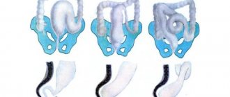
Types of dropsy
The classification of hydrocephalus of the brain is quite complex, with blurred diagnostic boundaries. Until recently, this disease belonged exclusively to children, but today it has been proven that it is possible to develop dropsy in adults.
Clinical manifestations of dropsy are divided into groups:
- The mixed form is severe, the volume of fluid increases inside and outside the brain. The prognosis is unfavorable. Violations cause epileptic seizures, convulsions, and paralysis of the limbs.
- Atrophic hydrocephalus of the head develops against the background of traumatic brain injury. The ventricles increase symmetrically, the volume of gray and white matter decreases. A month after the injury, changes become noticeable. This is a natural reaction of the body. The prognosis of the post-traumatic form of the disease is unfavorable.
- Hydrocephalus vicarious - the ventricles become enlarged, but the anatomical structure of the brain does not change. The prognosis is favorable if detected at an early stage. In the vast majority of cases, the development of the disease can be stopped and the situation stabilized.
- Hypotrophic hydrocephalus develops as a reaction to a malnutrition of the brain, when soft tissues lack nutrients. A person has a headache and vestibular functions are impaired. The condition is aggravated by nausea and vomiting.
- The compensatory form appears when the acute stage of the disease has passed. The volume of fluid in the ventricles stabilizes, but their volume remains increased. There is no treatment.
- Non-occlusive form - changes do not cause hypertension. The nature of the disorders is not clear, the inflow and outflow of cerebrospinal fluid remains at the same level, the circulation of cerebrospinal fluid is within acceptable limits.
- Partial form - an increase in fluid volume causes neurological changes. A characteristic symptom of the disease is epileptic seizures.
- Discirculatory hydrocephalus - a lack of cerebral circulation is diagnosed, often accompanied by atrophic processes.
Whatever the etiology, dropsy remains a dangerous disease, the consequence of which is disruption of brain function, deterioration of thinking ability and perception of information. It is important to identify the disease at an early stage and stop its development.
Treatment of hydrocephalus
The option of surgical intervention for hydrocephalus is quite effective. Two types of operations are used: neuroendoscopy or liquor shunting. They are performed by a neurosurgeon, a specialist who can correct problems in the brain or spinal cord, as well as the peripheral nerve ending system.
Bypass surgery
In this type of surgery, a thin tube is inserted into the brain ventricle. Excess cerebrospinal fluid flows through it to another anatomical zone of the body. If the second end of the drainage system is placed in the peritoneum, the shunt is called ventriculoperitoneal. Then the excess cerebrospinal fluid is absorbed by the abdominal cavity and then enters the blood.
When the draining end is directed into the cardiac chamber, the shunt is called ventriculoatrial. This option is practiced during surgery for children, because their increase in height does not affect the functioning of the organ as much, so the drainage does not have to be changed as often as the ventriculoperitoneal system.
Occasionally, lumboperitoneal shunts are also used. With the help of such drainage, cerebrospinal fluid is drained from the lumbar region into the peritoneal cavity.
Any drainage system contains a valve that is set to a certain capacity. It is selected for a specific patient, taking into account the patient’s cerebrospinal fluid pressure. The doctor performing the surgery determines these values in advance by installing a suitable valve, which then looks like a “bump” protruding under the skin on the head.
Currently, neurosurgeons have two types of valves for drainage systems:
- With predefined characteristics that define a specific throughput.
- Adjustable magnetic fixtures. When choosing them, the doctor remotely, using special equipment, without performing additional incisions, changes the pressure in the device to achieve an optimal result. Thanks to this, it is possible to neutralize adverse side effects in the form of insufficient or excessive cerebrospinal fluid pressure.
Bypass surgery is performed under general anesthesia. The procedure lasts 1–2 hours and after it the patient is kept in the hospital for several days. If the outflow through the shunt is disrupted or the patient becomes infected, a repeat procedure is sometimes performed.
Neuroendoscopy
This operation is an excellent alternative to bypass surgery. Without taking steps to install drainage, the neurosurgeon forms a hole directly in the wall of the cerebral ventricle, after which he creates a bypass path from the vessel through which cerebrospinal fluid flows freely to the surface. There it is absorbed by the brain tissue without obstacles.
This procedure is not universal - it cannot be done on all patients, although this method is often used if the natural cerebrospinal fluid pathways are blocked. In this case, the cerebrospinal fluid flows through the created hole, successfully bypassing all blocked vessels.
The endoscopic procedure is done under general anesthesia. First, the neurosurgeon makes a burr hole with a diameter of 1 cm on the skull. Then, using an endoscope, he examines the inside of the cerebral ventricles. Through the internal channel of the instrument, surgical instruments are inserted, which are used to perform an operation inside the brain. When the endoscope reaches the third ventricle, the doctor creates a hole in its lower wall through which the cerebrospinal fluid penetrates into the subarachnoid cavity. After removing the device, first the aponeurosis is sutured, and then the skin on the skull. The duration of the operation is approximately an hour.
The risk of infection with such an intervention is significantly lower than with cerebrospinal fluid bypass. Neuroendoscopy does not have significant advantages over drainage surgery over long periods of patient observation. After such an intervention, hydrocephalus can also develop again after a few years.
Actions for normal pressure hydrocephalus
When normal pressure hydrocephalus (a common occurrence in older people) is diagnosed, the patient's condition can be improved by performing a cerebrospinal fluid shunt. Although surgery is not effective for every patient. Due to the considerable risks that accompany all operations, certain tests are required to assess how much the potential benefit outweighs the risk of negative consequences.
Almost 80% of patients diagnosed with normal pressure hydrocephalus experienced significant improvement after preliminary testing, so ventriculoperitoneal shunting was recommended for them. Significant clinical postoperative improvement occurs after a few weeks, sometimes even months. Only a timely correct diagnosis guarantees successful recovery even with many years of development of hydrocephalus.
Reasons for the development of pathology
The human brain consists of soft tissues located in the skull. To protect against damage, cerebrospinal fluid circulates in the cavity - the liquid fills the internal ventricles and grooves. When a person is healthy, the inflow and outflow of cerebrospinal fluid is balanced. The performance of its functions does not affect the patient’s health. If disturbances occur due to the development of tumors, injuries and infections, intracranial pressure increases and the process of cerebrospinal fluid movement is disrupted, causing changes in brain function and neurological manifestations.
Hydrocephalus can develop due to a stroke or tumor development.
Classification
Based on the openness of the cerebrospinal fluid outflow pathways, hydrocephalus occurs:
- closed;
- communicating;
- substitutive.
According to the characteristics of the deformation of the outflow tract:
- internal;
- external;
- mixed.
By type of cerebrospinal fluid pressure:
- hypotensive;
- hypertensive;
- normotensive.
By etiology:
- congenital;
- after the inflammatory process;
- tumor;
- vascular;
- idiopathic.
Other types of classification:
- with the flow;
- by compensation phase;
- by activity;
- according to the speed of development.
How does dropsy manifest itself?
With moderate development of the process, minor neurological disorders appear. You can't ignore:
- Nausea;
- Headache;
- Vomiting;
- Deterioration of vision;
- Changes in the appearance of the eyeballs;
- Problems with the vestibular system;
- Mental abnormalities.
The open external form has manifestations similar to mental disorders. If the diagnosis is made incorrectly, the patient will be treated in a psychiatric clinic, and the underlying disease will go unnoticed. To make a correct diagnosis, the neurologist prescribes additional tests:
- MRI of the brain is clear - the results of tomographic diagnostics will show the localization of the pathology and the catalyst for the disorders (infants may be prescribed neurosonography instead of MRI);
- puncture - children are tested using general anesthesia;
- fundus examination.
At the initial stages of the development of the process, an accurate diagnosis can only be made using instrumental methods.
What is the danger of the disease?
The consequences of dropsy largely depend on the age at which the disorders occurred and the development of complications:
- Infants experience high excitability, sleep disturbances, and increased muscle tone. The most severe manifestation is developmental delay and mental deviations.
- Preschoolers show aggression, stutter, develop strabismus, and experience developmental delays.
- School-age children experience memory loss and headaches. The learning process is difficult.
- In adults, epilepsy, increased excitability, and hallucinations appear.
The danger of the disease in adulthood is the development of mental disorders, motor functions and motor skills. If urgent measures are not taken, the patient becomes disabled.
How to treat dropsy?
This brain pathology is practically incurable. Existing medications are designed only to slow down the process.
The most effective treatment method today is surgical, when the patient undergoes bypass surgery or endoscopic surgery.
Massage plays an important role in the treatment of hydrocephalus. Dropsy helps to increase muscle tone, and stroking and rubbing movements have a relaxing effect, helping to restore motor functions.
Another type of treatment, manual therapy, is used as an addition to the use of medications. Manual influence is aimed at activating the human body’s own reserves. The use of manual therapy for secondary dropsy is especially effective.
Recommendations for the use of treatment methods are given by a neurologist or neurosurgeon.
Prognosis and prevention
It is quite difficult to predict anything in those who suffer from hydrocephalus, although there is a certain connection between the outcome of the disease and the reasons that caused it. Several factors influence the likelihood of pathology:
- presence of concomitant diseases;
- the period between the appearance of the first signs and the establishment of an accurate diagnosis;
- adequacy of the treatment.
Until now, experts have not been able to figure out by what amount it is necessary to reduce ICP during surgery in order to minimize complications or prevent brain damage. Patients, together with their relatives, must understand that pathology harms both mental and physical development. Although after rehabilitation treatment, most children can be brought to a state where they are able to study educational programs and have a minimum of restrictions regarding the quality of life.
The treatment process, which involves doctors of various specialties, and then effective rehabilitation measures coupled with classes conducted by teachers, are extremely important for achieving a positive result. However, you must always remember that the lack of treatment contributes to the progression of hydrocephalus, which is why the disease can be fatal. Only early diagnosis and then adequate treatment guarantee the child’s recovery.
Signs of hydrocephalus, where the cerebrospinal fluid has normal pressure, often progress with age if not properly treated. But sometimes patients suddenly experience unexpected improvements. The effectiveness of organizing liquor shunting measures is not always guaranteed: it depends on the severity of the pathology, the age of the patient, and the reasons that provoked the development of the disease.
Sometimes, thanks to a complex of timely treatment, it is possible to achieve complete restoration of lost neurological functions. Therefore, the prognosis of how the pathology will proceed further when a shunt system is installed depends only on the patient’s body’s perception of such intervention. The main complications are blockage, infection or disconnection of the system:
- The first complication develops due to the entry of body particles into the drains. Blockage can also be caused by a malfunction of the valve device. Typically, liquor shunt systems operate for 5–10 years. If a blockage occurs, a complete replacement of the drainage is required.
- Infection develops when bacteria enter the tubes. Such a complication is likely during surgery, at the time of installation of the system, and also due to the migration of pathogenic microorganisms. Liqueur bypass systems are made of silicone. This material itself is foreign to the body, which makes it impossible to cure the infection with antibiotics. If such a complication is detected, immediate removal of the shunt system and a further course of therapy are required. Only after complete recovery is a new system implanted. To reduce the risk, it is recommended to use silver-impregnated equipment.
- Disconnection can occur when the seal in the tubes is broken. The cause is the movement of the shunt inside the patient's body, which often occurs in children due to natural growth. When system disconnection occurs, it is necessary to organize immediate surgical intervention, which involves checking the functionality of the shunt system and correcting areas that have lost their tightness.
Complications such as cerebral hemorrhage occur much less frequently (they are likely at the time of catheter installation), as well as disconnection of the catheter, after which it moves into the cavity of the cerebral ventricle. When such an event occurs, it is not always possible to remove the foreign object, so the associated infection becomes the cause of epilepsy or convulsive seizures.
Most patients who have undergone surgery for such an unpleasant reason lead an almost comfortable lifestyle. Almost 10% of them get rid of foreign implanted systems over time.
Nowadays, prevention of pathologies that can provoke hydrocephalus is carried out everywhere. To prevent meningococcal infection, which leads to infectious meningitis, active vaccination is used, and antibacterial agents are prescribed to those who have been in contact with an infected person to minimize the risk of infection. To prevent the possibility of rupture of identified arterial aneurysms in the brain, which cause subarachnoid hemorrhage, surgical interventions are organized to help exclude the aneurysm from the general blood flow.
Diet
The patient's nutrition should be aimed at improving the water-salt balance. Foods that cause fluid accumulation should be excluded from the diet. Instead, vegetables and fruits, steamed meat, and cereals are introduced into the menu.
It is important to lead a healthy lifestyle, exercise with moderate exercise, and walk a lot. Following these recommendations will help maintain the patient's mental and mental fitness.
Dropsy is a serious disease that does not go away on its own. The patient requires qualified specialist assistance for life. The disease is in an advanced stage and cannot be treated.
Which doctor should I contact?
Hydrops is treated by neurosurgeons. When you contact the RAS clinic in Moscow, you will be provided with qualified, effective assistance: they will conduct an examination, prescribe tests, make a diagnosis, and determine an effective treatment strategy.
If any symptoms of neurological diseases appear, you must immediately make an appointment with a neurologist to prevent the situation from worsening. We invite you to a consultation with a neurologist in Moscow at the Central Clinical Hospital of the Russian Academy of Sciences. Registration can be made by phone or using the feedback form on the website.
Diagnostics
When a patient enters the neurology clinic, the doctor conducts an examination, checks motor reflexes, muscle and joint reactions. To clarify the diagnosis, additional examinations are carried out:
- Ultrasound examination of the head and neck for a preliminary assessment of the condition of the vascular bed;
- Computed tomography, which helps to establish the degree of damage to brain tissue, assess the degree of expansion of the subarachnoid fissures, determine the presence of neoplasms in the skull that impede the outflow of cerebrospinal fluid;
- Magnetic resonance imaging, which allows you to see changes in brain tissue with maximum accuracy;
- X-ray of the skull to determine changes characteristic of liquor hypertension and space-occupying formations;
- An ophthalmological examination to determine the presence of congestion, swelling of the optic nerve, and atrophy of eye tissue.
Patients undergo a lumbar puncture, which is used to determine the level of cerebrospinal fluid pressure. Improvement in the condition of a patient with hydrocephalus after removal of 40 ml of cerebrospinal fluid indicates a good prognosis after surgery.