Reasons for appearance
Dacryocystitis occurs in the presence of physiological pathologies, namely congenital narrowing of the duct (stenosis). Sometimes doctors detect a complete blockage of the tear duct.
- Trauma to the eyes or paranasal sinuses.
- Inflammatory process of the nose, which provokes swelling of the tissues around the eye.
- An infectious process caused by bacteria and viruses, which leads to blockage of the duct.
- Getting foreign particles into the eye or working in dusty and smoky rooms. As a result, the channel becomes clogged.
- Allergy to exposure to an irritant.
- Reduced protective properties of the body.
- Overheating and hypothermia.
- Presence of diabetes mellitus.
Very often this pathology occurs in newborn babies. This is due to the structural features of the tear ducts. When the baby is in amniotic fluid, the tear duct is closed with a special membrane, which must rupture during or after childbirth. This process does not occur if pathology occurs. Tears collect in the canal and this provokes an inflammatory process. It mainly develops in women. Men are also no exception, but this pathology is detected very rarely in them. The reason is differences in the structure of the lacrimal canal. Women use cosmetics, most of which cause inflammation.
Sources
- Grassiri B., Zambito Y., Bernkop-Schnürch A. Strategies to prolong the residence time of drug delivery systems on ocular surface. // Adv Colloid Interface Sci - 2021 - Vol288 - NNULL - p.102342; PMID:33444845
- Zhu WX., Zhang YY., Sun ZP., Gao Y., Chen Y., Yu GY. Differential diagnosis of immunoglobulin G4-related sialadenitis and Kimura's disease of the salivary gland: a comparative case series. // Int J Oral Maxillofac Surg - 2021 - Vol - NNULL - p.; PMID:33384237
- Wang Y., Zhuo W., Hei Y., Tong QZ., Li T.Y. . // Zhonghua Yan Ke Za Zhi - 2021 - Vol56 - N11 - p.832-838; PMID:33152841
- Tao H., Wang YS., Wang F., Wang HB., Dong WL., Bai F., Wang P., Zhou XB., Wang LH., Liu C. Diagnosis of lacrimal punctum lesions using optical coherence tomography: a preliminary study. // Int J Ophthalmol - 2021 - Vol13 - N6 - p.902-906; PMID:32566500
- Nagendran S., Alsamnan M., Strianese D., Malhotra R. Ectopic Lacrimal Gland Tissue: A Systematic Review. // Ophthalmic Plast Reconstr Surg - 2021 - Vol36 - N6 - p.540-544; PMID:32205779
- Singh S., Sharma A., Mittal V., Ali MJ. Lacrimal drainage anomalies in Tessier cleft 3 with unilateral anophthalmos. // Eur J Ophthalmol - 2021 - Vol - NNULL - p.1120672119891475; PMID:31771345
- Jang JK., Lee SM., Lew H. A histopathological study of lacrimal puncta in patients with primary punctal stenosis. // Graefes Arch Clin Exp Ophthalmol - 2021 - Vol258 - N1 - p.201-207; PMID:31713749
- Tanaboonyawat S., Idowu OO., Copperman TS., Vagefi MR., Kersten RC. Dacryops - A review. // Orbit - 2021 - Vol39 - N2 - p.128-134; PMID:31512543
- Kubota T., Katayama M., Nishimura R., Moritani S. Long-term outcomes of ocular adnexal lesions in IgG4-related ophthalmic disease. // Br J Ophthalmol - 2021 - Vol104 - N3 - p.345-349; PMID:31272957
- Kojima T. Contact Lens-Associated Dry Eye Disease: Recent Advances Worldwide and in Japan. // Invest Ophthalmol Vis Sci - 2018 - Vol59 - N14 - p.DES102-DES108; PMID:30481813
Symptoms of the disease
Tears are necessary for the normal functioning of the visual organs. They moisturize the cornea of the eye, protect against mechanical irritants, and perform an antibacterial function. Sometimes tears stop flowing, this is the first sign of obstruction of the lacrimal canal. Treatment is one way to cope with the problem and prevent the development of canaliculitis. Sometimes massage of the tear duct helps.
- painful and unpleasant sensations in the eye area;
- redness of the skin around the eye;
- feeling of squeezing and bursting;
- swelling of the skin;
- lacrimation;
- edema;
- vision problems;
- increased secretion of mucus, which smells bad;
- formation of pus;
- high body temperature;
- intoxication of the body.
The acute stage of dacryocystitis appears as an inflammatory process affecting one eye. In the chronic stage, the tear duct swells, the eye turns red and the number of tears increases.
If you experience such symptoms, you should consult a doctor. Blockage can occur in both acute and chronic stages. The accumulation of tear fluid increases the likelihood of infectious processes.
Lacrimal gland disorders
Disorders of the lacrimal gland manifest themselves in increased tear production ( hyperfunction ) or insufficient production of tear fluid ( hypofunction ).
Increased lacrimation may occur due to bright light, strong wind, cold and other external irritants, or as a result of disturbances in the innervation of the eye. A characteristic sign of lacrimal duct pathology is increased lacrimation (epiphora).
Hypofunction of the lacrimal glands (or “dry eye syndrome”) is one of the manifestations of Sjögren’s syndrome. It also occurs in endocrine and autoimmune diseases, in patients on hormone replacement therapy, in people who work for a long time behind a monitor, in smokers.
Diagnostics
Dacryocystitis is detected without much difficulty. At the appointment, the doctor performs a visual assessment of the eye and palpation of the lacrimal sac.
- Test using paint. The eye is instilled with a solution containing a dye. If pigment appears in the eye after a few minutes, this indicates a blockage of the tear ducts.
- Probing. Using a probe with a needle, the ophthalmologist penetrates the duct, which helps to expand it and get rid of the problem.
- Dacryocystography. Carrying out an X-ray examination with the introduction of a dye. In the picture you can see the structure of the eye system and identify the problem.
- Patency can also be checked with the West test. A cotton swab is placed in the nasal passage, on the affected side. Collargol is instilled into the eye. The condition is considered normal when after 2 minutes the tampon turns dark. If the swab remains clean or stained after 10 minutes, there is a problem.
Symptoms of phlegmon of the lacrimal sac
Phlegmon of the lacrimal sac is externally manifested in redness of the skin around the eye, eyelid, cheek, nose and significant swelling, so strong that it can completely close the palpebral fissure. A painful, hot lump is felt above the lacrimal sac. In this case, the patient’s temperature rises, weakness and headache occur. Sometimes a feverish state occurs.
After two or three days, pus accumulates in the lump and it becomes soft to the touch. The surrounding skin turns yellow. The process progresses and opens through the surface of the skin; after the complete outpouring of pus, the inflammation subsides. In some cases, a fistula appears at the site of the lesion, secreting pus and tear fluid. It is possible to open the abscess into the nasal cavity through the nasolacrimal canal. In this case, a new artificial opening appears that drains fluid from the eye into the nasal cavity.
Treatment
Eyes are the mirror of the soul. When an eye problem arises, there is no need to take risks. Treatment should be prescribed by a doctor after preliminary diagnosis. The treatment method is selected depending on the form and cause of the pathology that provoked it, and age characteristics.
- Rinsing the eyes with antibacterial and disinfectant solutions.
- The use of special drops and ointments.
- Massage procedures and compresses to help clean the canal.
Eye rinsing with antiseptic solutions is carried out several times a day. The procedure is performed by an ophthalmologist in a hospital setting.
Ointments and drops that have an antibacterial effect:
- Floxal is a broad-spectrum antibacterial drug. Fights the inflammatory process. The course of treatment is 10 days, two drops twice a day.
- Dexamethasone - drops with an antibacterial effect. Effective against infectious processes. Instill 5 times a day. The required dosage and course of treatment are selected by the doctor individually for each patient.
- Levomycetin is a hormonal drug. Used for allergic reactions and inflammations.
- Ciprofloxacin - prescribed for tear duct infections. Buried every three hours.
The drugs have contraindications and side effects. Drug therapy is carried out under the supervision of the attending physician.
If the treatment does not have a positive effect, bougienage is performed - cleaning the tear duct from purulent contents; The disease can be quickly dealt with only if treatment is started in a timely manner. If symptoms are negative, you should visit an ophthalmologist.
Radical ways to fight
If there is no positive effect from drug treatment, or if the cause is a tumor or cyst, surgical treatment is performed.
Surgical intervention happens:
- Endoscopic dacryocystorhinostomy. A device with a camera is inserted into the duct. A puncture or incision is made using an endoscope. A special valve is created, the main purpose of which is drainage. The recovery period is 7 days. In parallel, antibiotic therapy is carried out to prevent the risk of developing an inflammatory process. The main advantage is the absence of visible marks after the operation.
- Balloon dacrycytoplasty is an intervention that, due to its safety, is performed even on newborns. A conductor with a reservoir filled with liquid is inserted into the channel. Allows you to expand the area, thereby breaking through it. The procedure is carried out under local anesthesia. During the rehabilitation period, special drops and antibacterial drugs are prescribed.
Book "Eye Diseases"
Chapter 8
- Pathology of the tear-producing apparatus
- Pathology of lacrimal organs
DISEASES OF LACRIMAL ORGANS.
Pathology of tear-producing organs.
The pathology of tear-producing organs can be represented by developmental anomalies, inflammatory processes, tumors, atrophies and degenerations, as well as post-traumatic changes. Developmental anomalies are more common in children. Anomalies of the lacrimal gland are manifested by its absence, underdevelopment, prolapse, and hypertrophy. The absence or insufficient development of the lacrimal gland leads to xerosis and loss of vision, and treatment in such cases is only surgical. It consists of transplanting the Stenon's duct of the parotid salivary gland into the outer part of the conjunctival cavity. There is a great similarity between the physicochemical composition of the secretion of the lacrimal and salivary glands, and this ensures a satisfactory condition of the eye.
<< CONTENTS
In case of prolapse of the lacrimal gland and disruption of its function, surgical treatment is also indicated - suturing the lacrimal gland to the periosteum in the area of the outer part of the eyebrow.
In case of hypersecretion, it is partially removed and sclerotherapy is used to reduce tear production.
Inflammatory diseases of the lacrimal gland - dacryoadenitis - are not so common. They are divided into acute and chronic. Acute dacryoadenitis (dacryoadenitis acuta) (Fig.) is more common in children and young people, and can be unilateral or bilateral. The disease occurs with influenza, scarlet fever, sore throat, rheumatism, pneumonia, typhoid fever, gonorrhea, and most often with mumps. With mumps, acute dacryoadenitis is always bilateral and is observed simultaneously with inflammation of the parotid and submandibular salivary glands, i.e. the same infection affects the salivary and lacrimal glands. This is explained by the fact that there is a certain unity in the histological structure of the lacrimal and salivary glands, as well as their general innervation. However, the inflammatory process in the parotid gland may end even before the development of dacryoadenitis or may be mildly expressed. This is especially important to keep in mind in children to avoid diagnostic errors. The clinical picture of acute dacryoadenitis manifests itself in swelling and soreness of the outer part of the upper eyelid. The swelling can spread from the gland area to the temporal region and the entire half of the face, and then the palpebral fissure closes. In severe cases, in weakened children, an abscess or phlegmon may occur, which can also invade the retrobulbar space. The eyeball shifts downward and inward, double vision appears with a slight protrusion of the eyeball from the orbit. Eye movements are limited upward and outward. The outer part of the conjunctiva is sharply hyperemic, swollen, and may have chemosis. The general condition suffers (headaches appear, sleep and appetite are upset, the temperature rises), regional lymphadenitis of the pre-auricular lymph nodes occurs.
Treatment consists of local application of various thermal procedures, UHF, vitamin drops and ointments with sulfonamides and antibiotics, subcutaneous injection of antibiotics into the area of the lacrimal gland. General treatment is aimed at combating the underlying disease. Antibiotics and sulfonamides, symptomatic drugs (analgesics, sleeping pills, etc.) are indicated. In case of suppuration or abscess formation, an incision is made and the cavity is washed with antibiotics.
For prolonged dacryoadenitis, radiotherapy is used. Usually the disease ends in recovery. Chronic dacryoadenitis can develop from acute ones, but more often they occur independently. The process can be either unilateral or bilateral and can occur with tuberculosis, syphilis, Mikulicz syndrome, and also with trachoma. There are no subjective sensations with chronic dacryoadenitis. The clinical picture is characterized by the formation of a swelling in the area of the lacrimal gland, dense to the touch, painless, extending deep into the orbit, gradually increasing. The skin over it is not changed. With tuberculous dacryoadenitis, suppuration and cheesy disintegration do not occur.
Mikulicz syndrome. This disease was first described in 1892 by the German surgeon Johann Mikulicz and since then it has been called Mikulicz disease or syndrome. This is a symptom complex that develops slowly, begins at the age of 20-30 years and is characterized by a slowly progressive, symmetrical enlargement of the lacrimal and salivary glands (usually submandibular, less often parotid and sublingual), displacement of the eyeballs downward and inward, and their protrusion forward. Suppuration of the glands never occurs. The lymph nodes are also involved in the process. The cause of the disease is not fully understood. Histologically, in Mikulicz's disease, lymphomatous hyperplasia of the lacrimal and salivary glands is observed. Some authors believe that tuberculosis is sometimes the cause. However, the majority regards the disease as lymphomatosis of the lacrimal and salivary glands, caused by a systemic disease of the entire lymphatic system - leukemia and pseudoleukemia. When examining a patient with suspected Mikulicz syndrome, blood tests are required. For the diagnosis of tuberculous dacryoadenitis, a general clinical examination for tuberculosis and the Mantoux skin tuberculin test are important. Luetic chronic dacryoadenitis occurs in the form of gummous dacryoadenitis of the lacrimal gland. Chancre is less common. With congenital lues there may be congenital interstitial dacryoadenitis.
Treatment is aimed at the underlying cause of the disease. Locally - radiotherapy. In all cases of Mikulicz's disease, the use of subcutaneous or oral arsenic preparations is also indicated.
Differentiate - with chronic productive sialadenitis, with salivary stone disease, tumors of the salivary glands, with Sjögren's syndrome, infectious mononucleitis. Tumors may include mixed tumors of the lacrimal gland, cylindromas, sarcomas, and adenocarcinomas.
Adenomas and oncocytomas are described as very rare. Sometimes lacrimal gland cysts occur. Cysts occur in the palpebral part of the lacrimal gland and appear as a result of infection of the excretory ducts of the gland, resembling a granular cyst of the excretory duct of the submandibular salivary gland. Its sizes range from cherry pit to hazelnut. The tumor is painless, elastic, with unchanged skin over it. When the upper eyelid is everted, it is visible in the form of a translucent transparent bubble.
Treatment comes down to opening the cyst or creating a permanent connection with the conjunctival cavity.
Currently, it is believed that mixed tumors of the lacrimal gland belong to a variety of mixed tumors of the salivary glands. They arise from abnormal cellular elements that are split off at various stages of embryo formation. They are divided into three groups: the first consists of mixed tumors, the parenchyma of which consists of several tissues derived from one germ layer; in the second group, the parenchyma is built from derivatives of two germ layers; the third includes mixed tumors, the parenchyma of which contains elements of all three germ layers. They usually develop in older people and grow slowly, although there are cases of rapid growth. The main symptoms are neurological pain and proptosis. Diplopia and visual disturbances are less common. In the fundus there are frequent changes in the form of a congestive nipple, neuritis and optic nerve atrophy. In a certain percentage of cases, the tumor has malignant features - a tendency to relapse and metastasize.
Treatment is removal of the tumor along with the lacrimal gland and radiotherapy. Sarcomas that occur in childhood and adolescence have a difficult course, and the prognosis for life is poor. For adenomas and oncocytomas, treatment is also surgical, but the prognosis is favorable. Tumors must be differentiated from chronic dacryoadenitis. Due to surgical interventions, injuries, previous dacryoadenitis, scar processes in the conjunctival cavity, and general diseases, degenerative processes may appear in the lacrimal gland. All atrophic and degenerative changes lead to hyposecretion and the development of severe degenerative processes in the conjunctiva and cornea. Degenerative processes in the lacrimal gland also include Sjogren's syndrome, which is discussed in the section “Diseases of the cornea.”
PATHOLOGY OF LACRIMACY ORGANS
The main symptom of lacrimal duct disease is constant lacrimation, which intensifies in the cold and wind. With pathological lacrimation, lacrimation is observed in the area of the lacrimal duct (gutter, between the eyeball and the free edge of the lower eyelid), and the lacrimal lake, and if it is prolonged, then maceration and hyperemia of the skin of the edges of the eyelids appears.
In some cases, lacrimation under the influence of cold may be due to contraction of subcutaneous muscle fibers in individuals with increased skin sensitivity. Rarely, lacrimation is associated with increased function of the lacrimal gland, more often it is associated with irritation of the I and II branches of the trigeminal, sympathetic nerves, pterygopalatine ganglion, with focal infection and in particular with dental diseases (reflex lacrimation). Watery eyes can sometimes be the only sign of malignant tumors of the paranasal sinuses, as well as the first sign of the initial stage of pituitary tumors. Lacrimation occurs with osteomas of the upper jaw growing into the sinus. Pathological lacrimation is observed in conjunctivitis, blepharitis, keratitis, scleritis, foreign bodies in the cornea, acute and chronic rhinitis, inflammatory processes in the paranasal cavities, etc.
In 1928, neuropathologist F.A. Bogorad. a symptom is described in which tear production is stimulated by various foods, often regardless of its taste, warmth and hardness, and sometimes only by the smell of food. Chewing movements alone, as well as mental crying, are not accompanied by lacrimation from the sore eye. In congenital syndrome, pathological connections develop in the nuclei of the abducens, superior salivary and facial nerves. The development of the acquired syndrome depends, according to the authors who described this syndrome, on improper regeneration of the secretory fibers of the facial nerve after its paralysis, growing not into the salivary gland, but into the lacrimal gland.
For treatment, it is recommended to block the parasympathetic innervation of the lacrimal gland, restorative treatment, vitamins, and climate therapy. If acquired, surgical treatment is offered.
Lacrimation occurs with the syndrome of the pterygopalatine ganglion, nasociliary nerve. Increased lacrimation is observed with Graves' disease, tabes dorsalis, with some encephalitis, Parkinson's disease, and with hysteria. However, more often pathological lacrimation is associated with dysfunction of the lacrimal ducts, congenital or acquired. Acquired disorders are associated with pathology of the lacrimal openings and canaliculi, as well as with pathology of the lacrimal sac and nasolacrimal canal. Quite often, atonic eversion of the lacrimal openings occurs in older people. In this case, due to the weakness of the Horner muscle, the inner third of the eyelid, together with the lacrimal punctum, is not tightly adjacent to the eyeball.
Treatment is surgical. Narrowing and obliteration of the lacrimal openings are most often a consequence of chronic blepharitis and conjunctivitis, which last a long time, but can be caused by persistent spasm of m. sphincter puncti lacrimalis. In the latter case, expansion of the lacrimal opening with a conical probe helps. If the narrowing is cicatricial, bloody dilation or surgery is performed. Foreign bodies in the tubules - eyelashes, food particles, maybe even worms - are removed surgically.
Purulent inflammation of the tubules (canalliculitis) is an inflammation of its mucous membrane. When inflammation spreads to surrounding tissues, pericanalliculitis develops. Skin hyperemia and swelling appear in the area of the tubule; it is palpated in the form of a dense, painful cord. Mucopurulent discharge is released from the lacrimal openings. Lacrimation and lacrimation are pronounced. The causes of the development of canalliculitis may be tuberculosis or syphilitic infection, brucellosis, trachoma, blepharitis, as well as mycoses, etc. Fungal canalliculitis occurs over a long period of time.
Treatment is the main action on the cause. Rinsing with antiseptics, frequent instillation of drops and ointments of antibiotics and sulfonamides are indicated. For fungal canalliculitis - dissection of the lacrimal canaliculus and scraping of the contents with treatment with iodine solution.
Congenital anomalies include: atresia of lacrimal puncta, atresia of lacrimal canaliculi, atresia of lacrimal puncta and canaliculi, accessory lacrimal puncta and canaliculi (several lacrimal puncta and canaliculi), dislocation of lacrimal puncta and canaliculi (forward, backward, outward, inward).
Inflammatory diseases of the lacrimal sac
Inflammations of the lacrimal sac are divided into acute and chronic. Chronic inflammation of the lacrimal sac - chronic dacryocystitis (dacryocystitis chronica) occurs as a result of a violation of the patency of the nasolacrimal duct, when tears are retained in the sac and various microorganisms (usually pneumococci and staphylococci) find favorable conditions for reproduction in the stagnant tear. The disease can also be caused by long-term processes occurring in the vicinity of the sac, for example, in the maxillary cavity, in the bones surrounding the sac, resulting in stenosis of the lacrimal canal. Congenital stenosis is also observed. Chronic dacryocystitis can be associated with a fracture of the upper jaw, with syphilitic processes in the nose. There may also be tuberculosis of the lacrimal sac.
With chronic dacryocystitis, swelling in the area of the lacrimal sac, lacrimation, and then suppuration are noted. When pressing on the area of the lacrimal sac, mucous, mucopurulent or purulent contents are squeezed out from the lacrimal openings. Sometimes, while maintaining partial patency in the stenotic nasolacrimal canal, the contents of the lacrimal sac can be emptied into the nasal cavity. The lacrimal caruncle, semilunar fold and conjunctiva are irritated, hyperemic, and persistent conjunctivitis and blepharitis are often observed. If dacryocystitis persists for a long time, the sac can become very stretched and then a sharp protrusion the size of a walnut is noted in the area of the lacrimal sac. The skin over the swelling is somewhat thinned. The mucous membrane of the sac may atrophy, lose the character of the mucous membrane, cease to separate mucus and pus, and then the sac contains a clear, somewhat viscous liquid (this is the so-called hydrops sacci lacrimalis). In women, the disease is 6-7 times more common than in men. Chronic dacryocystitis always poses a risk of developing ulcers of the cornea and phlegmon of the lacrimal sac (phlegmonae sacci lacrimalis).
Acute dacryocystitis, or phlegmon of the lacrimal sac, is a purulent inflammation of the walls of the lacrimal sac and the surrounding tissue. It is usually caused by coccal flora, but can also be gram-negative bacilli, primary tuberculosis infection, viruses and fungi. Most often, acute dacryocystitis develops as a complication of chronic, and in some cases due to the transition of the inflammatory process to the tissue surrounding the lacrimal sac from the adjacent adnexal cavities (maxillary, ethmoidal labyrinth), bones surrounding the lacrimal sac or from the nasal cavity. In the area of the lacrimal sac, severe redness and dense painful swelling appear, spreading to the adjacent areas of the nose and cheeks. The palpebral fissure is narrowed or closed. After a few days, the tumor becomes softer, an abscess forms, which then opens, often outward, forming a fistula. Such a fistula may not heal for a long time; a fistula appears, through which tear fluid is constantly released. A so-called internal fistula can also form when pus breaks into the nasal cavity through the nasolacrimal canal. If left untreated, especially in sick children, acute purulent dacryocystitis can lead to a diffuse phlegmonous process. If treatment is insufficient, relapses may occur. Treatment is dry heat, UHF in combination with intramuscular antibiotics, oral sulfonamides, local use of antibiotics in the form of injections, as well as 3% collarhead ointment. The nasal mucosa can be lubricated with cocaine and adrenaline. If an abscess has formed, open it through the skin and drain the cavity. When acute phenomena subside, surgical intervention is performed - dacryocystorhinostomy.
Dacryocystitis of newborns - develops due to the fact that after birth a mucous-gelatinous membrane is preserved, which during intrauterine development closes the lumen of the nasolacrimal canal from below (normally it bursts with the child’s first breath). In such cases, in the first days of newborns, discharge of mucous and moderate hyperemia of the conjunctiva are observed in the inner corner of the eyes. In the subsequent weeks of life, lacrimation appears, and then constant lacrimation. You may also notice swelling in the area of the lacrimal sac. When pressing on the area of the lacrimal sac (in small children it is better with the ball end of a glass rod), mucous, mucopurulent contents are squeezed out from the lower lacrimal opening. Acute dacryocystitis is, as a rule, a complication of congenital dacryocystitis; in rare cases, it can occur during the transition of the inflammatory process from adjacent tissues, for example, with purulent osteomyelitis of the frontal process of the upper jaw. In children it occurs with high fever and general anxiety. The outcome can be an external or internal fistula. Diagnosis of congenital dacryocystitis is not difficult, but there are mistakes when conjunctival hyperemia and discharge in newborns are mistaken for conjunctivitis and treated for a long time, unsuccessfully, with antibiotics. Treatment of dacryocystitis in newborns consists of using a jerky massage of the lacrimal sac area from top to bottom, followed by instillation of solutions of albucid (30%), chloramphenicol (0.25%), etc. into the conjunctival sac. and vitamin drops. The massage should be carried out for 1-2 weeks and at the same time, 2 times a day, it is necessary to squeeze out the contents of the lacrimal sac and instill a 0.25% solution of chloramphenicol and a 1:5000 solution of furacillin into the conjunctival sac. For purulent dacryocystitis, massage is contraindicated due to the possible development of phlegmon of the lacrimal sac. If there is no positive result, after 7-10 days the lacrimal ducts are washed with sterile saline solution after preliminary squeezing out the contents of the bag. Repeated washing can be carried out with a 0.5% solution of chymotrypsin and lidase at a concentration of 32 units per 1 ml of physiological solution, in equal concentrations. If patency is not restored, immediately proceed to conventional probing with a Bowman probe No. 1, if necessary, it is combined with retrograde. Probing should be carried out at an age later than 3 months. Conservative treatment is also carried out - washing the lacrimal ducts with a cocktail: 0.5 ml of a 2.5% suspension of hydrocortisone, 32 units of lidase and 5 ml of a sterile 0.5% solution of novocaine. Before surgery, a contrast X-ray examination is advisable.
Operation dacryocystorhinostomy (Dupuy-Dutan). 6-10 ml of a 2% solution of novocaine with the addition of a 0.1% solution of adrenaline is injected under the skin of the back of the nose in the area of the lacrimal sac. Add 8-10 drops of adrenaline to a five-gram syringe to novocaine and insert a needle under the skin approximately 2 cm below the internal ligament of the eyelids, moving inwards in the direction of the ligament. The second injection, without adrenaline, is made above the internal ligament, directing the end of the needle back and down. To anesthetize the nasal mucosa, a tampon moistened with a 5% solution of cocaine or a 2% solution of dicaine with adrenaline is inserted into the nose towards the superior concha. The incision with a scalpel begins 2-3 mm above the internal ligament of the eyelids, retreating from the place of its attachment by 2-3 mm and slightly outward and then continues along the lower edge of the orbit. The length of the incision is 2-3 cm. The edges of the skin wound are separated slightly to the side and a dilator is inserted into the wound. Using a small raspator, soft tissues (the orbicularis eyelid muscle) are moved apart, and if an angular vein or another vessel is encountered, they are carefully moved to the side and brought under the branch of the retractor. The soft tissues are pulled apart until the internal ligament of the eyelids appears. After its isolation, the periosteum is cut from top to bottom by 0.75-1 cm and peeled off along with the sac from the bone of the lateral dorsum of the nose and the lacrimal fossa down to the nasolacrimal canal. It is moved outward so that the anterior internal wall of the lacrimal sac, covered with exfoliated periosteum, is clearly visible, and in the inner part of the wound there is exposed bone (the lacrimal bone, the nasal process of the upper jaw and the nasal process of the frontal bone). Mark the boundaries of the area on the bone to be removed. A piece of bone up to 1.5 cm high and 1.0-1.2 cm wide is removed using a drill using a drill or a milling cutter. Do not damage the nasal mucosa! The bone flap is removed with tweezers. A swab is removed from the nasal cavity and a grooved probe or spatula is inserted into the nose with the left hand, and the mucous membrane is cut from top to bottom with the right hand using a narrow knife. The same vertical incision is used to dissect the lacrimal sac along its inner side along with the periosteum. A conical probe is inserted into the sac through the lacrimal opening, with the help of which the inner wall of the sac is protruded to ensure that the wall will be cut through. After the incision, the probe is visible in the wound cavity. Three sutures with a curved Ohm needle are placed on the posterior lip of the nasal mucosa and lacrimal sac. The seams are tied tightly and the ends of the threads are cut off.
Sutures are placed on the front lips in the same way. The skin wound is sutured with silk, the seams are cut, and the wound is lubricated with iodine tincture or brilliant green. A gauze pad and a pressure bandage are placed on the wound. To prevent bleeding, a strip of gauze is inserted into the nasal cavity until tamponade is obtained. During the first hours, the patient is prescribed bed rest; during the day, one should not blow his nose or consume hot food or drink. After 48 hours, the first dressing is done. If there is no bleeding, then the tampon is removed completely; if there is, a fresh one is inserted. If necessary, a solution of 10% calcium chloride 5.0-10.0 ml is administered intravenously, Vicasol or blood transfusion is given. After 6-7 days, the skin sutures are removed and the lacrimal ducts are washed with some disinfectant solution.
The positive or negative effect is judged only in the first 1-2 months; in case of relapse of dacryocystitis, repeated washing of the lacrimal ducts, probing or repeated surgery. If it is not possible to sew the lips, then you can remove the nasal mucosa within the hole made and cut out the walls of the sac, with the exception of the outer one (Toti operation). Otherwise, the operation is performed as just described, which is essentially a modification of the Toti operation.
Tumors of the lacrimal sac. They are rare. They can develop from any tissue that forms it: the epithelial layer, submucosa, muscle fibers of the fascial covering.
Benign tumors – papilloma. Adenoma, fibroma. Malignant degeneration of papillomas is possible.
Malignant - cancer of the lacrimal sac, sarcoma and very rarely melanoma. Cancer of the lacrimal sac is most often columnar cell, related to tumors of the nasal cavity and adnexal cavities since the epithelium lining the lacrimal sac and the epithelium of the respiratory apparatus are of the same origin. Sarcomas of the lacrimal sac occur mainly in children and adolescence, are very malignant and result in the rapid death of patients. For tumors of the lacrimal sac of various natures, the clinical picture is similar. First, lacrimation appears, then swelling and swelling forms in the area of the lacrimal sac. At this stage, a dense and elastic tumor is palpable. The correct diagnosis can be made if the tumor, which is palpated in the form of a dense formation fused to the skin, quickly increases in size, when pressing on the area of the lacrimal sac, blood discharge or droplets of blood appear from the lacrimal openings - a symptom of bloody tears, filling defects are detected with contrast dacryocystography and there are changes detected by CT and NMR.
In the third stage, the tumor grows into the nasal cavity, through the cribriform plate into the paranasal sinuses and into the upper jaw.
Treatment in all cases of tumors of the lacrimal sac is surgical. The nature of the tumor in the initial stages is judged only after opening the lacrimal sac. For malignant cases, treatment is surgical up to orbital exenteration and radiation. With sarcomas and melanomas, the prognosis is poor because the process generalizes and in the coming months after the onset of the disease, patients die.
Complications during and after surgery: in case of bleeding, apply a Pean clamp to the bleeding vessel and pack the wound. Late nosebleeds, as indicated, are stopped with nasal tamponade, intravenous administration of calcium chloride, and blood transfusion.
Questions:
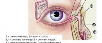
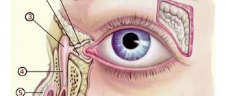
- Name the anatomical structure of the tear-producing lacrimal drainage apparatus?
- What disease causes dacryoadenitis most often?
- What is Mikulicz's syndrome characterized by?
- What is the main sign of lacrimal duct pathology?
- What symptom was described by F.A. Bogorad in 1928?
- What is the most common cause of canalliculitis?
- What are the differences between chronic and acute dacryocystitis?
- What surgery is performed for dacryocystitis?
- What causes dacryocystitis in newborns?
- Where should you start treating dacryocystitis in newborns?
- Name benign and malignant tumors that can develop in the lacrimal sac?
Share on social networks!
0
Massage
Prepare your hands before the procedure: wash, disinfect or wear gloves.
Massage scheme:
- Press on the outer corner of the eye, turn your finger towards the bridge of the nose.
- Gently press and massage the lacrimal sac, removing purulent masses from it.
- Instill a warm solution of furatsilin and remove the discharge.
- Carry out pressing and massaging movements along the tear ducts.
- Jerky movements along the nasolacrimal sac with some force to open the canal and extract the discharge.
- Instill a solution of chloramphenicol.
Dacryocystitis
The tear ducts connect to the lacrimal sac, which is located next to the nose. When this area becomes inflamed, the patient is diagnosed with dacryocystitis. It can occur both in adults and in infants who have only recently been born. Factors that provoke dacryocystitis are acute respiratory viral infections, rhinitis, sinusitis and similar diseases, in which the nasal mucosa swells and becomes inflamed. This leads to disruption of the high-quality outflow of moisture, and an active process of microbial reproduction begins in the lacrimal sac. Dacryocystitis in newborns is usually caused by the anatomical structure of the nasolacrimal duct in babies.
To restore the outflow of fluid, special drops, massage techniques are prescribed, and in more complex cases, probing or surgical treatment.
Folk remedies
After prior approval from a doctor, traditional medicine is successfully used at home.
Folk remedies:
- Aloe. For inflammation, it is good to instill freshly prepared aloe juice, half diluted with saline solution.
- Eyebright. Cook in the same way. Use for eye drops and compresses.
- Chamomile. Has an antibacterial effect. You need to take 1 tbsp. l. collection, boil in a glass of boiling water and leave. Use to wash eyes.
- Thyme. Due to its anti-inflammatory properties, the infusion is used for dacryocystitis.
- Kalanchoe. Natural antiseptic. Cut off the leaves and keep in the refrigerator for two days. Next, extract the juice and dilute it in a 1:1 ratio with saline solution. This remedy can be used to treat children. Adults can instill concentrated juice into the nose, 2 drops each. The person begins to sneeze, during which the tear duct is cleared of pus.
- Leaves from a rose. Only those flowers that are grown on your own plot are suitable. You will need 100 gr. collection and a glass of boiling water. Boil for five hours. Use as lotions.
- Burda ivy-shaped. Brew a tablespoon of herb in a glass of boiling water, simmer for 15 minutes. Use for rinsing and compresses.
- Bell pepper. Drink a glass of sweet pepper fruit every day. adding a teaspoon of honey.
Consequences and complications
Dacryoadenitis can be complicated by:
- Orbital phlegmon.
- Abscess of the upper eyelid.
- Formation of an external or internal fistula with spontaneous resolution of the abscess.
- A long-existing chronic form can develop into a lacrimal gland cyst - it forms gradually.
In the chronic course of inflammation of the lacrimal tubules, pronounced structural changes in the tubules are observed. Its narrowing (stricture) or obliteration (complete occlusion of the tubule) often develops, as a result of which the outflow of tears is significantly impaired or stops altogether. It is possible that surrounding tissues may be involved in the process with the development of orbital cellulitis and subperiosteal abscess.