Breast cancer is the most common malignant tumor in women. This type of cancer ranks first in mortality from cancer among the female population. The risk of developing it increases with age. Breast cancer in young girls (under 35 years of age) is rare, but has a worse prognosis.
The most common sign of breast cancer is the presence of a painless lump that a woman notices during a self-examination. Other symptoms include skin changes (orange peel), nipple discharge, and breast pain. According to statistics, only 10% of tumors in the mammary glands are malignant.
By the time breast cancer can be felt, the tumor already exceeds 1 cm. Earlier detection of the disease is carried out using mammography.
Treatment for breast cancer depends on the stage of the disease, and the most important information is the size of the tumor and its spread to the lymph nodes. Early detected breast cancer (by chance, on mammography, not yet palpable) has a better prognosis.
Treatment for breast cancer consists of surgery to remove the breast (mastectomy), radiation therapy (radiation therapy), chemotherapy, and hormone therapy. The choice of treatment method depends on the stage of the disease.
Signs of breast cancer
Signs of breast cancer in most cases are detected within the breast itself (in the early stages), with the exception of aggressive manifestations of cancer that have spread to distant organs, lymph nodes and tissues (late stages). If the tumor has penetrated beyond the breast, the clinical picture will look different. Migration of cancer cells is possible through the human lymphatic and circulatory system . It should be noted that many of the symptoms below are not unique to breast cancer. However, in any case, if you have one or more of these signs, you should immediately contact an oncologist for an in-depth diagnosis. When diagnosed with breast cancer, symptoms and treatment will be determined by the time the patient seeks medical help.
Definition of disease. Causes of the disease
The term “breast cancer” combines different types of tumors. Modern researchers note that breast cancer is the most commonly diagnosed type of cancer in women, and they distinguish several dozen varieties of the disease, which differ in the nature of the course and the likelihood of a favorable outcome for the patient.
Like other cancers, breast cancer develops as a result of uncontrolled division and growth of malignant cells. They deplete the body and poison it with the products of their vital activity, which can ultimately lead to associated disorders and death. Breast cancer is often a very aggressive tumor. Without treatment, it grows rapidly and spreads metastases - branches that affect other organs. Therefore, it is very important to start treatment in a timely manner, and for this it is necessary to identify the disease at an early stage.
According to modern research, breast cancer occurs due to a complex of reasons:
- genetics;
- unfavorable environmental conditions;
- disturbances in the production of female hormones (progesterone and estrogen);
- age over forty years;
- abortions;
- severe stress;
- obesity;
- lack of physical activity, etc. [1].
Thus, a significant part of the factors that cause breast cancer can be controlled. Although a family history of the disease increases a woman's risk of developing breast cancer, genetic predisposition is only one factor, and cases of familial cancer are uncommon [2]. Today, hereditary predisposition can be identified by analyzing the most common mutations: BRCA 1, BRCA -2, CHEK. This can be done in most large laboratories; all you need to do is donate blood.
At the same time, many other factors can be controlled. It is important to understand that if several factors are present simultaneously, the likelihood of cancer increases. A woman can reduce it if she undergoes regular examinations with a mammologist and leads a healthy lifestyle.
Symptoms of breast cancer
You should do a regular home breast exam between your doctor's visits every month, paying special attention to the symptoms listed below.
The most common symptom of breast cancer is the appearance of a new lump or lump. A painless, firm lump with an irregular outline is more likely to be cancerous, but malignant breast tumors can also be tender, soft, or round. Some women complain of obvious pain. Accordingly, if you have a new lump, lump or other change in the condition of the mammary glands, you need to be examined by an experienced specialist.
Other potential symptoms of breast cancer include:
- swelling of the whole mammary gland or part of it (even in the absence of lumps);
- the appearance of dimples on the skin (the skin looks like an orange peel);
- pain in the breast or nipple;
- nipple retraction;
- redness , dryness, flaking, or thickening of the skin of the breast or nipple;
- nipple discharge (other than breast milk);
- enlarged lymph nodes (sometimes breast cancer spreads to the lymph nodes in the armpit or around the collarbone and forms a lump there before the primary tumor in the breast grows large enough to be felt).
Although any of these symptoms may indicate a disease other than breast cancer, unusual symptoms should prompt you to see a doctor. Only a specialist can identify the real cause of your symptoms.
Remember that breast self-examination for signs of cancer does not replace regular mammograms and other screening procedures. Screening can detect breast cancer before symptoms appear. Early detection of cancer increases the chances of successful treatment.
Breast cancer classification parameters
The main elements of the breast are lobules with glands that produce mother's milk, as well as ducts that deliver it to the nipple. The tissues are penetrated by blood and lymphatic vessels responsible for supplying the mammary gland (MG) with nutrients and removing waste products. The disease begins when normal cells in a woman's body begin to divide uncontrollably, and the process of their death is disrupted. Types of breast cancer are classified according to different criteria.
Based on the location of the primary node, oncological pathologies are divided into neoplasms:
- pacifier;
- areolas;
- axillary area.
Depending on the quadrant of location, malignant nodes are:
- upper internal;
- upper-outer;
- lower internal;
- lower outer.
Up to 50% of primary tumors in women are formed in the upper outer quadrant of the mammary gland, about 20% - in the areola or central part of the breast.
Experts classify breast cancer of unspecified location and beyond the boundaries of individual regions into separate groups.
Tumor primary nodes can be ductal or lobular, based on the tissue of which element of the organ structure began to degenerate.
In addition to location, the classification of the disease according to parameters such as:
- histological structure;
- stage of development;
- form;
- molecular taxonomy.
Malignant neoplasms are also differentiated by the degree of aggressiveness of the impact and the speed of spread.
Metastasis
Regardless of the location of the primary node, cancer can be:
- non-invasive - the tumor grows, but does not leave the borders of the affected part of the organ;
- invasive - a malignant neoplasm grows beyond the boundaries of the lobule (or milk duct), its metastases first “capture” healthy parts of the breast, then other organs and systems.
Malignant cells “spread” throughout the body from the gland in two ways:
- lymphogenous - through lymphatic vessels;
- hematogenous - with the bloodstream.
First, metastases affect regional lymph nodes under the shoulder blades, armpits, sternum or collarbone area. Then they appear in distant areas of the lymphatic system, grow into the skin, soft and bone tissues.
At the terminal stage, malignant cells are found in almost all internal organs of the human body. Metastases from the primary node often spread hematogenously to:
- liver;
- brain;
- spine;
- pleura;
- lungs;
- ovaries;
- bones of the pelvis, hips.
When the development of breast cancer ends with the abnormal tissues being separated from the “maternal” node and transferred to other parts of the woman’s body, the disease is called metastatic.
If the primary pathogenic node forms again after treatment in the second mammary gland, in the same place, nearby tissues or organs, we are talking about a recurrent type of cancer.
Both forms of pathology are late-stage diseases; metastases can appear months or even years after the end of treatment. Relapse occurs in approximately 30% of women treated in the early stages of the disease. The average time for local “return” of oncology is five years. The risk of reappearance of the tumor is especially high in the first 24 months.
Non-invasive breast cancer can become invasive as the size of the tumor increases. It is very important to monitor the intensity of its growth, start treatment on time, and stop metastasis.
Experts distinguish three degrees of doubling in the size of carcinomas in women:
- high – the abnormal primary node grows twice in three months or less;
- average – doubles over a period of time from 3 to 12 months;
- low – doubling lasts more than a year.
Tumor cells undergo separate differentiation. In medical documents, the parameter is designated by the Latin letter “G”. Doctors distinguish three degrees of danger of breast cancer:
- highly differentiated (G1) - malignant cells are similar to natural ones, divide slowly, and have a low potential for metastasis;
- moderately differentiated (G2) - pathologically altered structures retain the characteristics of normal ones, multiply at an average speed, and are more prone to spread than the previous group;
- low-differentiated (G3) - cells are practically devoid of healthy signs, quickly divide, and have a high potential for the formation of metastases. They are a sign of cancer with an aggressive, rapid, life-threatening course.
Determining the type of malignant cells and the location of the primary tumor helps predict the possible spread of metastases.
Heredity factor as a sign of cancer
One of the mandatory actions in relation to a patient with breast cancer is to build his family tree and determine the need for genetic testing for the presence of BRCA genes . BRCA1 and BRCA2 are autosomal dominant genes that can be a sign of the development of breast cancer in up to 80% of relatives. There are several reasons to suspect the presence of these genes in the family:
- cases of cancer at a young age ;
- bilateral breast cancer , when after one breast is affected, the tumor forms in the second after some time;
- cases of breast cancer in men.
All of the above factors are a reason to conduct genetic testing to build a family tree for three generations and determine the mathematical probability of carrying the BRCA genes. Siblings and children of a carrier woman have a 50% chance of inheriting the gene . Thus, genetic testing is an effective method for preventing breast cancer.
Breast tumor: benign breast tumors, classification
Benign tumors develop in all breast tissues. The following types of benign neoplasms are found:
- Breast cyst.
- Fibroadenoma.
- Lipoma.
- Mastopathy.
- Intraductal papilloma.
Mastopathy is a general name for a large number of diseases associated with the proliferation of connective tissue. The type of nodular mastopathy is intraductal papilloma. Fibroids affect young women, the tumor has clear outlines. A lipoma is a fatty tumor, which is predominantly adipose tissue enclosed in a capsule. A breast cyst is a capsule filled with fluid. The classification of benign breast tumors contains six main groups:
- Epithelial tumors.
- Mixed type of neoplasms.
- Another type of neoplasia.
- Unclassified benign neoplasms.
- Tumor-like formations.
- Breast dysplasia.
Local cancer: symptoms
The clinical situation in this case can develop in four different directions:
1. The tumor is located in the middle part of the chest , in its glandular part. It is usually first discovered when a woman takes a bath or shower and notices a lump in one of her breasts. Immediate contact with a breast oncologist in such cases allows you to diagnose cancer even before the first clinically significant symptoms appear.
2.
The tumor is located near the nipple .
This localization of the tumor has two characteristic symptoms: deformation and retraction of the nipple (this is especially noticeable in comparison with the other nipple) and bloody discharge from the nipple. 3. The tumor has formed under the skin . In such cases, it manifests itself as a non-healing and unpleasant-smelling (due to bacterial infection) ulceration on the skin or “orange peel”, the appearance of which is caused by the accumulation of fluid due to compression of the lymphatic vessels.
4. The tumor lies deep in the thickness of the glandular tissue near the pectoral muscle, partially growing into it. A sign of this location of the tumor is a distinct asymmetrical shape in a bent position at the waist.
Check the price with a specialist
Breast cancer by lymphatic system
Spreading through the lymphatic vessels, the tumor can penetrate the axillary lymph nodes. In this case, the following symptoms are observed:
- enlargement of lymph nodes up to clearly palpable nodules
- accumulation of cancer cells in the armpit area
Less commonly, the tumor may migrate through the internal thoracic duct, which runs under the breastbone. Symptoms: since the tumor affects deep-lying lymph nodes, it can only be detected using instrumental diagnostic methods .
An even rarer case is a lesion of the supraclavicular lymph node, which manifests itself as a palpable lump in the area between the lower part of the neck and the upper rib. As it grows further, it can put pressure on the brachial plexus, causing loss of sensation in the fingers or shooting pain in the shoulder.
Stages of breast cancer
The stage of the disease also depends on the size of the tumor. If the nodule is no more than 2 cm in diameter and no cells are affected in the lymph nodes, doctors diagnose stages 0 and 1 of cancer. Three out of 4 women survive if treated in a timely manner.
At the next stage, the disease can develop in two ways.
1st way The tumor does not grow, but cancer cells appear in the 4–5 lymph nodes closest to the tumor, and the lymph nodes themselves change in size.
2nd way The tumor does not affect the lymph nodes, but increases to 20–50 mm.
Every 2nd woman has a chance of recovery in stage 2.
The 3rd stage is distinguished by: • damage to the lymph nodes next to the tumor; • tumor size over 5 cm; • penetration into the chest and bone tissue of metastases.
The survival threshold is 10–15%.
At stage 4, the prognosis for cure is unfavorable, since metastases penetrate into distant organs and affect the entire body.
Symptoms of breast cancer in the circulatory system
If the tumor spreads through the blood vessels, then most often metastases are found in the following organs and tissues:
- bones. Symptoms: large metastases can disrupt the integrity of the periosteum, causing severe pain. Bone mineral density decreases, leading to fractures. A special case is a fracture of the vertebrae, which results in the so-called. compression (radicular)
- lungs. Symptoms: when they enter the lungs, tumor cells usually form several separate groups that form multiple metastases. Their development causes depression of pulmonary function, manifested by shortness of breath (dyspnea). When the bronchi are damaged, a painful dry cough develops (if nearby blood vessels rupture - cough
- brain. Symptoms: as metastases grow, they begin to put pressure on neurons, which causes their irritation (resulting in seizures) or partial shutdown (resulting in loss of mobility, loss of vision, sensitivity, etc.). Since the skull is a limited space, there comes a time when it becomes too crowded for neurons and tumor cells. A sign of this is headaches with gradually increasing intensity.
- liver. Symptoms: once in the liver, tumor cells begin to grow rapidly, destroying hepatocytes, as a result of which the level of transaminases in the blood sharply increases (this is clearly visible in a laboratory blood test). When the tumor compresses the bile ducts, bile accumulates, and the patient’s skin acquires a yellow tint.
Diagnostics
In approximately 70% of breast tumors, suspicious lesions were initially discovered by the patients themselves rather than detected during a medical examination. Any woman should make it a rule to conduct an independent examination of her mammary glands. This procedure is simple and should be performed every month after the end of menstruation.
During the examination, priority attention should be paid to the following parameters:
- breast symmetry;
- their size;
- color of the skin;
- skin condition.
If a suspicious symptom or formation of unknown nature is detected, you should consult a mammologist. He will conduct a manual examination of the breast and may prescribe additional procedures such as ultrasound, mammography (x-ray of the breast area), ductography (mammography with a contrast agent).
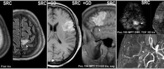
All women from 40 to 49 years old should undergo mammography once every 1-2 years, and doctors strongly recommend that women over 50 years old be examined annually. If suspicions about the malignancy of the formation still remain, then a biopsy is performed followed by examination of the cellular material. A blood test for tumor markers is also performed. If the oncological diagnosis is confirmed, then an immunohistochemical study, or a study of the molecular characteristics of the tumor, is performed at the oncology clinic of the Yusupov Hospital.
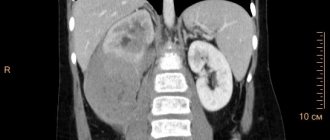
To assess the spread of the disease in the body, the oncology clinic of the Yusupov Hospital may prescribe:
- Ultrasound;
- CT scan;
- osteoscintigraphy;
- PET-CT;
- MRI.
How the quality of medications affects the cure for breast cancer
The treatment protocol for breast cancer includes chemotherapy and sometimes hormonal and targeted drugs. At the same time, the quality of drugs is crucial for the prognosis of the disease. To avoid counterfeiting, it is best to purchase drugs for cancer treatment in Israel.
- In Israel you will not be sold counterfeit medicines.
The Israeli Ministry of Health is the guarantor of the authenticity and high quality of medicines. It organizes inspections of pharmacies and control purchases of drugs. At the same time, pharmacies and pharmacists bear liability (including criminal liability) for the sale of low-quality drugs. - It does not take long for new drugs to be approved in Israel.
Licensing of drugs in this country is organized in such a way that all effective drugs appearing in the world are quickly licensed and introduced into widespread clinical practice. - The Israeli company TEVA is a leader in the global pharmaceutical industry.
Its products are used in 60 countries.
Comparison of similar symptoms with other breast diseases
In the breast, dishormonal diseases can coexist with a malignant process, because all pathological changes in the glandular parenchyma have a common beginning - proliferation of glandular cells under the influence of inadequate production of sex hormones or perverted sensitivity to them.
Ultrasound, mammography and, best of all, MRI can distinguish cancer from a benign process. Mammography is not always able to visualize pathology in dense, “full” breasts; ultrasound in young hormonally active women is more sensitive and accurate.
Mastopathy causes discomfort and pain, but these symptoms are not constant and are associated with fluctuations in the levels of sex hormones in the blood. Diffuse compaction is more common, but local compaction is also possible - focal, which differs from carcinomas in the alternation of dense areas with smooth small cysts or strands. The tissue density approaches normal, and on the MRI image it looks like normal parenchyma with cysts and connective tissue nodules.
Fibroadenoma of the mammary gland does not hurt, is softer to the touch, has smooth walls and is clearly demarcated from the rest of the parenchyma.
Cysts are asymptomatic, characterized by fairly rapid growth, dense but elastic, with a smooth surface; X-ray examination reveals a thick capsule and liquid.
How can I purchase Israeli-made medicines?
- After examination in Israel.
If you undergo diagnostics at the Ichilov Cancer Center, the doctor will prescribe medications for you, which you can purchase at an Israeli pharmacy. - In the country where the patient lives.
The telemedicine program developed at the Ichilov Cancer Center allows a patient from abroad to receive a video consultation with an Israeli doctor and a treatment protocol without leaving home. At the patient’s request, the oncology center will send him the prescribed medications (home delivery is possible).
Why is it worth treating breast cancer in Israel, at the Ichilov Cancer Center?
- Accurate diagnosis.
The latest diagnostic methods, including PET-CT and sentinel lymph node biopsy, allow Israeli specialists to accurately determine the stage of the disease and select the correct treatment program. - World-famous doctors and a personalized approach to treatment.
At the Ichilov Oncology Center, breast cancer treatment is carried out by leading Israeli oncologists, including Professor Moshe Inbar, a doctor with 40 years of experience and the author of more than 20 scientific papers. The professor selects individual treatment protocols for patients that correspond to the characteristics of the disease. - The latest equipment.
The oncology center uses the following for the treatment of breast cancer:
- linear accelerators Synergy S and Novalis TrueBeam STX, which allow delivering high doses of radiation to the tumor, while protecting healthy tissue from radiation;
- Marginprobe is a device that makes it possible to remove a tumor with “clean edges.”
Molecular taxonomy
Modern medicine divides breast cancer into four molecular forms:
- luminal, subtype A - hormone-dependent, characterized by low aggressiveness, occurs more often than others (approximately 40% of all cases). Amenable to conservative treatment;
- luminal, subtype B - has a pronounced dependence on estrogen synthesis, is diagnosed in 15% of patients. The outcome of treatment depends on what stage of the disease it began;
- HER2-positive is a hormone-independent form of malignant breast tumors, characterized by high aggressiveness and mortality of patients. It accounts for about 10% of the identified types of breast cancer;
- triple negative is a very dangerous neoplasm. It spreads rapidly, is difficult to treat, and has an extremely negative survival prognosis. The primary node lacks estrogen and progesterone receptors and Her2. Hormones do not affect the growth of tumors in size, therefore targeted hormone therapy is meaningless. This type of disease is the second most common. It is registered in 35% of cases.
The listed molecular subtypes, due to the huge number of differences, are actually separate pathologies and require an individual approach to treatment.
Where to go?
The best option is to get diagnosed in Israel. Thanks to the skill of doctors and the latest equipment, Israeli oncologists often revise the diagnoses made by their colleagues from abroad. As a result, some of our patients are surprised to learn that they do not have breast cancer at all - it also happens that the symptoms indicate another disease, much less dangerous.
We suggest you contact oncology and undergo diagnostics in Israel , with leading oncologist professors. Examination methods may vary. First, you will be examined by a doctor, then a mammogram will be scheduled. A biopsy determines how the tumor has spread. Next, an MRI is performed to clarify the data from other examinations. Then the final diagnosis is made.
just 4 days you will receive an accurate answer as to whether you have breast cancer, and if not, you will be told what the characteristic symptoms and signs indicate. We will draw up a treatment program that can begin in our clinic within a few days.
Relationship between cancer and age
There is a myth that breast cancer occurs only at a certain age. Alas, this is not true. Perhaps this opinion arose due to the difference in the results of diagnostic methods for different age groups. For example, the older the patient, the easier it is to see a tumor during mammography. With ultrasound diagnostics, everything is exactly the opposite: this method has proven to be most effective for people under 35 years of age.
It is safe to say that all age groups are at risk for breast cancer, and concomitant factors are more important than age. According to Woods' confirmed hypothesis, the tumor appeared more often in young women with two or more full-term pregnancies than in women without children. Also, the development of the tumor is influenced by changes in hormonal levels, which explains the high percentage of detected diseases in postmenopausal age and in cases where menstruation began before 12 years.
The age of the patient is of great importance for carrying out preventive measures related to ovarian cancer. According to research from the University of Carolina, if breast cancer was detected at age 50 or younger, this increases the risk of ovarian cancer by 4.3 times. However, in women over 50 years of age, this risk tends to zero.
Cost of breast cancer treatment
The duration of treatment at Ikhilov for breast cancer on an outpatient basis is 3-4 working days.
Prices for cancer treatment set by the Israeli Ministry of Health at the Ichilov Cancer Center:
| Breast cancer treatment procedures | Price |
| Breast biopsy | 1761$ |
| Examination by the leading oncologist of the oncology center | 512$ |
| Mammography | 246$ |
| PET-CT | 1488$ |
| Ultrasound of the mammary glands | 408$ |
| Chemotherapy course | 1270$ |
| Segmental mastectomy | 5308$ |
| Sectoral mastectomy | 13211$ |
| Bilateral mastectomy | 10231$ |
| Mastectomy unilateral | 7750$ |
| Radiation therapy | 17007$ |
You can take the first step towards recovery right now. To do this, fill out an application and one of our doctors will contact you within 2 hours.
Or call: +972-3-376-03-58 in Israel and +7-495-777-6953 in Russia.
Causes and risk factors
Every year in Russia alone, malignant tumors in this organ are detected in 50 thousand women. And worldwide this number exceeds a million. And the survival statistics are also disappointing so far. Almost half of the cases in women are fatal. Risk factors that can trigger tumor development include:
- hormonal imbalance;
- childlessness and having many children;
- very late first birth;
- frequent abortions;
- early onset and late cessation of menstruation;
- presence of mastopathy;
- age over 40 years;
- addiction to smoking and alcohol;
- obesity, as well as frequent stress;
- hypertonic disease;
- inflammation of the uterus and ovaries;
- atherosclerosis;
- hypothyroidism;
- liver diseases.
But it happens that cancer is discovered in patients who are not included in any of these risk groups.