Lymphocytic leukemia is a malignant disease, as a result of which the bone marrow begins to produce a large number of immature lymphocytes that are unable to fully perform their functions. Lymphocytes are a type of white blood cell that is responsible for immunity. If leukocytes are malignant, they not only do not perform a protective function, but also suppress the formation of normal blood cells and disrupt the functioning of other human organs. There are many varieties of this disease. Lymphocytic leukemias differ in the speed of development of the pathological process, the types of damaged lymphocytes (B- and T-lymphocytes), and the degree of their maturity. But most often there are two main types of this disease: chronic lymphocytic leukemia and acute lymphoblastic leukemia.
Lymphocytic leukemia disrupts the formation of lymphocytes, which is why the patient has many immature cells that are unable to cope with their work. This, in turn, causes serious disruptions in the immune system, due to which a person becomes more susceptible to various diseases - in particular, candidiasis and tuberculosis. The patient may experience serious complications even from routine vaccinations. Patients often experience autoimmune reactions: when the immune system begins to fight healthy cells of the body - say, red blood cells. This causes anemia.
In addition, malignant lymphocytes are able to penetrate the spleen and lymph nodes, enlarging them, damaging bones, lungs, brain and liver.
In the bone marrow of a patient suffering from acute lymphocytic leukemia, lymphoblasts predominate, which divide very quickly, displacing other cells from the bone marrow and blood, actively populating the spleen and lymph nodes. The most common are acute B-cell leukemias, in which the patient begins to produce many immature B-lymphocytes. Acute B-cell leukemia is one of the most common types of this disease among children.
With chronic lymphocytic leukemia, more mature forms of lymphocytes are found in the patient’s blood: for some time they are even able to perform their functions. This type of leukemia is typical for people aged 50-55 years.
Symptoms of lymphocytic leukemia
As a rule, acute lymphoblastic leukemia develops very quickly: within a few weeks. Its main symptoms are:
- headache;
- malaise and weakness;
- pain in the abdomen, in the bones;
- frequent nosebleeds, bleeding gums;
- pallor;
- enlarged lymph nodes in the neck, groin and axillary areas;
- fever.
Chronic lymphocytic leukemia often does not manifest itself at all in the initial stages. It can develop over years: during the development process, the following symptoms gradually appear:
- increased sweating (especially at night);
- heaviness in the stomach;
- pallor;
- malaise and weakness;
- frequent infections;
- dyspnea;
- weight loss for no reason;
- hemorrhages in the mucous membranes and skin.
Who is at risk?
In acute lymphocytic leukemia, the risk group includes:
- people who have undergone chemotherapy or radiation therapy as part of treatment for another form of cancer);
- people who have been exposed to radioactive radiation;
- people suffering from Down syndrome and other genetic disorders;
- people whose sisters or brothers have been diagnosed with acute lymphocytic leukemia.
In case of chronic lymphocytic leukemia, the risk group includes:
- people whose relatives suffered from leukemia;
- people over 60 years of age;
- representatives of the Caucasian race.
Lymphocytic leukemia is a malignant disease in which the bone marrow produces a large number of immature lymphocytes that are unable to perform their functions. Lymphocytes are a type of white blood cell and are responsible for immunity. Malignant leukocytes do not cope with the protective function, while suppressing the formation of normal blood cells and disrupting the functioning of other organs.
There are many varieties of this disease. Lymphocytic leukemia is divided depending on the speed of development of the pathological process, the degree of maturity of lymphocytes, and the type of damaged lymphocytes (T- and B-lymphocytes). However, most often there are 2 main types: acute lymphoblastic leukemia and chronic lymphocytic leukemia.
Treatment of leukemia is usually complex. At the moment, there are several treatment options, and every year new, increasingly effective methods for treating lymphocytic leukemia appear. The prognosis for acute lymphocytic leukemia is favorable - 95% of patients are completely cured. The prognosis for chronic lymphocytic leukemia depends on the speed of development of the disease and associated pathologies; it progresses steadily, but appropriate treatment often leads to remission and significant improvement in the patient’s condition.
Synonyms Russian
Acute lymphocytic leukemia, acute lymphocytic leukemia, acute lymphoblastic leukemia, chronic lymphocytic leukemia, chronic lymphocytic leukemia.
English synonyms
Pediatric acute lymphoblastic leukemia, childhood acute lymphoblastic leukemia, acute lymphocytic leukemia, ALL, chronic lymphocytic leukemia, CLL.
Symptoms
Acute lymphoblastic leukemia develops quite quickly - usually within a few weeks. Its symptoms are:
- fever,
- weakness, malaise,
- bleeding gums, frequent nosebleeds,
- stomach ache,
- bone pain,
- headache,
- enlarged lymph nodes in the neck, axillary and groin areas,
- pallor.
Chronic lymphocytic leukemia usually does not manifest itself in any way at the initial stage. It develops over years, and the following symptoms gradually appear:
- frequent infectious diseases,
- increased sweating, especially at night,
- hemorrhages in the skin and mucous membranes,
- heaviness in the stomach,
- weakness, malaise,
- causeless weight loss,
- pallor,
- dyspnea.
General information about the disease
Lymphocytes are a type of white blood cell. Like all other blood cells, lymphocytes are formed from a single stem cell, which is found in the bone marrow and gives rise to lymphoid and myeloid stem cells. Lymphocytes come from the lymphoid stem cell, and other types of leukocytes, erythrocytes and platelets come from the myeloid stem cell.
In order for a lymphoid stem cell to develop into mature T and B lymphocytes, it must go through a series of successive divisions. First, lymphoblasts are formed from a lymphoid stem cell, which then give rise to 2 types of cells - T-lymphocyte precursors and B-lymphocyte precursors. As cells divide, they become more mature and specialized. The last stages of lymphocyte maturation no longer take place in the bone marrow, but in the lymphoid organs: the thymus, lymph nodes and spleen. As a result, mature T and B lymphocytes are formed.
Lymphocytes are essentially immune cells. This means that they are involved in the recognition and destruction of foreign bodies (viruses, bacteria) or pathologically altered tissues of one’s own body (for example, tumor cells). T and B lymphocytes do this differently. B lymphocytes fight foreign cells (antigens) with the help of immunoglobulins - proteins that bind antigens and trigger the process of their destruction by special proteins. T lymphocytes recognize antigens, destroy them independently or in interaction with other blood cells, and activate the production of immunoglobulins by B lymphocytes.
In lymphocytic leukemia, the formation of lymphocytes is disrupted. A large number of immature cells appear that are not able to do their job. This leads to serious immune failures. A person becomes more susceptible to infectious diseases such as tuberculosis and candidiasis. Serious complications from routine vaccinations can occur. So-called autoimmune reactions often appear - that is, the immune system fights normal cells of the body, such as red blood cells. The result is anemia.
Malignant lymphocytes penetrate the lymph nodes and spleen, causing them to enlarge, and can damage the liver, lungs, brain, and bones.
In acute lymphocytic leukemia, lymphoblasts predominate in the bone marrow. They divide very quickly, displace other cells from the bone marrow and blood, and actively populate the lymph nodes and spleen. The most common are acute B-cell leukemias, which produce a large number of immature B-lymphocytes. Acute B-cell leukemia is the most common type of leukemia among children.
In chronic lymphocytic leukemia, more mature forms of lymphocytes are found in the blood, which are able to perform their functions for some time. This type of leukemia is typical for people over 50-55 years old.
Who is at risk?
In case of acute lymphocytic leukemia at risk:
- people exposed to radioactive radiation,
- people who have undergone chemotherapy or radiation therapy for another form of cancer,
- patients with Down syndrome and other genetic disorders,
- people whose siblings have been diagnosed with acute lymphocytic leukemia.
In case of chronic lymphocytic leukemia at risk:
- representatives of the Caucasian race,
- people over 60 years old,
- people whose relatives have had cases of leukemia.
Diagnostics
- Complete blood count (without leukocyte formula and ESR) with leukocyte formula. This study provides the doctor with information about the quantity, ratio and degree of maturity of blood elements.
- Leukocytes. In acute lymphocytic leukemia, leukocytes may be increased, normal, or decreased. The leukocyte formula (the ratio of individual types of leukocytes) is determined from a blood smear. To do this, a thin smear is applied to a glass slide, stained with special dyes, and then examined under a microscope. In this way, the doctor can not only determine the ratio of leukocytes, but also identify pathological, immature cells that differ in appearance from normal ones. In acute lymphocytic leukemia, lymphocytes of varying degrees of maturity can be found in the blood - from lymphoblasts to mature cells.
- In chronic lymphocytic leukemia, lymphoblasts are usually absent from the blood. A characteristic sign of chronic leukemia is the detection of Botkin-Gumprecht cells (or shadows) in a blood smear. They are the remains of destroyed lymphocytes. Botkin-Gumprecht shadows are absent in liquid blood and are formed during the preparation of a smear. Their number determines the intensity of destruction of lymphocytes in the blood.
- Platelets may be low.
- Red blood cells and hemoglobin. Can also be reduced.
- Flow cytometry, immunophenotyping. For complex variants of lymphocytic leukemia, these techniques make it possible to accurately determine the type of malignant cells. Flow cytometry measures cell parameters using a laser beam. Immunophenotyping involves the detection of proteins specific to different cell types on the surface of the lymphocyte membrane.
- Cytogenetic studies. Usually venous blood is taken. Blood cells are fixed and stained, after which a specialist examines their karyotype under a microscope - a complete set of chromosomes that is identical in any human cell. Used to identify chromosomal abnormalities characteristic of lymphocytic leukemia. With lymphocytic leukemia, the 11th, 13th, 17th chromosomes can be damaged - a certain part of them falls out, and an extra 12th chromosome (trisomy) can also appear. The prognosis of the disease largely depends on the type of chromosomal abnormalities. For example, the loss of a section of the 13th chromosome or trisomy 12 in the absence of other changes in the chromosomes is a favorable sign, and the loss of a section of the 11th or 17th chromosomes determines a worse prognosis.
- Bone marrow biopsy is the removal of a sample of bone marrow from the breastbone or pelvis using a fine needle. It is carried out after preliminary anesthesia. Leukemia cells are then identified under a microscope.
- Spinal puncture to detect leukemia cells in the cerebrospinal fluid that bathes the spinal cord and brain. A sample of cerebrospinal fluid is taken with a thin needle, which is inserted between the 3rd and 4th lumbar vertebrae after local anesthesia.
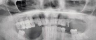
- Chest X-ray. May show enlarged lymph nodes.
- Ultrasound of the abdominal organs. Helps identify enlarged liver and spleen.
Treatment
- Chemotherapy is the use of special drugs that destroy leukemia cells or prevent them from dividing.
- Radiation therapy is the destruction of leukemia cells using ionizing radiation.
- Targeted therapy is the prescription of drugs that have a targeted effect on certain types of malignant cells. These drugs interact with certain proteins on the surface of leukemia cells and cause their destruction.
- Bone marrow transplant - normal bone marrow cells are transplanted into the patient from a suitable donor. A course of chemotherapy or radiation therapy in high doses is first administered to destroy all pathological cells.
- Radiation therapy is the destruction of leukemia cells using ionizing radiation. Can be used in acute lymphocytic leukemia to completely destroy leukemia cells before bone marrow transplantation.
The prognosis for acute lymphoblastic leukemia is favorable, especially in children. Most patients are completely cured. The prognosis of chronic lymphocytic leukemia depends on the rate of disease progression and sensitivity to chemotherapy. The average life expectancy of patients with chronic lymphocytic leukemia is 3-5 years.
Prevention
There is no specific prevention of lymphocytic leukemia. It is necessary to undergo timely preventive examinations, during which blood diseases are often detected.
Recommended tests
- General blood analysis
- Leukocyte formula
- Hemoglobin
- Cytological examination of punctates, scrapings of other organs and tissues
Diagnosis of lymphocytic leukemia
To diagnose lymphocytic leukemia, a clinical blood test is performed (code 2 and code 5). It allows the specialist to obtain information about the ratio of blood elements, their quality and quantity.
Leukocytes in acute lymphocytic leukemia can be elevated, decreased, or normal. To determine the leukocyte formula (the ratio of certain types of leukocytes), a blood smear is used, which is applied to a glass slide, stained with special dyes and examined under a microscope. This is how the doctor determines the ratio of leukocytes and identifies immature, pathological cells that differ from normal ones in appearance.
List of sources
- Savchenko V.G., Parovichnikova E.N. Acute leukemia//Clinical oncohematology: a guide for doctors. Ed. Volkova M.A.. 2nd ed., revised. and additional 2007
- Blindar, Valentina Nikolaevna Algorithm for laboratory diagnosis of acute leukemia. Guide for doctors / Blindar Valentina Nikolaevna. — M.: Medical Information Agency (MIA), 2013
- Gavrilov, O.K. Bone marrow and peripheral blood cells / O.K. Gavrilov, G.I. Kozinets, N.B. Chernyak. — M.: Medicine, 2014
- Davidenkova, E.F. Clinic and genetics of leukemia / E.F. Davidenkova, S.I. Sherman, N.N. Kolosova. — M.: Medicine, 2010
Treatment of lymphocytic leukemia
As a rule, treatment of leukemia is complex. Now there are several effective treatment options - moreover, every year specialists develop new methods for treating lymphocytic leukemia, which are becoming more and more effective. In acute leukemia, the prognosis is favorable: approximately 95% of patients are completely cured. In chronic leukemia, the prognosis depends on the presence of concomitant pathologies and the speed of development of the disease: despite the fact that it is steadily progressing, properly selected treatment can in most cases lead to remission and significantly improve the patient’s condition.
Stages
The following stages of the disease are distinguished.
- Start. It is characterized by a general deterioration in well-being, the appearance of the first nonspecific signs - pallor, fatigue, pain in the muscles and bones.
- The height of Symptoms characteristic of oncopathology are pronounced, the patient’s condition sharply worsens.
- Remission. The patient’s well-being improves, clinical and hematological parameters return to normal.
- Relapse. After remission, the number of blast lymphocytes increases again, and pathological symptoms return.
- Terminal stage. The disease progresses rapidly and vital organs are affected. The most common outcome is death.