- Causes of pulmonary edema
- What diseases are there a risk of edema?
- Types of pathology
- Symptoms of pulmonary edema
- Diagnostics
- Treatment of pulmonary edema
- Predictions and prevention
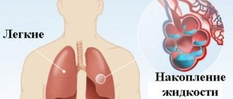
Pulmonary edema is a condition in which gas exchange is disrupted and respiratory failure develops. Swelling occurs due to an imbalance of pressure in the pulmonary capillaries and the release of a large amount of edematous fluid into the lung tissue without its reabsorption.
Causes of pulmonary edema
A mixture of edematous fluid (transudate) and a liquid substance that “lines” the alveoli of the lungs from the inside (it is called “pulmonary surfactant”) enters the lumen of the alveoli, where it combines with air. This leads to the formation of foam, which creates an obstacle to the flow of oxygen. Lack of oxygen provokes the development of shortness of breath, due to which the pressure inside the chest decreases and blood flow to the right side of the heart increases.
Pulmonary edema causes infiltration of the alveoli and is manifested by symptoms that indicate oxygen starvation, which is dangerous for tissues. The patient requires emergency care due to the high risk of death due to suffocation.
Questions and answers
What causes pulmonary edema?
There are dozens of factors that contribute to the development of this pathology. They are associated with other diseases of different body systems. Clinical recommendations for pulmonary edema are largely determined by the situation.
Can pulmonary edema lead to death?
Yes, the risk of death in the case of such a pathology is very high, and for the fulminant form it is the only possible outcome of events. This is an extremely dangerous condition that requires immediate professional help.
How to treat pulmonary edema?
This is done exclusively in medical institutions, most often in intensive care. Only doctors know everything you need to know about the symptoms and treatment of pulmonary edema in adults or children - the pathology cannot be eliminated without special knowledge and skills.
What are the features of pulmonary edema in children?
Unlike adults, this pathology occurs much less frequently in children due to other diseases. The underlying cause is usually allergies or exposure to toxins. Or the cause may be congenital anomalies of various body systems.
What diseases are there a risk of edema?
Pathology indicates a complication of the underlying disease, increases its duration and worsens the prognosis of treatment. The most dangerous from the point of view of pulmonary edema include:
- Heart diseases. Respiratory failure often occurs against the background of pathologies of the heart and blood vessels. Cardiogenic pulmonary edema is a life-threatening complication that develops with myocardial infarction, aneurysm, endocarditis and myocarditis, severe hypertension, and pathological conditions caused by congenital or acquired heart defects.
- Pulmonary diseases. At risk are patients with injuries and severe diseases of the respiratory tract. Respiratory edema develops against the background of exacerbation of chronic bronchitis and bronchial asthma, tuberculosis, and lobar pneumonia. The risk is increased by tumor lesions of the lung tissue, pleurisy, and chest injuries, which are accompanied by pneumothorax. If a person is connected to a ventilator for a long time and receives a mixture with a high concentration of oxygen, this also leads to an imbalance of pressure in the pulmonary capillaries and the release of exudate.
- Infectious diseases. In severe cases or inadequate treatment of whooping cough, scarlet fever, influenza, diphtheria and other infectious diseases, edema develops, followed by acute pulmonary failure. In young children, this clinical syndrome can cause acute laryngitis and the proliferation of adenoids.
- Pathologies of intrauterine development and difficult childbirth. Severe pregnancy, prematurity, oxygen hypoxia, fetal bronchopulmonary dysplasia - these factors increase the risk of developing pulmonary failure in newborns. In pregnant women, pulmonary failure can develop due to eclampsia. The same phenomenon occurs with ovarian hyperstimulation syndrome against the background of stimulation of ovulation by hormonal drugs.
- Foreign bodies entering the respiratory tract. Mechanical asphyxia is one of the possible causes of acute respiratory failure. Pathology occurs when foreign objects enter the respiratory tract, after drowning or attempted hanging, or blockage of the respiratory tract with vomit.
- Diseases of the urinary system and gastrointestinal tract. Nephrogenic pulmonary edema can be complicated by acute inflammatory diseases, which are accompanied by renal dysfunction and the development of severe renal failure. In gastroenterology, patients with liver cirrhosis, severe pancreatitis, and intestinal obstruction have to deal with respiratory failure.
- Neurological diseases. Traumatic brain injuries, meningitis, encephalitis, neoplasms, cerebral hemorrhages, complications after brain surgery - all this leads to neurogenic pulmonary edema.
- Allergies, injuries, poisoning. Acute respiratory failure can be caused by the ingestion of various toxic substances into the body. Pulmonary edema is encountered by patients with alcohol and drug intoxication, poisoning with drugs, salts of heavy metals, and toxic gases. Pulmonary insufficiency is diagnosed in case of blood poisoning, burns with a large affected area, acute allergic reactions.
Hospital treatment
Treatment of the pathology is carried out in a hospital setting and includes the following measures: oxygen therapy, the prescription of diuretics and cardiac glycosides, intravenous administration of albumin, heparin, atropine, aminophylline. In some cases, hormones are prescribed. All prescriptions are strictly individual and are adjusted taking into account the patient’s condition.
The prognosis for treatment of cardiogenic pulmonary edema depends on the type of pathology, severity, presence of concomitant diseases, as well as the quality of medical care provided.
Types of pathology
Depending on the cause of development, pulmonary edema can be cardiogenic or non-cardiogenic. In the first case, the cause of respiratory failure is cardiovascular disease. The second category includes all other pulmonary edema caused by diseases of the gastrointestinal tract, nervous system, and injuries.
Pulmonary edema is also classified according to the nature of its course. There are four types of pathology development:
- With fulminant development, severe respiratory failure occurs within a few minutes and leaves no chance of survival.
- Acute swelling can take up to four hours to develop. This form is considered less dangerous than the previous one, but in the absence of timely assistance, the risk of death is extremely high. Most often, acute pulmonary edema develops with myocardial infarction, traumatic brain injury, and acute allergic reactions.
- In case of intoxication, renal and liver failure, subacute pulmonary edema may occur. The intensity of symptoms is constantly changing, so continuous medical monitoring of the patient's condition is required.
- Prolonged swelling may take several days to develop. Symptoms are mild compared to the acute and subacute forms. Prolonged pulmonary edema usually occurs against the background of various chronic diseases, including heart failure.
Symptoms
It is possible to describe the symptoms of pulmonary edema only in general terms, since certain types of pathology occur with blurred characteristics. Signs include the following:
- severe weakness;
- shallow, very rapid breathing;
- dry cough;
- dry wheezing;
- severe shortness of breath;
- puffiness of the face and neck;
- bubbling breathing and moist wheezing;
- foam at the mouth with a pink tint;
- lethargy, confusion;
- shallow breathing;
- thready pulse.
Some signs of pulmonary edema contradict each other for the reason that everything can start with one condition and end with another. For example, rapid breathing occurs for several minutes or hours, and then it weakens. In the fastest and most dangerous forms of edema, the patient's death occurs from suffocation (asphyxia).
Are you experiencing symptoms of pulmonary edema?
Only a doctor can accurately diagnose the disease. Don't delay your consultation - call
Symptoms of pulmonary edema
Before the main symptoms of pulmonary failure appear, patients experience weakness and dizziness, a feeling of tightness in the chest, headache, and dry cough. A few hours after the first symptoms of the pathology, an attack of cardiac asthma develops - the first stage of pulmonary edema. It manifests itself:
- severe lack of air;
- severe dry cough;
- increased heart rate;
- hoarse breathing;
- profuse sweating;
- pale skin, blueness of nails and lips;
- feelings of fear and anxiety.
As a rule, the attack occurs at night or before dawn. In patients with cardiac diseases, cardiac asthma can be caused by physical or emotional stress and hypothermia.
As edema develops, the alveoli become involved in the pathological process. This leads to increased suffocation. The patient experiences severe shortness of breath, the face becomes burgundy-blue and puffy, the veins in the neck swell, and mental retardation appears. Breathing becomes hoarse, foam mixed with blood comes out of the mouth.
In the absence of medical care, the patient loses consciousness and falls into a coma. In this state, blood pressure decreases critically, breathing becomes shallow, and the pulse weakens. The cause of human death is asphyxia, in which the body experiences an acute lack of oxygen with an excess of carbon dioxide.
Clinical manifestations in adults
Pathology does not always develop rapidly. In some cases, it is preceded by weakness, dizziness, chest tightness, dry cough, headache and tachypnea. These symptoms can appear a few minutes or several hours before swelling develops.
Primary manifestations: compressive chest pain, increasing tachycardia, dry wheezing turning into wet wheezing, increased breathing.
Signs of progression of pathology:
- shallow breathing;
- bubbling wet rales;
- earthy skin color;
- increased suffocation;
- cold sweat;
- the appearance of foamy sputum with a pink tint from the mouth.
On our website Dobrobut.com you can learn more about pathology and, if necessary, make an appointment with a specialist. The doctor will answer questions and tell you the difference between interstitial and alveolar pulmonary edema.
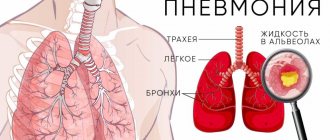
Diagnostics
A doctor can determine the development of pulmonary edema by numerous external symptoms. In addition, the patient is prescribed instrumental and laboratory examinations. Among them:
- measurement of blood oxygen levels, determination of carbon dioxide content;
- blood chemistry;
- electrocardiogram and ultrasound of the heart;
- chest x-ray to evaluate the condition of the lungs.
Inserting a catheter into the pulmonary artery allows you to determine the type of edema: cardiogenic or non-cardiogenic.
If pulmonary edema is suspected, it is important to carry out the necessary tests and examinations as soon as possible, identify the cause and type of pathology, and begin treatment. Saving the patient's life directly depends on this.
Diagnostic methods in Medscan
The doctor determines the pathological condition using physical methods - percussion and auscultation. With increasing edema, a phonendoscope can be used to hear wheezing over the entire surface of the lungs.
The Medscan diagnostic center uses the following methods to determine excess fluid in the lungs due to oncology:
- X-ray of the chest cavity. The examination will show a significant amount of fluid.
- CT, MRI are more accurate methods. They allow you to assess the condition of the lung tissues and identify the affected lymph nodes near the bronchi or in the mediastinum.
- Immunohistochemical analysis. It is carried out to study the pleural effusion in detail. Immunohistochemistry helps identify and characterize malignant cells based on the interaction of the immune complex of antibodies and antigens.
- Thoracoscopy is a method that allows you to examine the chest using a special tube (thoracoscope). In combination with an immunohistochemical study, it diagnoses a malignant neoplasm in almost 100% of cases.
The Medscan diagnostic center has expert-level equipment. We offer our patients modern and highly accurate laboratory and instrumental diagnostic methods.
Treatment of pulmonary edema
People with acute respiratory failure are treated in the intensive care unit. First aid consists of measures that reduce venous return to the heart. To do this, the patient must take a semi-sitting position. Applying tourniquets to the limbs and hot foot baths have a positive effect.
It is imperative to monitor blood oxygen saturation levels. Oxygen therapy is used to compensate for its deficiency. If pulmonary edema has developed against the background of mechanical asphyxia, it is necessary to clear the airways of foreign objects. If it is impossible to breathe independently, the patient is connected to a ventilator.
A combination of medications is used to eliminate pulmonary edema. Among them:
- morphine and other narcotic analgesics;
- drugs to stimulate diuresis;
- nitroglycerine;
- drugs to lower blood pressure.
Depending on the underlying disease, patients with pulmonary edema are prescribed antihistamines, antibiotics, hormonal and antiarrhythmic drugs, and cardiac glycosides. The task of the resuscitator is to stop the attack and prevent acute hypoxia, which leads to cell death and death of the patient. After the symptoms of respiratory failure are relieved, treatment of the disease that caused it begins.
It is very important to eliminate the root cause that led to the clinical syndrome. Otherwise, a relapse of the disease is possible.
Classification of the disease
Depending on the cause, the following types of pulmonary edema in a patient can be distinguished:
- Membranous. Occurs as a result of toxic effects on capillary and alveolar walls, which are subsequently destroyed.
- Hydrostatic. Formed when intravascular hydrostatic pressure increases. The cause is cardiovascular failure.
Forms of complications of pulmonary edema:
- Interstitial. Excellent treatment. However, untimely medical care provokes its transition to the alveolar stage.
- Alveolar. The most dangerous. Its consequences are the death of the patient.
Classification by severity of symptoms:
- First or pre-edema. It is characterized by a disorder of the rhythm and frequency of breathing, and the presence of slight shortness of breath.
- Second. Shortness of breath intensifies, wheezing appears.
- Third. Symptoms increase: wheezing and shortness of breath are heard at a distance from the patient.
- Fourth. All the characteristic features of pulmonary edema are present.
Predictions and prevention
Pulmonary edema is a deadly condition; Mortality with this clinical syndrome is up to 50%. Patients with anaphylactic shock most often develop fulminant or acute pulmonary edema, which in more than 90% leads to death.
The consequences of acute pulmonary failure are no less serious than the disease itself. Many patients who have suffered pulmonary edema experience ischemic damage to internal organs, the appearance of areas of pneumosclerosis, and congestive pneumonia. This affects the functioning of the respiratory system, in the organs of which irreversible changes occur. The pathology is manifested by shortness of breath, chest pain, and cough. Pneumosclerosis and other consequences of pulmonary edema require long-term treatment, can lead to disability and shorten the patient's life expectancy.
The likelihood of a favorable prognosis and outcome of the disease with pulmonary edema increases if the diagnosis was made in a timely manner and doctors began to stop the attack at the initial stage.
Main complications after emergency care
Such complications include:
- development of a fulminant form of edema;
- intensive foam production can cause airway obstruction;
- depression (depression) of breathing;
- tachyarrhythmia, asystole;
- angiotic pain. Such pain is characterized by an unbearable pain syndrome; the patient may experience pain shock, worsening the prognosis;
- impossibility of stabilizing blood pressure. Pulmonary edema often occurs against a background of low and high blood pressure, which can alternate within a large amplitude. The vessels will not be able to withstand such a load for long and the patient’s condition worsens;
- increase in pulmonary edema against the background of high blood pressure.