Cervical osteochondrosis is a disease that affects the vertebrae and intervertebral discs. Cervical osteochondrosis refers to deforming dorsopathies. Involutive changes in the discs are observed as early as 20 years of age. At the same time, they become more sensitive to stress, less elastic, and lose lubricating fluid.
Most often, the pathology occurs in the elderly, but currently there is a significant increase in incidence among children and young people. Neurologists at the Yusupov Hospital identify cervical osteochondrosis using the latest diagnostic tests. After clarifying the diagnosis, complex therapy is carried out with the most effective medications, physiotherapeutic procedures and innovative methods of physical rehabilitation.
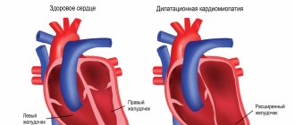
The name of the disease consists of two Greek terms “osteon” (bone) and “chondros” (cartilage). Cervical osteochondrosis begins with changes in the central part of the disc. The intervertebral disc loses moisture and decreases in size, this leads to the convergence of the vertebral bodies and pinching of the nerve roots and blood vessels. The vertebrae receive nutrients from surrounding tissues, which causes harm to the body. Compression of nerves and blood vessels leads to a protective muscle spasm, which, as the disease progresses, becomes a cause of pain.
Which doctor treats this disease?
At the Yusupov Hospital, the treatment of osteochondrosis is the field of activity of neurologists. However, if symptoms of neck osteochondrosis appear, you may contact a general practitioner. A neurologist will select medications for cervical osteochondrosis that have the least burden on the body, which is important during drug therapy.
To determine the presence of a pathological process in cartilage tissue and cervicobrachial osteochondrosis, the patient is sent for a comprehensive examination to the diagnostic center of the Yusupov Hospital. Tactics on how to treat cervical osteochondrosis are being developed in accordance with research results.
Interdisciplinary collaboration also makes it possible to treat the patient's comorbidities. In addition, when contacting the Yusupov Hospital, the patient receives full information support: a treatment plan, an extract on the cost of services, information about consultations with specialists and diagnostic measures.
Make an appointment
What is the danger of cervical osteochondrosis?
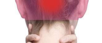
Vital arteries and capillaries are located in the cervical areas. There are complaints of headaches and shoulder pain, chronic fatigue and decreased performance. These signs should not be ignored. Treatment not started on time is often accompanied by many unpleasant syndromes:
- lesions of the spine: pinched nerve, resulting in the appearance of hernias, protrusions of a chronic nature;
- disturbances in the functioning of the vegetative-vascular system: high blood pressure, rapid heartbeat, tachycardia;
- hypertension and hypotension: headaches, dizziness, nervous system disorders;
- disturbances of the vestibular apparatus: deterioration of visual and auditory functions, disorientation in space, noise and darkening in the eyes, nausea and vomiting;
- laryngeal syndrome and voice changes;
- endocrine pathologies.
To avoid irreversible consequences, you need to consult a doctor as soon as possible. In the neurosurgical department of Dr. Zavalishin E.E. They use modern methods of diagnosis and treatment of such dangerous diseases as osteochondrosis and intervertebral hernia.
Causes
Cervical osteochondrosis develops under the influence of various provoking factors. No specific cause of cervical osteochondrosis has been identified. Often the disease is associated with metabolic disorders and aging of the vertebrae.
Researchers suggest that cervical osteochondrosis develops for the following reasons:
- Excessive load on the spine. A high load on the spine is observed when wearing incorrect shoes, flat feet, obesity, and prolonged sitting;
- Metabolic disorders. Deficiency of vitamins, minerals, and calcium metabolism disorders can cause degenerative processes in the vertebrae;
- Congenital and acquired anomalies of the development of the spine and ligamentous apparatus (thickening of the ligaments, lumbarization, sacralization);
- Pathologies of the gastrointestinal tract leading to insufficient absorption of nutrients;
- Infections, intoxication;
- Injuries, bruises, spinal fractures, as a result of which the blood supply and innervation of the spinal column are disrupted, which causes their degenerative disorders;
- Stress;
- Wearing shoes with heels;
- Pregnancy, especially multiple pregnancy;
- Autoimmune connective tissue lesions, pathological structure of collagen types 1 and 2;
- Occupational hazards (lifting heavy loads, prolonged vibration, working in a sitting position with constant head tilt);
- Atherosclerotic and other changes in the vertebral arteries;
- Curvature of the spine (kyphosis, scoliosis, kyphoscoliosis).
An important risk factor for the development of cervical osteochondrosis is family history. This fact proves the presence of osteochondrosis in children when the spine is not yet overloaded.
Symptoms of cervical osteochondrosis in women
As a rule, symptoms of cervical osteochondrosis in women appear 5-10 years later than in men. First of all, this is due to the later onset of menopause and anatomical features (including a lighter bone structure).
Before the mass distribution of computers and the demand for “sedentary” professions, the average age of onset of symptoms of cervical osteochondrosis in women ranged from 50 to 55 years. But now the disease has “rejuvenated” to 40-45 years.
Unlike a similar problem in men, symptoms of cervical osteochondrosis in women often include:
- sudden surges in pressure, especially due to the weather;
- pallor of the face, blue discoloration of the lips and skin;
- decreased sensitivity, up to numbness, of the skin of the face and cervical-collar area, which is accompanied by tingling or goosebumps;
- a feeling of nausea, which is especially intensified in stressful situations.
It should be remembered that cervical osteochondrosis is a disease of the whole body, so it can negatively affect conception and the course of pregnancy. Also, symptoms of cervical osteochondrosis in women can cause ovarian dysfunction and disruption of the monthly cycle.
Degrees
Thanks to the special structure of the spine, it is able to perform its functions. The main structural unit is considered to be the spinal motion segment (SMS). It consists of two adjacent vertebrae, an intervertebral disc and a muscular-ligamentous apparatus. Osteochondrosis leads to dystrophic-degenerative processes, first in the intervertebral disc, then in the vertebra. When one vertebra is damaged, its functions are provided by adjacent ones. This leads to increased load and loss of mobility of the affected segment.
Doctors distinguish several stages in the development of cervical osteochondrosis:
- First degree of cervical osteochondrosis. Since the intervertebral disc is deprived of its own blood supply and receives nutrients from surrounding tissues, it is susceptible to degenerative changes. Osteochondrosis at the 1st stage of development is characterized by destruction of the nucleus pulposus and cracks in the fibrous ring. Clinically, this is manifested by acute or persistent local pain in the neck (cervicalgia) and stiffness;
- Osteochondrosis of the second degree of the cervical spine. At this stage, the destruction of the fibrous ring continues, pathological mobility and instability of the vertebrae appear. Patients complain of pain in the neck, aggravated by physical activity, tilting the head or in a certain position;
- The third stage of the disease is characterized by complete destruction of the fibrous ring. The nucleus pulposus is not fixed. Intervertebral hernias may occur, which cause severe pain. At this stage, due to poor fixation of the SMS, spinal curvature may form;
- At the fourth stage of the disease, the intervertebral disc is replaced by connective tissue, and other adjacent segments are affected. Spondyloarthrosis and arachnoiditis develop. The joints become completely immobile - ankylosis develops. Bone tissue grows around the affected area - osteon is formed. With the fourth degree of cervical osteochondrosis, clear symptoms are observed: severe pain that radiates to the arm, sternum, to the area between the shoulder blades, sensitivity disorders.
Symptoms of cervical osteochondrosis in men
Symptoms of cervical osteochondrosis in men usually appear at the age of 40-45 years. Patients often notice them during physical activity (for example, working out in the gym or lifting weights). Delayed self-diagnosis of symptoms of cervical osteochondrosis in men is associated not only with a reluctance to go to the doctor, but also with physiological prerequisites. Men have stronger and thicker vertebrae than women, and the vessels are stronger. Therefore, they may notice discomfort only when changes have already affected the intervertebral discs. Characteristic symptoms of cervical osteochondrosis in men include:
- decreased overall strength and endurance;
- deterioration of libido and erectile dysfunction;
- irritability, sometimes neuroses;
- deterioration of motor skills (“awkward fingers”);
- headaches that do not go away after taking analgesics.
Vertebral hernias with symptoms of cervical osteochondrosis are more common in men than in women.
Symptoms and signs
Signs of cervical osteochondrosis in the initial stages may be nonspecific: dizziness, headaches, weakness, crunching when moving the head. As the disease progresses, the following symptoms develop:
- Severe pain in the neck and shoulders;
- Numbness of the hand;
- Dizziness;
- Increased blood pressure;
- Impaired coordination of movements;
- Increased sweating.
There are several syndromes that appear with the development of a pathological condition of the muscles of the back and cervical spine:
- Cervical migraine syndrome.
- Vertebral artery syndrome.
- Hypertension syndrome.
- Cardiac syndrome.
- Radicular syndrome.
They occur when nerve endings are injured, arteries and veins are compressed during the development of the disease. The most dangerous complication is considered to be vertebral artery syndrome. There is a disruption of blood flow through the artery supplying the brain and spinal cord. The patient's hearing decreases, vision decreases, and constant dizziness develops. The patient may lose consciousness while moving due to a sudden disruption of blood flow.
As a result of compression of the nerves responsible for the innervation of the muscles of the chest and diaphragm, pain appears in the heart area, not associated with heart disease, but at the same time tachycardia, arrhythmia and hypotension may develop. Compression of the veins leads to the development of hypertensive liquor syndrome. Intracranial pressure increases, nausea, vomiting, and severe headache appear due to impaired blood flow from the brain.
As a result of compression of the neck, radicular syndrome develops - severe pain appears in the neck, shoulders, shoulder blades, and back of the head. With this syndrome, the arms and neck area become numb. With cervical migraine syndrome, the patient experiences severe pain in the back of the head, which is often accompanied by nausea and vomiting.
Reflex syndromes occur when the spinal roots are not yet affected. Patients complain of pain in the neck, head (especially the back of the head), and arms on one or both sides. Reflex pain, unlike radicular pain, is not combined with sensory disturbances. Cervicalgia can be dull and aching. Acute sharp “shoots” of pain are called cervicago. There is muscle spasm and pain, pain in the paravertebral points. Signs of cervical osteochondrosis intensify in an uncomfortable position, when tilting the head, coughing, or physical activity. Signs of epicondylosis, glenohumeral periarthrosis and shoulder-hand syndrome appear due to nerve impulses from the annulus fibrosus of the affected segment, which causes compensatory muscle spasm.
Radicular syndromes are accompanied by impaired motor activity and sensitivity. In this case, nerves and blood vessels are infringed, venous and lymphatic outflow in the pathological focus is disrupted as a result of a decrease in the intervertebral canal. The pain with radicular syndrome is acute and intense. A common cause of pinched spinal nerves is the formation of a hernia. In the area of the pathological focus, muscle tone decreases. With radiculoischemia, in addition to nerves, blood vessels are compressed.
If the phrenic nerve is involved in the pathological process, cardiac syndrome occurs. It manifests itself as a burning, acute pain in the left half of the chest with radiation to the arm and interscapular region. The name of the syndrome is due to the fact that the nature of the pain is similar to an attack of angina. The main difference between pain during angina pectoris is that it is relieved after taking nitroglycerin, can occur at rest and is combined with interruptions in heart rhythm (tachycardia, arrhythmia).
Signs of cervical osteochondrosis depend on the location of the pathological process. When the upper cervical vertebrae are affected, the blood supply to the brain is disrupted due to compression of the cerebral arteries. This leads to headaches (especially in the occipital region), dizziness, fainting, and high blood pressure. Dizziness with cervical osteochondrosis is caused by a decrease in blood flow to the inner ear. Patients also experience nausea and vestibular and ocular symptoms.
With combined damage to the vertebrae, they speak of cervicothoracic osteochondrosis. The disease is manifested by the following symptoms:
- Dizziness;
- Pain in the neck and arm;
- Tingling, crawling sensation on the upper limb;
- Intercostal neuralgia.
Make an appointment
Forecast
If you ignore the first manifestations of cervical osteochondrosis and, without sparing the spine, continue to lead the same lifestyle, the disease can progress2, causing various disorders. Among them are vertebral artery syndrome2.
The vertebral arteries pass through openings in the transverse processes of the cervical vertebrae and provide blood supply to the brain5. With osteochondrosis, the vertebral artery may become pinched in its orifice; sometimes it spasms in response to irritation, which often impairs cerebral circulation2,5.
A typical manifestation of vertebral artery syndrome is a burning, throbbing headache2 that:
- is felt in the back of the head and “gives” to the temporal, frontal and parietal regions5;
- usually occurs or worsens when turning the head or throwing it back5;
- accompanied by other unpleasant symptoms - redness or pallor of the face, nausea, dizziness and tinnitus5.
to come back to the beginning
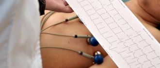
Diagnostics
Cervical osteochondrosis is a chronic disease that can lead to the formation of hernias and compression of the spinal cord. Therefore, it is important to establish an accurate diagnosis in a timely manner and begin therapy. To identify cervical osteochondrosis, the following types of instrumental diagnostics are used:
- Spondylography or radiography of the spine. This research method is painless, highly informative and does not require special training. An X-ray of the spine allows you to evaluate its anatomical and functional features. In the picture, attention is paid to the structure of the vertebrae, their relationship to each other, the distance between them, the lumen of the spinal canal;
- Computed tomography - provides information mainly about the condition of bone tissue, allows you to identify narrowing of the spinal canal and disc herniation;
- Magnetic resonance imaging - allows you to determine changes in soft tissues. The MRI image clearly shows changes in the intervertebral discs and spinal cord.
At the Yusupov Hospital, the patient undergoes a comprehensive examination. Doctors take into account the individual characteristics of his body and concomitant diseases. An important advantage of the neurology clinic is the availability of modern, high-quality equipment and specialized specialists: neurologists, neurosurgeons, oncologists.
Diagnosis of osteochondrosis of the cervical spine
If you experience one of the symptoms listed above, it is advisable to immediately seek medical help. The sooner a problem is diagnosed, the higher the likelihood of it being completely eliminated. A neurologist is involved in the diagnosis and treatment of cervicothoracic osteochondrosis of the spine.
After the patient contacts, the neurologist will interview the patient and, based on his complaints, make a conclusion about the presence of the disease. Most often, patients complain of pain, discomfort, and problems with movement of the upper limbs. After the interview, the doctor will conduct a visual examination of the patient. Will be assessed:
- Correct posture;
- At what angle are the shoulder blades, shoulders and ilia bones located?
- The location of the line of the spinous processes along the length of the spine.
A visual examination allows you to determine what pathological processes began in the spine and how much they affected the spine and surrounding tissues.
The final diagnosis is not made solely on the basis of examination and questioning of the patient. Additional examinations are required. Most often, the patient is sent for radiography and computed tomography. If for one reason or another it is impossible to make a final diagnosis, the patient is referred for magnetic resonance imaging. Additional examinations include Doppler sonography; the procedure allows you to find out the state of the circulatory system in the cervical region.
Drug treatment
Treatment of osteochondrosis of the cervical spine consists of drug and non-drug therapy. Even after complete recovery, to exclude relapses of the disease, neurologists at the Yusupov Hospital carry out preventive measures. In the acute period, for the treatment of cervical osteochondrosis, doctors prescribe medications to patients from the following pharmacological groups:
- Non-narcotic analgesics (analgin, baralgin, trigan). They are taken orally or administered intramuscularly to quickly achieve an effect;
- Nonsteroidal anti-inflammatory drugs (diclofenac, ibuprofen, ketanol);
- B vitamins in large doses.
In order to reduce fluid retention in the area of the spinal root and surrounding tissues, diuretics (furosemide, Triampur, Lasix) are used. Antihistamines (diphenhydramine, suprastin, pipolfen) potentiate the effect of analgesics. Muscle spasms are eliminated by muscle relaxants (sirdalud, miorix, mydocalm, flexen). For prolonged severe pain, neurologists perform a nerve block.
To improve metabolic processes in the intervertebral disc, chondroprotectors (alflutop, evalar) are used. These drugs increase the content of glycosaminoglycans, increase the firmness, elasticity and shock absorption of the intervertebral discs.
Anti-dizziness pills
Patients often experience dizziness with cervical osteochondrosis. To reduce them, doctors prescribe non-steroidal anti-inflammatory drugs. NSAIDs belonging to different groups differ in their mechanism of action and effect, so only a qualified specialist can determine the appropriate drug.
It is important to remember that medications for cervical osteochondrosis cannot be taken without a doctor’s prescription. Nonsteroidal anti-inflammatory drugs have side effects, so before prescribing them, the neurologist determines the presence of contraindications in the patient and the required dosage. Drugs for dizziness in cervical osteochondrosis can improve the patient’s quality of life.
Injections for osteochondrosis
Injections for osteochondrosis of the cervical spine help relieve pain during an exacerbation. With this method of drug administration, the effect occurs quickly. Neurologists use various injections.
Nurses administer drug solutions subcutaneously, intramuscularly or intravenously. During the period of exacerbation of the disease, drugs that are administered by injection for cervical osteochondrosis have an exclusively symptomatic effect. To treat the disease, contact the neurology clinic of the Yusupov Hospital.
Make an appointment
Headache treatment
Headache is a symptom that occurs with various disorders. However, cervical osteochondrosis is characterized by attacks of intense headaches. Head movements increase symptoms, so to eliminate them, doctors prescribe analgesic tablets and non-steroidal anti-inflammatory drugs.
Physiotherapy
Physiotherapeutic procedures pursue a number of goals:
- Localization of the inflammatory process;
- Relieving muscle spasm;
- Pain relief;
- Launch of regenerative processes;
- Increasing general and local immunity;
- Restoring the normal position of nerve fibers, eliminating compression and pinching.
Most often, the following procedures are prescribed for cervical osteochondrosis:
- Shock wave therapy. Using a special device, the acoustic wave is directed directly to the cartilage tissue of the spine that has been damaged. As a result, metabolic processes are launched, salt and calcium deposits are destroyed, which interfere with the normal movement of joints and vertebrae. The procedure is characterized by a cumulative effect, often the first results become noticeable only 2-3 months after the start of treatment.
- Acupuncture. Acupuncture is often used to treat and prevent cervical osteochondrosis. It is important that the procedure is performed only by a qualified doctor, otherwise you may not only experience a lack of effect, but also a worsening of the current condition. The essence of the procedure is that special needles are installed on biologically active points, forcing the body to start metabolic processes and stimulate the production of natural painkillers.
- Massage. The main goal is to reduce pain and improve blood circulation in the damaged area of the cervical spine. With proper massage, the muscles acquire the lost tone, and as a result, it is possible to eliminate the risk of relapse of osteochondrosis in the future. When attending the first massage sessions, the patient encounters severe pain; it is important not to stop treatment due to pain, but to go through all the procedures prescribed by the doctor.
Surgery
It is mainly prescribed in advanced stages of the disease, when the use of medications and visits to physiotherapeutic procedures does not bring any results. The indication for surgical intervention is catastrophic narrowing of the spinal canal.
Modern surgical techniques allow the patient to be discharged from the hospital within 3-5 days and begin outpatient treatment of the symptoms of cervicothoracic osteochondrosis. Over the next three months, the patient undergoes rehabilitation.
Physiotherapy
A correctly chosen set of exercises for osteochondrosis can not only improve the patient’s general condition, but also speed up the process of treating the disease. There are several effective exercises:
- Turns and tilts the head in different directions. The exercise is performed in a sitting position, it is important not to jerk, all movements should be smooth with a gradual increase in the number of repetitions and the amplitude of the inclination.
- Tilt the head to the sides with resistance. Body position - sitting at the table, one elbow stands on the table, while the palm presses on the temple. Tilt your head towards your hand, creating slight resistance.
- Shoulder lift. Raise your shoulders as high as possible and hold in this position for a while.
- Self-kneading of the back of the head and neck with your fingertips. It is important that the movements are soft and do not cause pain. You can perform self-massage in any comfortable position.
It is important not to treat cervical osteochondrosis at home without consulting a neurologist; a set of exercises must be agreed upon with the attending physician.
Non-drug therapies
Complex non-drug therapy for cervical osteochondrosis of the spine includes:
- Protective mode - if the roots are pinched, patients lie on a hard surface,
- Massage;
- Physical therapy;
- Spinal traction;
- Physiotherapeutic procedures.
Massage for cervical osteochondrosis is used to reduce pain and swelling, improve peripheral blood supply, and eliminate muscle spasms. A contraindication to performing this procedure is the presence of acute pain. Massage the neck and back in the direction of lymph outflow. Particular attention is paid to the interscapular and paravertebral zones.
Therapeutic exercises for osteochondrosis of the cervical spine are aimed at eliminating muscle spasms and strengthening the muscular frame. Since instability of the vertebrae often occurs in the cervical spine, the exercise therapy instructor conducts individual classes, during which he teaches the patient how to safely perform exercises. Some authors recommend conducting physical therapy classes in a Shants collar.
To improve the mobility of the cervical vertebrae, rehabilitation experts recommend performing the following exercises:
- Flexion and extension of the neck. Bend your head forward toward your sternum without pulling your shoulders forward and then back. Hold the incline for 3 seconds, repeat each exercise 8-10 times;
- Neck turns. Turn your neck first to the left until it stops, then to the right, without changing the position of your shoulders and the level of your chin;
- Lower your head all the way down. Then tilt your head back without changing the level of your shoulders. Hold the position for 5 seconds.
The following exercises have been developed to strengthen the neck muscles:
- Place your hand on the back of your head. Tilt your head back, resting on your hand;
- Place your hand in the temporal region. While tilting your head, resist with your hand;
- Place your hand on your forehead, resisting it, tilt your head forward;
- With your right hand, tilt your head to the side, your left hand should be behind your back. Repeat the exercise on the other hand.
Autogravity therapy is the exact name for the spinal traction procedure. It is carried out using special devices. The goal of therapy is to reduce muscle spasm and restore the correct position of the vertebrae. To avoid complications, spinal traction is performed by a doctor.
To improve blood supply to the pathological focus, relieve swelling and eliminate pain, the following physiotherapeutic procedures are used in the Yusupov Hospital:
- Diadynamic currents. During this procedure, low-frequency currents are applied using a special device, which stimulate the muscles, relieve spasm and pain. They have a positive effect by improving tissue trophism;
- Ultraviolet irradiation. Under the influence of UV radiation, vitamin D metabolism improves, calcium content increases, bone tissue becomes stronger;
- Exposure to ultrasound - used to accelerate blood flow, antispasmodic and reparative effects. Ultrasound is capable of penetrating deep into tissues; sometimes it is used for better absorption of medicinal substances;
- Amplipulse therapy - allows you to relieve pain by blocking nerve impulses from the source of pain.
In the acute period of the disease, which lasts 4-7 days, painkillers, antispasmodics, and irritants are used to reduce pain. The patient is provided with rest. Immobilization of the cervical spine is carried out using a Shants collar. Exercise therapy and massage are contraindicated. Ultraviolet radiation is used.
The duration of the subacute period is 29 days. After complete recovery, the patient should rest for several days. Then you can begin a course of rehabilitation therapy. In the chronic course of the disease, the patient is prescribed muscle relaxants, chondroprotectors, B vitamins, and for pain - analgesics, NSAIDs. Physical therapy classes and massages are provided. The patient is given physiotherapeutic procedures (amplipulse, alternating current exposure), and spinal traction is performed.
At the Yusupov Clinic, doctors have extensive experience in successfully treating cervical osteochondrosis. Patients are given the opportunity to undergo a full course of rehabilitation: physiotherapeutic procedures, massage, spinal traction. The neurology clinic employs specialists of the highest medical category, professors, who use proprietary methods for the treatment of osteochondrosis.
Treatment and prevention of symptoms of cervical osteochondrosis
Comprehensive treatment of the symptoms of cervical osteochondrosis includes drug therapy, exercise therapy, physiotherapy, massage and maintaining a healthy diet.
Please note that at the 1st, preclinical, stage, a complete cure for cervical osteochondrosis is possible! This requires a change in lifestyle to a more active one, daily therapeutic exercises, and a balanced diet.
In advanced cases, surgical treatment may be required (if a vertebra is displaced, a hernia forms, or a severe narrowing of the lumen of the spinal column).
Massage for cervical osteochondrosis
Massage of the cervical-collar area is an excellent way to prevent the development of cervical osteochondrosis and protect against its exacerbations. It helps relieve pain, strengthens muscles and improves tissue trophism (after a session, patients often “clear their heads”). Some massage techniques are only available to qualified professionals. They should not be repeated at home, since the neck contains a huge number of nerve bundles and blood vessels. Clamping them is fraught with loss of consciousness, the development of inflammation in the nerve endings, and disruption of brain trophism.
But a simple “household” massage with warming and pain-relieving ointments, creams and balms can be performed at home. It helps relax muscles and eliminate “tightness”
Remember that if there is inflammation (swelling, increased sensitivity of the skin, local increase in temperature), massage is strictly prohibited! It will only worsen the patient's condition! Also, “home” massage is contraindicated for hypertonicity of the muscles of the cervical-collar area. In this case, you should contact a specialist or start with special therapeutic exercises.
Classic massage techniques for symptoms of cervical osteochondrosis include:
- stroking the skin from the back of the head to the subclavian area to warm the skin and relax the muscles;
- squeezing - performed by a kind of “grab” between the thumb and index finger. In this case, it is necessary to clasp the neck, gently pressing on the muscles and stretching them;
- rubbing - affects the skin and muscles, warming them up, relaxes the so-called. “protective” tension, restores impaired blood circulation;
- kneading - affects the deep layers of tissue, so it must be performed strictly by a specialist.
Please note that massage techniques, intensity and localization of massage effects vary depending on the stage of cervical osteochondrosis. If the patient is bothered by pain on only one side, the massage is performed symmetrically, starting with the healthy half. Before a neck massage, it is advisable to warm up the entire back, since the position and nutrition of the vertebrae with osteochondrosis can be disrupted according to the domino principle.
Therapeutic exercises for cervical osteochondrosis
Therapeutic exercises for cervical osteochondrosis can be performed at home and at work. An important condition: the entire set of exercises should be done daily, ideally 3-4 times a day. If you spend a lot of time at the computer or in one position, every 1.5-2 hours you should take a 5-minute break to warm up.
Exercise therapy for cervical osteochondrosis helps strengthen the neck muscles, eliminate tension, and restore blood circulation.
- Sit straight on a chair. Without sudden movements, turn your head left and right, making a 180° turn.
- Lower your head down, trying to reach your chin to your chest, but without pulling your shoulders forward.
- Tuck your chin in, trying to bring it behind your chest line.
- Sit at the table and place your elbows on it. Place your palms on your temples and turn your head, overcoming moderate resistance from your own hands. Repeat for head tilts (with your palm on your forehead).
- Pull your shoulders toward your earlobes and then lower them.
- Do a self-massage of the back of your head.
For patients with cervical osteochondrosis, water activities are recommended: swimming, water aerobics, hydromassage and others.
In addition to therapeutic exercises, patients with symptoms of cervical osteochondrosis are advised to wear special orthoses-collars, which help reduce compression of nerve endings, serve as the prevention of hernias, and combat sore throat and trophic disorders.
Physiotherapy for symptoms of cervical osteochondrosis
Physiotherapy for symptoms of cervical osteochondrosis in women and men is carried out in courses several times a year. It allows you to slow down the progression of the disease and its complications, and relieve symptoms.
For pathology of the cervical spine it is recommended:
- magnetic therapy;
- laser therapy;
- shock wave therapy;
- mud therapy and balneotherapy;
- hydromassage;
- manual therapy;
- traction therapy;
- mechanotherapy.
Medicines for cervical osteochondrosis
To relieve symptoms of cervical osteochondrosis in women and men, the following groups of drugs are used:
- Non-steroidal anti-inflammatory drugs (Nimesil, Etoricoxib) - help relieve pain, inflammation and swelling, but are taken in symptomatic courses (about 10-12 days) and do not affect the causes of the disease.
- Glucocorticoids (Prednisolone, Diprospan) are indicated to eliminate pain and decompress nerve roots in severe cases when simple analgesics and NSAIDs are ineffective. They have significant side effects and should be used strictly as prescribed by your doctor.
- Blood microcirculation correctors, angioprotectors (Pentoxifylline, Actovegin) - help improve blood circulation and protect brain vessels from damage.
- Chondroprotectors (Artracam) - affect the very cause of the disease (deterioration in the quality of intervertebral discs). This group of drugs promotes the structural normalization of cartilage tissue, its rapid regeneration and normal nutrition. Of all the medications, only chondroprotectors are truly capable of slowing down the destruction of cartilage and improving their shock-absorbing qualities (all others only relieve symptoms).
Nutrition
Proper nutrition for osteochondrosis is an important condition for achieving remission. The progression of cervicothoracic osteochondrosis stops with diet and therapeutic measures. Neurologists at the Yusupov Hospital know how to treat osteochondrosis of the cervical spine, so they create a complex of treatment measures, including procedures, exercise therapy, proper nutrition and lifestyle changes.
Many patients turn to neurologists with the question of how to treat osteochondrosis of the cervical spine and whether there are any dietary restrictions. Specialists at the Yusupov Hospital create individual nutrition programs that take into account the patient’s preferences. The diet for osteochondrosis is based on balanced, low-fat foods that are rich in nutrients. The patient's daily diet includes foods high in calcium.
Features of the pathology
Of all the parts of the human body, the neck is considered the most mobile. The cervical spine includes 7 individual vertebrae. Between these vertebrae there are elastic discs, which become damaged and deformed when osteochondrosis develops . As a result, the distance between the vertebrae gradually decreases, causing pinched nerves. The main reason for the development of pathology is a violation of metabolic processes in the cervical spine.
Osteochondrosis of the cervical spine
Cervical osteochondrosis can affect anyone, regardless of gender or age. But there are certain factors that contribute to the development of the disease:
- mechanical damage to the spine in the neck area;
- improper or unbalanced diet;
- disruption of metabolic processes in the body;
- passive lifestyle.
Orthopedic chair
Why is cervical osteochondrosis dangerous?
On a note! At an early stage of development, cervical osteochondrosis, as a rule, does not manifest itself, so the patient can simply not pay attention to the pathology. As a last resort, he will take a painkiller tablet if the illness does manifest itself. But such ignorance will not lead to anything good.
Stages of osteochondrosis
To avoid serious complications, at the first signs of cervical osteochondrosis, you should immediately seek help from a doctor. As a rule, the disease is accompanied by attacks of vomiting, loss of consciousness, general weakness of the body, increased fatigue, decreased sensitivity in the hands, pain in the neck, and dizziness.
How to sleep with cervical osteochondrosis
For patients with diseases of the musculoskeletal system, the question of how to sleep properly with cervical osteochondrosis is relevant. Sleeping on your stomach provokes further development of the disease, so it is better to avoid sleeping in this position. The most optimal positions are on the back and side.
Cervical osteochondrosis progresses while resting on a bed with a soft mattress. Therefore, experts recommend giving preference to elastic mattresses, as well as moderately soft pillows. If a patient is diagnosed with cervicothoracic osteochondrosis, experienced specialists at the Yusupov Hospital will tell you which bedding is safe for sleeping.
Types of cervical chondrosis
Cervical chondrosis is classified according to degrees, each of which is characterized by certain symptoms and special treatment is applied:
- 1: is asymptomatic, minor changes are observed in the intervertebral space, they can only be noticed with x-ray examination
; - 2: at this stage of the disease, nerve endings and blood vessels are squeezed, so pain appears when moving;
- 3: the deformity of the cervical spine is significant, therefore visible during a medical examination, this degree is characterized by constant pain at rest;
- 4: at this stage, cervical chondrosis is almost irreversible, since it can no longer be completely cured; with grade 4 chondrosis, the pain is intense at the slightest movement.
Unfortunately, due to the absence of symptoms, cervical chondrosis is diagnosed at degrees 2 and 3, when serious treatment is necessary. Chondrosis of the 1st degree is usually detected by chance.
Prevention
To prevent the occurrence or progression of cervical osteochondrosis, doctors recommend:
- Maintain correct posture;
- Lead an active lifestyle, take breaks at work;
- Do physical therapy exercises regularly;
- Sleep on a hard and flat surface, orthopedic mattress and pillow;
- Get rid of bad habits, especially smoking;
- Choose shoes taking into account the physiological structure of the foot;
- Do not carry bags on one hand, this leads to curvature of the spine;
- Lead a healthy lifestyle, eat right, eat plenty of fruits and vegetables;
- Do not sit for a long time with your head bowed;
- Go swimming.
In order to improve blood circulation, you should regularly undergo therapeutic massage.
Reasons for development, stages
The area of the cervical vertebrae is the most mobile part of the spine, which experiences constant stress, supporting the head and holding its weight.
Important! When the head is tilted forward, the load on the intervertebral discs increases significantly, this accelerates their wear and provokes the development of osteochondrosis. That is why many experts consider the passion for smartphones and other gadgets to be one of the factors in the rapid spread of this disease.
Loads on the spine are absorbed and absorbed by elastic pads that are located between the vertebrae and are called intervertebral discs. They consist of connective (collagen) tissue and contain a large amount of fluid. Collagen provides elasticity and shock absorption, and water provides resistance to compression.
If the blood supply to the intervertebral discs is disrupted, this leads to a slowdown in the regeneration of connective tissues and at the same time to dehydration. As a result, the discs lose their shock-absorbing properties and resistance to stress.
The main reason for the development of cervical osteochondrosis is muscle tension, hypertonicity and muscle spasms in the upper back and cervical-collar area.
The development of the disease is largely facilitated by poor posture (stooping), a sedentary lifestyle, and prolonged stay in a static position. Muscle spasms prevent blood flow to the spine, disrupt blood circulation and blood supply to the intervertebral discs. This leads to disruption of metabolic processes and tissue regeneration.Intervertebral discs receive less and less collagen (the building material of connective tissue) and oxygen. The process of their cellular renewal slows down.
As a result, wear of the intervertebral discs occurs faster than their restoration - osteochondrosis develops.
Chichkov Mikhail Yuryevich Reflexologist, neurologist, surgeon Experience 28 years
Starvation of the intervertebral discs leads to their degeneration and, as a consequence, to degenerative changes - they become increasingly flattened, dry, and thin. When the nucleus pulposus dries out, radial cracks form in it, and the rigid fibrous ring of the disc becomes loose and loose.
Since the cervical spine experiences constant stress from the weight of the head and its movements (tilts, turns to the right, left), the process of degenerative changes in it develops especially quickly. The thickness of the discs becomes less and less, the height of the gaps between the vertebrae decreases, and they move closer to each other.
Each vertebra consists of a body, in which the spinal canal is located and the spinal cord passes, and processes. When the vertebrae come together, their processes close and, like pincers, capture and pinch the roots of the nerves extending from the spinal cord.
Pain when pinched spreads along the nerve, radiating to the heart, arm, shoulder, and under the shoulder blade.
Important! Nerve roots are called “radiculi,” and the pain that occurs when they are pinched is called radiculopathy. Long-term pinching of nerve roots often leads to their inflammation - radiculitis.
Pain from a pinched nerve causes additional muscle spasm, which compresses the vertebral artery. This artery carries blood to the brain. When it is compressed, the blood supply to the brain deteriorates, oxygen starvation (hypoxia) develops, and this becomes the cause of vertebrobasilar insufficiency syndrome.
To compensate for the loads against the background of degenerative-dystrophic changes in the intervertebral discs, bone outgrowths - osteophytes - appear at the edges of the vertebrae. Their growth limits the range of movements in the cervical region and creates a feeling of stiffness.
At a late stage of the disease, the physiological lordosis (the curvature of the spine in the cervical region) is smoothed out.
In its development, cervical osteochondrosis goes through four stages:
- At the first stage, there is a progressive decrease in the height of the gap between the vertebrae against the background of increasingly thinning of the intervertebral disc. This causes pinching of the nerve root, disruption of the innervation of the hand. Pain syndrome develops, as well as vertebrobasilar insufficiency syndrome, associated with deterioration of blood supply to the brain and its hypoxia. Bone growths form along the edges of the vertebrae. On an x-ray and tomogram, degenerative-dystrophic changes in the spine are clearly visible.
- At the second stage, protrusion of the disc occurs against the background of weakening, fiberization and loosening of the outer, hard fibrous ring - protrusion. Most often, the protrusion is local in nature and directed in the posterior direction - towards the spine. Such protrusions are called dorsal. A protrusion in the lateral direction is called a lateral protrusion. Against the background of instability of the cervical spine and the growth of osteophytes, the development of spondyloarthrosis is possible.
- At the third stage, the outer fibrous ring cannot withstand the internal pressure and ruptures. In this case, part of the nucleus pulposus is squeezed out - an intervertebral hernia is formed. If disc prolapse occurs in the posterior direction, compression (stenosis) of the spinal cord is possible with the development of unilateral or bilateral paresis and paralysis.
- At the fourth stage, the intervertebral discs completely lose their functions, and the range of movements in the cervical spine decreases to a minimum. Osteophytes reach such a size that they make it impossible to turn the head.
Treatment in Moscow
At the Yusupov Hospital, doctors have been treating cervical osteochondrosis for many years. For the rehabilitation and restoration of performance of patients with osteochondrosis, the rehabilitation clinic has all the necessary equipment. You can find out the cost of treatment on the official website of the hospital.
Make an appointment with a neurologist online or call the contact center phone number. The neurology clinic employs the best doctors in Moscow, leading experts in the treatment of degenerative-destructive diseases of the spine. Patients are offered a comprehensive rehabilitation program, the cost of which is lower than the use of individual procedures.
Make an appointment
Are there any contraindications?
Despite the large number of positive properties, almost all drugs have contraindications, which must be taken into account when treating cervical osteochondrosis. In addition to medical recommendations and regulations, you must carefully read the instructions for the drug used. It may contain components whose use requires compliance with certain restrictions.
Always read the instructions before use
Achieving the maximum therapeutic effect in the treatment of cervical osteochondrosis is possible only if two main rules are observed. First, the patient must strictly follow all the doctor’s recommendations throughout the therapeutic course. Secondly, taking this or that drug is allowed only after the consent of a highly qualified doctor .
Effective treatment of cervical osteochondrosis
Also, upon completion of the course of treatment, it is necessary to carry out certain preventive measures, including regular exercise, giving up bad habits and following a therapeutic diet.
How to live with osteochondrosis
Osteochondrosis
- a diagnosis that can scare everyone.
After all, the disease is chronic and, accordingly, it is completely impossible to cure it. There is no need to be afraid of such a terrible “sentence”. It is possible to live with osteochondrosis. The main thing is to take measures to reduce the manifestation of its symptoms. If the main reason for the destruction of spinal tissue is a sedentary lifestyle, you need to change it: give up laziness and move more. Osteochondrosis is a disease of the century.
Over time, the discs lose their strength and elasticity, and the spine loses its flexibility and mobility.
The cause of degenerative changes in the musculoskeletal system is not only age, but also the lack of regular physical activity, hereditary predisposition, and poor nutrition. Active sports and massage procedures can prevent the impact of negative factors on the spine (they accelerate the flow of nutrients to the intervertebral discs and their absorption). Recently, there has been a trend toward “rejuvenation” of osteochondrosis. A sedentary lifestyle, neglect of healthy food, as well as a lack of desire and ability to change anything lead to the manifestation of the disease at the age of 25 years (cervical osteochondrosis). Most often it affects office workers and drivers. To prevent the occurrence of complications and reduce attacks of pain, one must remember the main causes of the development of degenerative processes in the bone and cartilaginous tissues of the spine. Daily moderate physical activity at home or directly at the workplace helps improve well-being with osteochondrosis (muscle spasms are reduced, blood circulation is normalized, and vertebral mobility is restored). Attacks of pain during osteochondrosis
The intensity of pain is highest during the acute period of osteochondrosis.
They bother the patient for a couple of days, but sometimes the duration of the pain syndrome is much longer: several weeks or months. In general, the severity of the main symptom of osteochondrosis decreases as the destructive processes in the intervertebral discs progress. As a rule, at the age of 60 years the pain completely disappears (the disease becomes chronic). The main task of people suffering from osteochondrosis is to reduce the frequency and severity of painful spasms during exacerbation. The use of medications in combination with therapeutic exercises and compliance with all recommendations of the treating doctor will allow you to restore the functions of the spine and avoid surgery. Lifestyle changes as a method of combating osteochondrosis
Osteochondrosis is a disease of the spine that requires not only immediate therapeutic measures, but also a change in the usual lifestyle. The first thing you need to do after making a diagnosis is to review your diet and set yourself a task: move more. Only by following the doctor’s recommendations can you achieve your goal: overcome the symptoms of osteochondrosis and live a full life. Lifting weights is a kind of taboo for patients with osteochondrosis, but sometimes you can’t do without it (buying new furniture, renovating an apartment, carrying groceries from the market). In situations where you have to neglect your health (lift a load), you need to remember the consequences and try to prevent their occurrence. The condition of the muscles of the back and spinal column is negatively affected by the following factors: • performing household and industrial work associated with increased load on the spine, with a bent back (it should be straight all the time); • improper lifting and carrying of weights (with outstretched arms). The load is lifted by squatting (the load is transferred to the legs and pelvic muscles), placing it as close to the body as possible. Lowering the object, take the original position; • careless turns of the body while lifting weights; • no breaks between loads. An exercise helps to reduce tension in the back muscles: we move our hands behind our heads, stretch our necks, and reach towards the top. Repeat up to 10 times; • staying in one position for a long time; • uneven load on the back muscles and spine. You need to carry heavy objects (for example, bags) in both hands, keeping your back straight. In this case, the outer part of the fist should “look” forward (fingers, respectively, turned in the opposite direction), the arm should be slightly moved away from the body, and the shoulders should be pulled back; • neglect of rest. In the evening, after hard work, it is recommended to take a body position in which complete muscle relaxation occurs. The spine will benefit from stretching your back in a chair or on a bed for a few minutes. The strength and functionality of the musculoskeletal system is determined by its structure: bone tissue (vertebrae) can withstand a load of 40-80 kg per square centimeter, and cartilaginous tissue provides mobility and flexibility. Lifting heavy objects entails the destruction of intervertebral discs, provokes the development of complications of osteochondrosis (protrusion, hernia of the spine) Lifting weights: how to reduce the load on the spine Reducing the pressure on the discs while lifting weights, you can prevent the further development of degenerative processes if you do the following: 1. Divide the load into several parts and gradually move it. 2. Use mobile bags (on wheels). If you need to enter a vehicle with luggage, first place the trolley on the step. Then we move it, keeping our back straight (when pulling the bag into the bus, the load on the spine increases several times). 3. Stop trying to carry the load alone: it’s much easier to do it together. 4. Evenly distribute the load on your back. The best option for carrying weight is in a bag with a wide handle, worn over the shoulder or in a backpack. 5. Carry objects, holding them tightly to the body. When carried with outstretched arms, the pressure on the discs increases 10 times. 6. Engage your torso muscles if you had to carry bags of food in your hands (turn the back of your hand forward). 7. Pay attention to shoes: they should be comfortable, without heels (otherwise, overstrain of the back muscles and spine is possible). 8. Use a stool (bench) if necessary to lift a small load above you. 9. Get plenty of rest. Timely rest helps restore spinal tissue, prevents the development of scoliosis and the progression of degenerative processes in the discs. In general, regular movements and moderate physical activity are necessary for the spine. Their absence can lead to deterioration of health and complications of osteochondrosis. How to sit correctly The consequence of prolonged sitting is muscle strain and pain in the neck and back. In order to reduce the negative impact of a sedentary lifestyle on the spine, you need to: 1. Buy a suitable chair: it must be hard and match the person’s height. The optimal length of the seat is no more than 2/3 of the thigh. 2. Warm up every hour of sedentary work (smooth turns of the body back and forth, back bends, walking, exercises to develop arm muscles). 3. Maintain the correct body position while sitting: your back is straight, your shoulders are straightened, your feet are on the floor (you can put them on a low chair, a makeshift step). 4. Buy a book stand (you won’t have to bend over while reading). 5. Adjust the car seat so that you feel comfortable in it. It is recommended to place a cushion under your lower back (the load on your back will be even), and get out of the car every 2-3 hours of driving to do a short warm-up. The figure demonstrates how to lift weights correctly. In general, regular movements and moderate physical activity are necessary for the spine. Their absence can lead to deterioration of health and complications of osteochondrosis. How to eat properly One of the possible reasons for the development of osteochondrosis is a lack of nutrients in the body. Eating the “right” foods and simultaneous use of medications contribute to the rapid recovery of the spine and reduction of symptoms of the disease. Compliance with the diet is necessary for: • supplying the bone, cartilage tissue of the spinal column and muscles with all the necessary elements; • normalization of metabolic processes; • loss or prevention of excess weight. With degenerative changes in the spine, it is important that enough protein reaches the tissues. It is better to reduce the amount of absorbed fats of animal origin by replacing them with vegetable ones. Consuming sugar in moderation is acceptable. An exception is made for obese patients: they need to completely give up sweets. Since salt prevents fluid from leaving the body, the optimal dose of the product is no more than 7 grams per day. Marinades, smoked meats, and salty foods should be replaced with fresh fruits and vegetables (they improve digestion and saturate tissues with vitamins and microelements). Water helps prevent the depletion of cartilage tissue: it is not recommended to limit its consumption. How to stand correctly When walking or standing for a long time, the muscles of the lumbar spine experience the greatest load. The following help to reduce back tension: • changing body position every 15 minutes: first, focus on one leg, then on the other; • raising your arms up while taking a deep breath. At the same time, we examine ourselves through the right and then through the left shoulder: we look at the heels, lower back, reach the cervical spine and direct our gaze at the imaginary treetops (the muscles of the back, neck, back of the head and shoulder girdle are developed); • walking in place, tapping the ground with your heels, small body movements (if possible). When washing dishes and ironing clothes, you must use a footrest. For osteochondrosis, low bends are not recommended, so it is better to iron in a sitting position. To clean your apartment, you should get a vacuum cleaner with an extended hose and a mop. If you need to clean under the closet, place the bed on your knee. Lifting objects from the floor is carried out on all fours, resting your hand on a nearby piece of furniture (this way it is possible to prevent an increase in the load on the back). How to lie correctly Patients with osteochondrosis should pay attention to their bed: sleeping on a surface that is too soft or too hard has a negative effect on the condition of the spinal column. This is why experts recommend purchasing an orthopedic mattress: it follows the contours of the back, preventing unnatural curves of the spinal column and increasing pressure on the intervertebral discs. Before going to bed, it is necessary to ensure complete relaxation of the spinal muscles. You can do an exercise aimed at stretching the spine: lie down on the bed, put your hands behind your head, pull your toes towards the ceiling (while keeping your legs straight, place your feet perpendicular to the bed). We stretch slowly, spreading our limbs in different directions (there should be no pain). Performing this exercise is important in the acute period of the disease after awakening. Before getting out of bed, we stretch our feet, then move to the edge of the bed (on our stomach), lower one leg and arm to the floor. Having felt a strong support, we bend the second leg at the knee and help, with the hand placed on the bed, to raise the torso. To avoid overloading the back, this method of getting out of bed should be used by all patients, regardless of the stage of the disease.
How to lie with osteochondrosis
What position is best to sleep in? If you have a spinal disorder, it is important that your spinal muscles relax as much as possible. A properly chosen sleeping position ensures proper rest for the body. Depending on the degree of increasing safety and relaxing effect, they can be presented in the following order: • lying on your side. It is recommended to fall asleep in this position only on an orthopedic bed. Despite the properties of mattresses, the likelihood of spinal deformation when constantly sleeping on the side is very high. • sleep in a prone position. According to some vertebrologists, it is better for patients to fall asleep in this position. Its main advantage is the absence of compression of internal organs. Disadvantage: discomfort in the neck. When you stay in a forced position for a long time (the head is turned to the right or left), tension occurs in the joints connecting the first and second motor segments of the spine. This is why patients with cervical osteochondrosis are not recommended to sleep on their stomachs (the pain may increase). • lying on your back. Sleeping in this position is considered safe (the risk of spinal deformation is low), but it also has a significant drawback: in the position on the back, the muscles of the lumbar region do not relax (lordosis remains with straightened legs). In order to ensure proper rest and healthy sleep, you need to place a small pillow under your lower back (knees): it smooths out the contours of the body, helping to relieve tension in the spinal muscles. The quality of sleep and well-being in case of spinal disease largely depends on the pillow: it should be of moderate hardness. It is best to purchase a pillow with orthopedic filling and a bolster (they support the neck, no matter what position the body is in). Physical education for osteochondrosis Changing lifestyle is the main condition for good health in case of spinal column disease. Engaging in active sports or even a short warm-up helps reduce or completely eliminate pain in the back and restore the mobility of the motor segments of the spine. Performing a set of exercises for osteochondrosis helps to: • strengthen the spinal muscles; • slowing down destructive processes in cartilage and bone tissues; • normalization of body weight. In addition to regular physical exercise, general physical activity is recommended for spinal diseases. You can do jogging (at a moderate pace), swimming, skiing and fitness (according to a specially developed program for patients with osteochondrosis). It is better to avoid exercises that place stress on the back, as well as jumping from heights. When skating, you need to be careful: awkward turns of the body, loss of balance, and falling can cause an exacerbation of the disease and a deterioration in overall health. To alleviate the condition of osteochondrosis, you need to get rid of bad habits and acquire new ones: replace a sedentary lifestyle with movement. Daily morning exercises for at least 15 minutes is exactly what the spine needs. Physical education classes should include exercises aimed at developing the back and abdominal muscles. A positive effect will be brought by regular lifting of the body from a lying position (the hands are held behind the head, the legs are fixed in one position, hooking them on a wall bars or bed). In order to reduce or prevent the occurrence of pain due to osteochondrosis, drivers are recommended to do the following every 2-4 hours of driving: 1. Get out of the car, rest your hands on its solid surface, place your feet shoulder-width apart. As you exhale, bend your torso down, lowering it as low as possible (while keeping your arms and legs straight). The effect of the exercise, known as the jackknife, is to stretch the spine, back muscles and legs. 2. We place our legs as wide as possible, turning the body towards any limb (the toe of one leg “looks” forward, the second foot is located perpendicularly). We smoothly bend towards the leg (to which we are facing), trying to reach the ground with our hands. If the exercise is performed correctly (straight legs, slow bends as you exhale), mild pain should appear in the back muscle of the leg and lumbar region. A few tips for office workers Osteochondrosis or joint diseases most often affect office workers, since they spend most of the day in a sitting position. The following can reduce the negative impact of a sedentary lifestyle on the back muscles and spine: - short walks or bike rides before work. If the office is too far away, you can get off the bus one stop earlier and walk to it at a fast pace; • performing work in a standing position. Even walking for a few minutes (for example, from one office to another) is beneficial for the musculoskeletal system, so you should not miss the opportunity to walk somewhere; • light warm-up during your lunch break or every 45 minutes of work (helps maintain normal muscle tone); • daily walks over long distances (at least 7000 steps). We really hope that our advice will help you avoid exacerbation of pain due to spinal osteochondrosis and that your quality of life will improve. Be healthy!